Bot. Bull. Acad. Sin. (1995) 36: 201-205

Wu and Chen — Identification of strain KS3-5

Identification of the Streptomyces strain KS3-5

Rong-Yang Wu1 and Ming-Ho Chen

Institute of Botany, Academia Sinica, Taipei, Taiwan 115, Republic of China

(Received May 9, 1995; Accepted June 22, 1995)

Abstract. An actinomycete, designated strain KS35, was isolated from a soil sample collected from Kaohsiung, Taiwan, ROC. This organism is capable of producing a series of antibiotics that strongly inhibit the growth of Grampositive and Gramnegative bacteria and yeast-like fungi. The spore morphology and cell wall chemotype suggest that strain KS35 is a streptomycete. Further cultural and physiological characterization and the DNA homology suggest that strain KS3-5 is identical to Streptomyces toxytricini.

Keywords: Streptomyces; Streptomyces toxytricini.

Introduction

The `strike-back' of pathogens has revitalized the search for new drugs (Lemonick, 1994; Jaroff, 1994). Novel antibiotics are required to counter drug-resistant bacteria, fungi, and viruses. Only about 10% of the estimated total number of microbial species are known—there is an extensive and diverse resource that can be tapped for useful products, such as antibiotics, and processes, such as novel mechanisms of action (Bull et al., 1992). In this respect, natural antibiotics (particularly those from the genus Actinomyces, the most abundant microbial source of anti-microbial compounds; Miyadoh, 1993) are as important as those, such as the b-lactam antibiotics, which are derived from chemical modification of existing antibiotics.

In our screening program for bioactive compounds, an actinomycete (which we designated strain KS35) was isolated from a soil sample collected from southern Taiwan. This actinomycete is capable of producing antibiotics that strongly inhibit the growth of Grampositive and negative bacteria, but appear to be non-toxic to experimental mice and tomato seedlings. These data suggest a use for these KS35 antibiotics in the treatment of animal and plant diseases. We present the identification of strain KS35 through a study of its biological properties.

Materials and Methods

Microorganisms and Culture Conditions

Strain KS3-5 was isolated from a soil sample collected at Tapehu, Kaohsiung, Taiwan. Streptomyces toxytricini ATCC 19813 was purchased from the American Type Culture Collection for comparison. Except where otherwise specified, both strains were cultured on tryptone-yeast extract-glucose (TYG) agar medium containing 10 g glu

cose, 3 g yeast extract, 5 g peptone, 1 g KH2PO4, 1 g K2HPO4, and 20 g agar in 1,000 ml of distilled water, and incubated at 28°C.

Cultural and Morphological Characterization

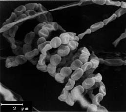
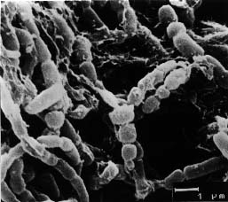
Cultural characteristics of strain KS3-5 were compared on the basis of observations made after 7, 14, and 21 days incubation on Czapek-Dox agar, nutrient agar, Sabouraud agar, and ISP media (Shirling and Gottlieb, 1966). Morphology was examined by light microscopy and scanning electron microscopy (Zeiss DSM model 950).

Physiological Characterization

Utilization of carbohydrates was investigated with a basal carbon nutrient medium (Pridham and Gottlieb, 1948; Waksman, 1967). Methods and media used for physiological tests were as described by Luedemann and Brodsky (1964), Luedemann (1971), Neyra et al. (1977) and Waksman (1967). All cultures were incubated at 28°C for 10 days, except for the gelatin liquefaction (15°C, 21 days). The assay for enzymatic activity was performed according to Hopwood (1967) and Hopwood and Wright (1973). The cultural broth was tested for its antimicrobial activity using the cup or the paper disc diffusion methods (Wu, 1984).

Cell Chemistry

Determination of the cellwall composition, including A2pm (diaminopimelic acid) isomers, sugars, phospholipids, fatty acids and menaquinones was based upon the methods of Becker et al. (1965), Boone and Pine (1968), Kawamoto et al. (1981), Lechevalier and Lechevalier (1970, 1980), and Pine and Boone (1967).

DNA-DNA Homology Study

The DNA-DNA relatedness was determined by the method of Ezaki et al. (1989).

1Corresponding author.


Botanical Bulletin of Academia Sinica, Vol. 36, 1995

Table 1. Cultural characteristics of strain KS35.

Characteristics

Medium Growth Vegetative Aerial mycelia Spore Soluble mycelia pigment

CzapekDox agar moderate moderate, light brown poor, white poor, white light-pink Glycerol tyrosine agar moderate moderate, brown moderate, light brown poor, white brown

Peptone yeast agar moderate moderate, brown moderate, white moderate, white brown

Oatmeal agar well, spreading abundant, brown abundant, white abundant, white pink

Glycerol asparagine agar well, spreading moderate, light brown abundant, light brown abundant, powdery pink

Yeastmalt extract agar well abundant, light brown abundant, grayish white abundant, white, powdery pink

Starch agar well, elevated moderate, light brown abundant, brown abundant, white, powdery pink

Minimal actinomycetes medium moderate moderate, light brown poor, white poor, white pink

Peptone agar moderate moderate, brown moderate, white poor, white pink

Tryptone yeast glucose agar well, elevated well, brown abundant, white abundant, white, powdery brown

Nutrient agar moderate moderate, brown poor, white poor, white pink

Potato plug moderate moderate, brown moderate, white moderate, white brown

Carrot plug moderate moderate, brown poor poor brown

Results and Discussion

Cultural Characteristics

The cultural characteristics of strain KS35 on various media are presented in Table 1. Strain KS35 grew well on most of the organic and synthetic media tested. Typically, the colonies were elevated, spreading, and covered with white aerial mycelia and spores. Diffused melanoid pigments were sometimes observed.

Morphological Characteristics

The scanning electron micrograph of strain KS3-5 revealed that aerial mycelia were monopodially branched with compact spirals of sporophore. Each spore chain consisted of 10 to 50 grayish white, oblong (0.8_1.0 µm by 1.2_1.5 µm), smooth-surfaced spores formed on a short (1.0 µm in diameter, 2.0 µm in length) conidiophore developed on the terminal of an aerial mycelium (Figure 1). Such smooth spore containing spirals are common to most Streptomyces sp. (Tresner et al., 1961).

Physiological Characteristics

Table 2 lists the physiological properties of strain KS3-5. It is capable of milk coagulation and peptonization, suggesting a proteolytic ability. The negative hemolysis result agreed with the lack of toxicity found in preliminary tests on mice (data not shown). Strain KS3-5 can also produce moderately active enzymes, e.g. a-amylase, gelatinase, protease and agarase (Table 3).

Carbohydrate Utilization

The utilization of various carbohydrates by strain KS3-5 suggests a very narrow pattern of carbon source assimilation (Table 4). Glucose and fructose were utilized well; rhamnose, sucrose, and xylose were poorly utilized, and other carbohydrates were not utilized.

Figure 1. Scanning electron micrograph (×10,000) of a spore chain of strain KS 3-5.

Table 2. Physiological characteristics of strain KS35.

Reaction Medium Response

Gelatin liquefaction gelatin medium _

Starch hydrolysis starch agar +

Milk coagulation litmus milk +++

Milk peptonization litmus milk +++

Nitrate reduction nitrate broth _

Tyrosinase reaction tyrosine agar +

H2S production peptone iron agar +

NaCl tolerance TYG slant with NaCl 3%

Growth temperature TYG slant 4_37°C

Melanin formation PY slant +

Growth on potato plug potato plug +

Growth on carrot plug carrot plug +

Blood hemolysis 5% blood agar _

pH range TYG agar slant pH 611

+: positive; _: negative.


Wu and Chen — Identification of strain KS3-5

Table 3. Enzyme production of strain KS 3-5 and Streptomyces toxytricini.

Enzymes Reaction

Strain KS3-5 S. toxytricini

a-Amylase + ++

Gelatinase + +

Lipase _ _

Pectinase _ _

Protease +/_ +

Agarase ++ ++

Cultures were grown on minimal agar medium (Hopwood, 1967; Hopwood and Wright, 1973) at 28°C for 3 weeks.

properties of S. toxytricini were tested for comparison. Streptomyces toxytricini was distinct from strain KS3-5 in having more-open spirals of spore chains and a red to gray spore mass (Figure 2). Both strains showed similar patterns of carbohydrate utilization and enzyme production (Tables 3 and 4), but S. toxytricini demonstrated a narrower antimicrobial spectrum than did strain KS3-5 (Table 5). The fluorometric, DNA-DNA hybridization experiment indicated a near-100% DNA similarity between strain KS3-5 and S. toxytricini, suggesting a significant genomic relatednesss. Consequently, strain KS3-5 belongs to the Streptomyces sp. and is identical to S. toxytricini.

Table 5. Antimicrobial activities of antibiotics KS35 and Streptomyces toxytricini.

Indicatora Inhibition zone (mm dia.)b

Strain KS3-5 S. toxytricini

Staphyloccus aureus ATCC 25923 + _

Sarcina lutea ATCC 9341 +(9.6) _

Escherichia coli ATCC 10536 + _

Escherichia coli NIHJ + _

Enterobacter coloacae ATCC 23355 + _

Salmonella typhimurium ATCC 14028 + _

Klebsiella pneumoniae ATCC 13883 + _

Bacillus subtilis PCI 219 +(8.9) +(26.5)

Bacillus subtilis ATCC 6633 + +

Bacillus cerius ATCC 11778 +(7.8) _

Pseudomonas aeruginosa _ _

Candida albcans ATCC 10231 _ +/_

Saccharomyces cervisiae ATCC 9763 +(7.4) _

Aspergillus niger _ _

Gibberella fujikuroi _ _

Penicillium digitatum _ _

a Bacteria were incubated on antibiotic medium 1 (Difco) at 37°C for 24 hours. Fungi were incubated on CzapekDox agar, and yeast on sabouraud agar at 28°C for 5 days.

b +: positive; _: negative.

Table 4. Utilization of carbon sources by KS35 and Streptomyces toxytricini.

Carbon source Response

Strain KS3-5 S. toxytricini

No carbon source _ _ Fructose + +

Dglucose + NA

Lglucose + NA

Raffinose _ _

Lrhamnose +/_ _

Salicin _ +/_

Sucrose +/_ +/_

Dxylose +/_ _

+: well utilized; +/_: poorly utilized; _: not utilized; NA: not available.

L-Arabinose, Cellulose, Dulcitol, D-Galactose, Glycerol, i-Inositol, Inulin, Lactose, D-Mannose, D-Maltose, D-Mannitol, Melibiose, Raffinose, S-Sorbitol, and Starch were not used by both KS 35 and Streptomyces toxytricini.

Antimicrobial Activities

Strain KS35 showed a broad antimicrobial spectrum against Gram (+) and (-) bacteria and yeast (Table 5). Many such broad-spectrum antibiotics have been produced by Streptomyces sp. (Korzybski et al., 1967).

Chemotaxonomy

Analysis of the whole-cell hydrolysate of strain KS3-5 showed the presence of a chemotype I cell wall characterized by LLA2pm. No diagnostic sugars were found. The main components of fatty acids were anteios-(anteios-C15:0), iso-(iso-C16:0) and normal (n-C16:0) acids. The menaquinones were MK-9 (H8) and MK-9 (H10). The phospholipid pattern was PII type containing phosphatidylethanolamine. Integration of this information suggests that strain KS3-5 belongs to the genus Streptomyces (Lechevalier, 1989).

Identification

A computerized database was used to compare the biological properties of strain KS3-5 with those of other Streptomyces sp. The results suggest that strain KS3-5 is a streptomycete strongly related to S. toxytricini (M. Tseng, personal communication). Subsequently, some biological

Figure 2. Scanning electron micrograph (×10,000) of aerial mycelia and spore chains of S. toxytricini.


Botanical Bulletin of Academia Sinica, Vol. 36, 1995

position as a criterion in the classification of aerobic actinomycetes. Int. J. Syst. Bacteriol. 20: 435_443.

Lechevalier, M.P. and H.A. Lechevalier. 1980. The chemotaxonomy of actinomycetes. In A. Deitz, and D.W. Thayer (eds.), Actinomycete Taxonomy. Special Publication No. 6, SIM, Arlington, pp. 227_291.

Lechevalier, H.A. 1989. A practical guide to generic identification of actinomycetes. In S.T. Williams, M.E. Sharpe, and J.G. Holt (eds.), Bergey's Manual of Systematic Bacteriology. vol. 4, Williams & Wilkins, Sydney, pp. 2344_2347.

Lemonick, M.D. 1994. The killers all around. Time 37: 40_47.

Luedemann, G.M. 1971. Micromonospora purpureo-chromogenes. Int. J. Syst. Bacteriol. 21: 240_247.

Luedemann, G.M. and B.C. Brodsky. 1964. Taxonomy of gentamicinproducing Micromonospora. Antimicrob. Agents Chemother. 1963: 116_124.

Miyadoh, S. 1993. Research on antibiotic screening in Japan over the last decade: A producing microorganism approach. Actinomycetologica 7: 100_106.

Neyra, C.A., J. Bobereiner, R. Lalande, and R. Knowles. 1977. Denitrification by N2fixing Spirillum lipoferum. Can. J. Microbiol. 23: 300_305.

Pine, L. and C.J. Boone. 1967. Comparative cell wall analyses of morphological forms within the genus Actinomyces. J. Bacteriol. 94: 875_883.

Pridham, T.G. and D. Gottlieb. 1948. The utilization of carbon compounds by some Actinomycetales as an aid for species determination. J. Bacteriol. 56: 107_114.

Shirling, E.B. and D. Gottlieb. 1966. Methods of characterization of Streptomyces species. Int. J. Syst. Bacteriol. 61: 313_340.

Tresner, H.D., M.C. Davies, and E.J. Backus. 1961. Electron microscopy of Streptomyces spore morphology and its role in species differentiation. J. Bacteriol. 81: 70_80.

Waksman, S.A. (ed.). 1967. The Actinomycetes. A Summary of Current Knowledge. Ronald Press Co., New York.

Wu, R.Y. 1984. Studies on the Streptomyces SC4. II. Taxonomic and biological characteristics of Streptomyces strain SC4. Bot. Bull. Acad. Sin. 25: 111_123.

Acknowledgments. This study was supported by Academia Sinica and by grant NSC 770211B00145 from the National Science Council of the Republic of China. The authors are grateful for the assistance of Ms Tseng of the Culture Collection and Research Center, Taiwan for her identification work.

Literature Cited

Becker, B., M.P. Lechevalier, and H.A. Lechevalier. 1965. Chemical composition of cell-wall preparation from strains of various form-genera of aerobic actinomycetes. Appl. Microbiol. 13: 236_243.

Boone, C.J. and L. Pine. 1968. Rapid method for characterization of actinomycetes by cell wall composition. Appl. Microbiol. 16: 279_284.

Bull, A.T., M. Goodfellow, and J.H. Slater. 1992. Biodiversity as a source of innovation in biotechnology. Annu. Rev. Microbiol. 46: 219_252.

Ezaki, T., H. Yasuhiro, and E. Yabuuchi. 1989. Fluorometric deoxyribonucleic acid-deoxyribonucleic acid hybridization in microdilution wells as an alternative to membrane filter hybridization in which radioisotopes are used to determine genetic relatedness among bacterial strains. Int. J. Sys. Bacteriol. 39: 224_229.

Hopwood, D.A. 1967. Genetic analysis and genome structure in Streptomyces coelicolor. Bacteriol. Rev. 31: 373_403.

Hopwood, D.A. and H.M. Wright. 1973. A plasmid of Streptomyces coelicolor carrying a chromosomal locus and its inter-specific transfer. J. Gen. Microbiol. 79: 331_342.

Jaroff, L. 1994. Counterattack: how drugmakers are fighting back. Time. 37: 46.

Kawamoto, I., T. Oka, and T. Nara. 1981. Cell wall composition of Micromonospora olivoasterospora, Micromonospora sagamiensis, and related organisms. J. Bacteriol. 146: 527_534.

Korzybski, T., Z. Kowszyk-Gindifer, and W. Kurylowicz. (eds.). 1967. Antibiotics, Origin, Nature and Properties. Translated by Paryski, E. Pergamon Press, Oxford, pp. 450_715.

Lechevalier, M.P. and H.A. Lechevalier. 1970. Chemical com


Wu and Chen — Identification of strain KS3-5