Bot. Bull. Acad. Sin. (1995) 36: 207-214

Yang et al. — Coat protein genes of phage Cf

Molecular cloning and expression of the coat protein genes of Cf, a filamentous bacteriophage of Xanthomonas campestris pv. citri

Mei-Kwei Yang1,4, Huei-Mei Huang2, Yen-Chun Yang1, and Wei-Chih Su3

1The Graduate School of Biology, Fu-Jen University, Taipei, Taiwan, Republic of China

2Institute of Life Science, National Defense Medical Center, Taipei, Taiwan, Republic of China

3Graduate Institute of Botany, National Taiwan University, Taipei, Taiwan, Republic of China

(Received April 11, 1995; Accepted August 18, 1995)

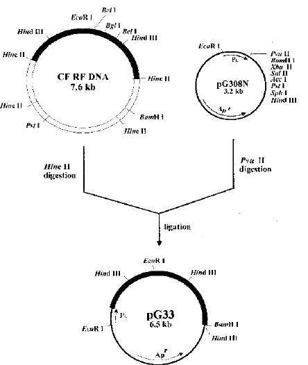
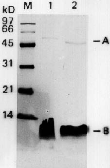
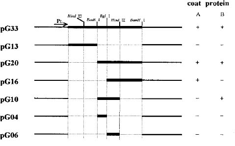
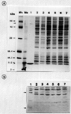
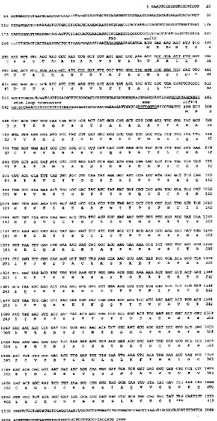
Abstract. Particles of the filamentous bacteriophage Cf contain a major coat protein, the B protein, with a molecular weight of approximately 6,000. In addition, a minor coat protein, the A protein, with a molecular weight of about 50,000, was also identified on sodium dodecyl sulfate-containing polyacrylamide gels. A 3.3 kbp HincII fragment derived from Cf genome was cloned into the expression plasmid pG308N, an E. coli plasmid which carries pL promoter. The recombinant plasmid pG33 and a series of deletion derivatives of pG33 were constructed and transformed into E. coli DG116 for expression of phage Cf genes. The genes coding for A and B proteins of Cf were found on the 2.0 kbp EcoRI-HincII fragment. The complete nucleotide sequences of the 2.0 kbp EcoRI-HincII insert were determined. The deduced amino acid sequence corresponds to a 62-amino acid-residue polypeptide that has a calculated Mr of 6070 was identified as the B protein by SDS/PAGE and immunoblotting. Another open reading frame (ORF419) downstream of the B protein gene (ORF62) was found, and was shown to code for a polypeptide of 419 amino acids with a calculated Mr of 44,676 that exhibits considerable identity to the A protein.

Keywords: Bacteriophage Cf; Coat protein genes; Gene expression; Host specificity; Nucleotide sequences.

Introduction

Cf, Xf, and FLf are filamentous bacteriophages that infect different pathovars of Xanthomonas campestris. All three phages contain a single-stranded circular form DNA surrounded by a long protein coat (Dai et al., 1980; Kuo et al., 1967, 1969; Tseng et al., 1990). Similar to other filamentous phages isolated in Escherichia coli, the infection of these phages results in the releasing of progeny phages into the medium without cell lysis (Hofschneider and Preuss, 1963; Marvin and Hohn, 1969). Although the phages are of comparable genomic size, studies of their host range have revealed a significant difference. Phage Cf, isolated from X. campestris pv. citri, can infect neither X. campestris pv. oryzae nor X. campestris pv. campestris. Another filamentous phage (Xf) isolated from X. campestris pv. oryzae, cannot infect X. campestris pv. citri and X. campestris pv. campestris. We have recently reported, however, that DNAs of Cf and Xf can be transformed into non-host X. campestris strains by electroporation and propagated to accumulate intracellular phage DNA. They then export phage particles at a constant rate (Yang et al., 1991). This demonstrates that adsorption to host cell is responsible for the infectivity of the phage particles. Our interest in the detail of host specificity of phage infection has lead us to examine how Cf adsorbs to X. campestris pv. citri.

The mechanism of adsorption in filamentous phages of E. coli has been inferred from genetic, electron microscopic, and molecular biological studies (Gray et al., 1981; Jacobson, 1972; Rasched and Oberer, 1986; Segawa et al., 1975). It was found that the A protein, specified by gene III of M13, has been implicated in the attachment of the phage to the host receptor (Henry and Pratt, 1969; Pratt et al., 1969), and it may also function in penetration (Goldsmith and Konigsberg, 1977). This adsorption protein comprises a minor fraction of the phage coat and is located only at one end of the phage (Lopez and Webster, 1982; Grant et al., 1981). After infection, the A protein remains with the phage, acting as a pilot protein to guide its DNA into the cell, and converts the single-stranded DNA to the double-stranded replicative form (RF) (Jazwinski et al., 1973; Lin and Bendet, 1976). The A protein also functions as a cut-off agent in the final stage of phage assembly (Crissman and Smith, 1984; Dotto and Zinder, 1983). The adsorption protein of the filamentous phage was found to have both very early and late functions.

Data concerning the structure and function of other coat proteins also exists (Rasched and Oberer, 1986; Simons et al., 1981). B protein encoded by gene VIII is the major protein constituent of the mature phage particles (Henry and Pratt, 1969; Luiten et al., 1983). Within the infected Escherichia coli cell, a large quantities of B protein is synthesized as precursor molecules with amino-terminal signal peptides of 18 to 23 amino acid residues. After insertion of the precursor coat protein into the membrane,

4Corresponding author.


Botanical Bulletin of Academia Sinica, Vol. 36, 1995

coli transformants, LB was supplemented with ampicillin (100 µg/ml) or tetracycline (10 µg/ml).

DNA Manipulation and Cloning

Plasmid DNA and replicative form DNA of the phage was isolated from the infected culture by the alkaline lysis procedure (Birnboim and Doly, 1979) and further purified by ethidium bromide/cesium chloride buoyant density gradient centrifugation. Restriction enzymes obtained from Bethesda Research Laboratories were used according to the manufacture's specifications. Restriction fragments were separated by agarose gel electrophoresis and then recovered from the gel by electroelution. The DNA cloning was performed in E. coli, and appropriate clones were selected as described by Sambrook et al. (1989).

Determination of Nucleotide Sequences

The 2.0 kbp EcoRI-HincII fragment of cf RF DNA was subcloned into M13 mp18 and mp19. DNA from both strands was sequenced by the modified dideoxy chain termination method (Hattori and Sakaki, 1986) by following the instructions of the supplier (U.S. Biochemical Corp.)

Polyacrylamide Gel Electrophoresis and Immunoblotting

Purified phage particles or soluble protein fraction of plasmid-containing strains were separated by electrophoresis through 15% sodium dodecyl sulfate-polyacrylamide gel as described (Weber and Osborn, 1969). Gels were either stained with Coomassie brilliant blue R250 or used for electrotransfer to a nitrocellulose membrane. Antibodies were raised against purified Cf particles in male New Zealand white rabbits by administering four injections, one month apart, of 1×10¹¹ pfu/ml phage plus Freund complete adjuvant into the backs of the rabbits. Approximately one

the amino-terminal peptides are removed by signal peptidase (Ohkawa and Webster, 1981). The mature B protein remains as an internal constituent of the cytoplasmic membrane until it is assembled into a phage particle. Besides these two proteins, two additional minor coat proteins, C protein and D protein, encoded by genes VI, VII, and IX of phage M13 have also been reported (Simons et al., 1981). Although data indicated that these minor coat proteins are located at one or both ends of the phage filament, their biological function is still unclear.

As mentioned above, the mechanism used by coliphage to infect E. coli cell has been well-studied. It appears that F pili of male cells play a major role in the process of filamentous phage infection, but only limited data exists concerning the process of phage adsorption in Xanthomonas campestris. Whether Cf uses F pili or another portion of the cell surface as a receptor has not yet been studied. Unlike other filamentous phages, it was demonstrated that Cf can incorporate its DNA into the genome of its host (Kuo et al., 1987b). The life cycle of filamentous phages isolated from X. campestris may depart significantly from that of the coliphages (Kuo et al., 1987a). To further characterize Cf, and to investigate the mechanism by which it infects host cells, we need to identify genes coding for coat proteins with regard to their biological function. In this communication, we report on polyacrylamide gel studies of Cf, with particular emphasis on the cloning and expression of coat protein genes in E. coli.

Materials and Methods

Bacterial Strains, Bacteriophages and Plasmids

The bacterial strains, bacteriophages, and all plasmids used in this study are described in Table 1. Escherichia coli and Xanthomonas campestris strains were routinely grown at 37°C and 28°C respectively. For selection of E.

Table 1. Bacteria and plasmids.

Strain and plasmid Relevant characterizations Source or reference

Xanthomonas campestris

X. campestris pv. citri Host of phage Cf. Our collection

X. campestris pv. oryzae Host of phage Xf. Our collection

Escherichia coli

DH5a F'lacZ M15recA1 (lacZYA-argF) Sambrook et al., 1989 DG116 F_ endA1 thi-1 hsdR17 supE44 lcI857 Huang et al., 1990

JM103 hsdR4D(lac pro)recA+/F'tra D36 proABlacIqM15 Messing et al., 1981

Plasmids

pG308N An expression vector, containing a pL promoter and the upstream untranslated region Huang et al., 1990

of the N gene of phage lambda; 3.2 kbp.

pUC18 Apr lacZ, 2.7 kbp. Norrander et al., 1983 M13mp18 M13 derivative contains multiple cloning sites. Yanisch et al., 1985

pG33 A 3.3 kbp HincII fragment of phage Cf inserted in the PvuII site of pG308N. This study

pG13 A 1.3 kbp HincII-EcoRI fragment of phage Cf inserted in the PvuII site of pG308N. This study

pG20 A 2.0 kbp EcoRI-HincII fragment of phage Cf inserted in the PvuII site of pG308N. This study

pG04 A 0.4 kbp EcoRI-BglI fragment of pG33 inserted in the PvuII site of pG308N. This study

pG06 A 0.6 kbp BglI-HindIII fragment of pG20 inserted in the PvuII site of pG308N. This study

pG10 Deletion of the 0.96 kbp HindIII fragment of pG20. This study

pG16 A 1.6kbp BglI-BamHI fragment of pG20 inserted in the PvuII site of pG308N. This study


Yang et al. — Coat protein genes of phage Cf

month after the last injection, the rabbits were bled (50 to 60 ml) and antisera were prepared. The anti-cf serum was diluted 100- to 500-fold and used for Western blotting. Immunological detection was performed according to the method of Burnett (1981).

Results

Proteins Present in Virions of the Filamentous Bacteriophage Cf and Xf

To obtain information about the protein composition of the filamentous phages isolated from Xanthomonas campestris, Cf and Xf virions were propagated in X. campestris pv citri and X. campestris pv. oryzae, respectively. The purified virions were analyzed and size-fractionated on sodium dodecyl sulfate-polyacrylamide gels. As shown in Figure 1, it was found that in both cases the phage particles were composed of two different polypeptide chains. The faster-migrating major coat protein that has a molecular weight of about 6,000 was found in Cf and Xf. In addition, a faint band with a molecular weight of about 46,000-50,000 was assigned as a minor coat protein component of the virions. In accordance with the nomenclature of other filamentous phages, the major and minor proteins have been denoted as B protein and A protein, and are encoded by gene VIII and gene III, respectively.

Cloning of Bacteriophage Cf Coat Protein Genes

Previous studies on the location of coat protein genes in Cf indicated that the 3.3 kbp HincII fragment contains the information for Cf A and B proteins (Kao, 1991). We have chosen a cloning strategy that should result in determining the entire nucleotide sequences for the coat protein genes. The 3.3 kbp HincII fragment derived from Cf DNA was cloned into the expression vector pG308N, an E. coli plasmid that carries the controllable pL promoter. The construction of the hybrid plasmid, pG33, is outlined in Figure 2. Cells transformed by this hybrid DNA were selected for growth in the presence of ampicillin. As shown in Figure 3, a series of deletion derivatives of pG33 was obtained by digestion with restriction enzymes and religation.

Expression of Cloned Cf Coat Protein Genes in E. coli

To test the expression of the cloned bacteriophage Cf coat protein genes in E. coli, a culture of E. coli DG116 transformed with plasmid pG33 was grown at 37°C in LB medium and induced by heating at 42°C for 15 min. After induction, cells were collected, washed, and analyzed by SDS-polyacrylamide gel electrophoresis. As shown in Figure 4A, the major products specified by pG33 plasmid DNA comigrated with that specified by intact Cf phage particle. From its electrophoretic mobility, it was identi

Figure 1. Polyacrylamide gel electrophoretic analysis of the coat proteins present in filamentous phages. Purified Cf (lane 1) and Xf (lane 2) phages were subjected to 15% SDS-polyacrylamide gel electrophoresis followed by staining with Coomassie brilliant blue.

Figure 2. Construction of the expression plasmid pG33. The 3.3 kilo base-pair (kbp) HincII fragment of Cf RF DNA was cloned into the PvuII site of pG308N, forming plasmid pG33.


Botanical Bulletin of Academia Sinica, Vol. 36, 1995

fied as Cf A and B proteins, whereas products specified by vector pG308N did not correspond with those of phage Cf. The identity of the supposed Cf coat proteins was further confirmed by means of an immunoblot employing antiserum raised against bacteriophage Cf (Figure 4B).

Among deletion derivatives of pG33, only pG20 gave the same result as pG33. In plasmid pG13, which contains a 1.3 kbp HincII-EcoRI fragment of Cf RF DNA, no protein band was detected. Genes coding for the A and B proteins of Cf were found to be located on the 2.0 kbp EcoRI-HincII fragment (Figure 3). Further deletion was carried out by digesting this DNA fragment with BglI and religating to produce plasmid pG16. Only A protein with Mr of 50,000 was detected. In contrast to this, a protein with Mr of 6,000 was made in cells carrying pG10 (Figure 4C). In cells carrying the deletion plasmids pG04 and pG06, which contain the 400 bp EcoRI-BglI fragment and 600 bp BglI-HindIII fragment, respectively, the synthesis of both A and B proteins can no longer be detected.

Nucleotide Sequence Analysis of the Cloned Coat Protein Genes

The complete nucleotide sequence of the 2.0 kbp EcoRI-HincII insert was determined and is shown in Fig

Figure 3. Structure and expression of pG33 and its derivatives. The 3.3 kbp HincII fragment of Cf RF DNA was inserted into pG308N and introduced into E. coli DG116. We isolated coat protein-expressing, ampicillin-resistant clones that carried pG33. Plasmid pG20, pG16, and pG13 were obtained from pG33 by digesting with HincII and EcoRI, HincII and BglII, and EcoRI and BamHI, respectively, followed by religation. Plasmid pG10, pG04, and pG06 were constructed by ligation of fragments digested by EcoRI and HindIII, EcoRI and BglI, and BglI and HindIII, respectively, with PvuII-cleaved pG308N DNA. The regions derived from Cf RF DNA and pG308N are indicated by darkened lines and open boxes, respectively. Dashed lines represent deletions. The expression of coat proteins was detected by SDS-polyacrylamide gel electrophoresis.

Figure 4. Expression of bacteriophage cf coat protein gene in E. coli cells transformed with recombinant plasmids. A, Samples of whole cells were subjected to SDS-polyacrylamide gel electrophoresis and the products were visualized by Coomassie brilliant blue staining. Lane 1, Cf particle; lane 2, pG33-transformed cells, induced at 42°C for 15 min; lane 3, pG33-transformed cells, not induced with heating; lane 4, E. coli DG116 induced at 42°C for 15 min; lane 5, E. coli DG116 not induced with heating; lane 6, pG308N-transformed cells, induced at 42°C for 15 min; lane 7, pG308N-transformed cells, not induced with heating; M1, high-molecular weight protein-size marker; M2, low-molecular weight protein-size marker. B, Immunoblot of a SDS-polyacrylamide gel employing antiserum raised against bacteriophage Cf. The lanes are as described for A. C, Cells transformed with various pG33 derivatives were also subjected to SDS-polyacrylamide gel electrophoresis and the proteins were stained with Coomassie Brilliant Blue. Lane 1, pG20, induced; lane 2, pG20, not induced; lane 3, pG04, induced; lane 4, pG04, not induced; lane 5, pG06, induced; lane 6, pG06, not induced; lane 7, pG10, induced; lane 8, pG10, not induced; lane 9, pG16, induced; lane 10, pG16, not induced; lane 11, pG308N, induced; lane 12, pG308N, not induced. M1 and M2 are high- and low-molecular weight protein-size markers, respectively. D, Immunoblot of a SDS-polyacrylamide gel employing antiserum raised against bacteriophage Cf. The lanes are as described for C.


Yang et al. — Coat protein genes of phage Cf

ure 5. The deduced amino acid sequences of two open reading frames contained within this sequence are presented below the DNA sequences. Open reading frame 62, starting at position 312 and ending at 498, encodes a polypeptide of 62 amino acids with a calculated Mr of 6,070. This is in good agreement with the Mr of the B protein of Cf, determined by sodium dodecyl sulfate-polyacrylamide gel electrophoresis and identified by

immunoblotting techniques. A putative Shine-Dalgarno ribosome-binding site is located 9 base pairs upstream of the open reading frame 62 (Figure 5). In addition, analysis of the downstream region of ORF 62 showed the presence of putative rho-independent transcription termination signal, which is identical to that of the Cf1c (Kuo et al., 1991). The transcript of this sequence may be characterized by a high potential for forming base-paired stem and loop structure similar to Ff (van Wezenbeek et al., 1980). From these analyses, we conclude that ORF 62 is the real coding sequence of the B protein of phage Cf.

Another open reading frame (ORF419) starts at base pair +663, ten nucleotides downstream from a putative Shine-Dalgarno ribosome binding sequence, TGGTG (Figure 5). The structural part of this gene is 1,257 nucleotides long and encodes a polypeptide of 419 amino acids with a calculated Mr of 44,676. This Mr coincides with the value for the minor protein (the A protein) estimated from SDS-polyacrylamide gel electrophoresis and identified by immunoblot analysis. These findings indicated that ORF419 contains the whole coding sequence of the A protein of phage Cf.

Discussion

In SDS-polyacrylamide gel electrophoresis of proteins from two filamentous phages isolated from Xanthomonas campestris, we found not only the major coat protein but also a minor coat protein. The rapidly moving major virion protein, B protein, was present in greater abundance than was the slower migrating minor coat protein, A protein. According to the reports (Goldsmith and Konigsberg, 1977; Woolford et al., 1977) on other known filamentous bacteriophages, a value of about 2,700 to 3,000 copies of B protein and approximately 5 copies of A protein per f1 and fd virion was estimated. Although the copy number of the A and B protein subunits of Cf had not been determined in the present study, our data presented here shows that B protein is the major constituents of the phage coat. But only one minor coat (A) protein associated with the Cf viral particle was identified in SDS-polyacrylamide gel. On the basis of composition of minor coat proteins in Ff coliphages, two additional capsid proteins were reported, but their presence was not revealed. Since they most probably existed in very small amounts, it was not possible to characterize them as phage-specific proteins. Until the application of radiochemical techniques to identify the appearance of protein, the actual composition of the coat protein in Cf will not be established with certainty.

To gain a better understanding of the biological function of two proteins associated with the Cf particle, molecular cloning techniques were used to study these proteins in more detail. We have shown that coat protein genes (gene III and VIII) of bacteriophage Cf can be cloned and successfully expressed in E coli under the control of the pL promoter in the plasmid pG308N. We determined the nucleotide sequences of a DNA fragment containing two coat protein genes of Cf. Two open read

Figure 5. Nucleotide sequence and derived amino acid sequence of the phage Cf coat protein genes. The nucleotides are numbered above the sequence starting from the ATG initiation codon of the A and B protein. The deduced amino acid sequence is shown below the nucleotide sequence. The putative Shine-Dalgarno sequences and stem-loop terminators are underlined. The amino acids are represented by one-letter symbols. Negatively charged residues are labeled `-', positively charged residues are labeled `+', and the hydrophobic regions are overlined.


Botanical Bulletin of Academia Sinica, Vol. 36, 1995

ing frames, encoding certain proteins of 62 and 419 amino acid residues, respectively, were found. The estimated Mr of these two proteins were in excellent agreement with those of the predicted mature polypeptides B and A. In comparing the amino acid sequences of ORF62 with that of gene VIII of E. coli filamentous phages, no significant homology was found between the two coat proteins. The major coat protein of some other filamentous phages not belonging to the Ff group have been investigated in more detail (Peeters et al., 1985; Putterman et al., 1984; Thomas et al., 1983). The secondary structure of B protein is basically the same for all phages examined, but there are differences in amino acid sequences. As has been pointed out by Nakashima et al. (1974, 1981), the primary structure of major coat (B) protein was segregated into three domains: acidic N-terminus, hydrophobic central part, and basic C-terminal region. This amino acid distribution determines the orientation of the mature B protein in its host cell membrane and provides specific biological function. In phage Cf, similar functional side chains were also found in the major coat protein, the product of ORF 62. The involvement of B protein in phage morphogenesis needs to be investigated.

Study of the major coat protein of the filamentous phages of E. coli and Pseudomonas has shown that B protein is synthesized as a precursor form with an N-terminal signal peptide and is correctly inserted in the membrane (Boeke et al., 1980 ; Greenwood and Perham, 1989; Luiten et al., 1983; William, 1988). The recognition and cleavage of precoat by signal peptidase takes place in the membrane. After processing to mature coat protein, its central hydrophobic region is retained in the membrane bilayer, while the C-terminal part will interact via its basic residues with the negatively charged DNA when progeny phage is assembled (Andreans and Willian, 1986). The data presented in the present study demonstrated the existance of gene coded for B protein of phage Cf, but the primary sequence of B protein was only deduced from nucleotide sequence data. The processing of this protein, although not shown in the present study, will be identified by direct sequence analysis. In an attempt to examine the hydrophilicities of ORF62, we found a striking internal hydrophobic sequence that is preceded by a positively charged amino acid and followed by two negatively charged residues (Figure 5). We suggest that the charged residues halts the passage of the polypeptide through the cytoplasmic membrane. If this interpretation is correct, we propose a similar mechanism of the insertion of the major coat protein of Cf into the cell membrane as Ff phage (Andreas and Willian, 1986 ; Greenwood and Perham, 1989 ; Boeke and Model, 1982; Dotto and Zinder, 1983 ; Boeke et al., 1980).

The product of ORF419 appeared to be the only protein of phage Cf besides the major coat (B) protein. In comparing our results from the amino acid analysis with sequences in the A protein of Ff, no possible homologies were found. These differences may be a result of differences in the bacteriophages, since adsorption protein of

the phage mediates recognition of and attachment to the cell surface of its specific host (Gray et al., 1981; Lopez and Webster, 1982; Segawa et al., 1975). Like the major coat protein, A protein is initially synthesized in a precursor molecules with 18 extra amino acods at its N-terminus (van Wezenbeek et al., 1980). The amino-terminal part of the protein has been shown to attach to the tip of F pilus on E. coli cell, whereas the C-terminal part anchors the protein to the host membrane (Boeke and Model, 1982). A central hydrophobic domain was also found, which may be essential to its aggregation to oligomers. Phage Cf, unlike Ff coliphages that require F pili as the host receptor, attach to certain receptor on the surface of X. campestris pv. citri by unknown mechanism. In analysis of the amino acid sequence that was deduced from the nucleotide sequence of ORF419, no distinct functional domains were found.

Although a substantial amount of information is now available concerning the morphogenesis of filamentous phage coat protein, relatively little is known about how Cf coat proteins are assembled at the molecular level. The data presented here shows that the products of ORF62 and ORF419 are major and minor constituents of the Cf coat. Examination of the sequences flanking these two ORFs reveals a potential regulatory signal for the genes. At 75 base pairs upstream of the start codon ATG of the ORF62 is a region of dyad symmetry capable of folding into a stem-and-loop structure followed by five U residues, if transcribed into RNA. This hairpin structure corresponds to a probable rho-independent transcription terminator. The existence of this terminator may halt the translation of the following gene and result in a very low level of A protein expression. It is of particular interest to investigate the coat protein synthesis in more detail. To this end, examination of the apparent promoter activity of sequences upstream of ORF62 and ORF419 will be carried out. DNA fragments, including upstream sequences of the B protein and A protein genes, will be placed on a promoter probe plasmid and their influence on the expression of reporter gene will be tested.

Acknowledgments. The authors thank Dr. Yu-Shen Chang for valuable advice, generous assistance, and helpful discussions. This research was supported by a grant from the National Science Council, Republic of China (NSC83-0211-B030-004).

Literature Cited

Andreas, K. and W. Willian. 1986. The cytoplasmic carboxyl terminus of M13 precoat is required for the membrane insertion of its central domain. Nature 322: 335_339.

Boeke, J. D., M. Russel, and P. Model. 1980. Processing of filamentous phage pre-coat protein. Effect of sequence variations near the signal peptidase cleavage site. J. Mol. Biol. 144: 103_116.

Boeke, J. D. and P. Model. 1982. A prokaryotic membrane anchor sequence: carboxyl terminus of bacteriophage f1 gene III protein retains it in the membrane. Proc. Natl. Acad. Sci. USA 79: 5200_5204.


Yang et al. — Coat protein genes of phage Cf

Birnboim, H. C. and J. Doly. 1979. A rapid alkaline extraction procedure for screening recombinant plasmid DNA. Nucleic Acids Res. 7: 1513_1523.

Burnett, W.N. 1981. Western blotting: electrophoretic transfer of unmodified nitrocellulose and radiographic detection with antibody and radioiodinated protein A. Anal. Biochem. 112: 195_203.

Crissman, J. and G. Smith. 1984. Gene 3 protein of filamentous phages: evidence for a carboxyl-terminal domain with a role in morphogenesis. Virology 132: 445_455.

Dai, H., K. S. Chiang, and T.T. Kuo. 1980. Characterization of a new filamentous phage Cf from Xanthomonas citri. J. Gen. Virol. 46: 277_289.

Dotto, G. and N. Zinder. 1983. The morphogenetic signal of bacteriophage f1. Virology 130: 252_256.

Goldsmith, M. E. and W. H. Konigsberg. 1977. Adsorption protein of the bacteriophage fd: isolation, molecular properties, and location in the virus. Biochemistry 16: 2686_2694.

Grant, R., T. Lin, W. Konigsberg, and R. Webster. 1981. Structure of the filamentous bacteriophage f1. Location of the A, C, and D minor coat proteins. J. Biol. Chem. 256: 539_546.

Gray, C., R. Brown, and D. Marvin. 1981. Adsorption complex of filamentous fd virus. J. Mol. Biol. 146: 621_627.

Greenwood, J. and R. N. Perham. 1989. Dual importance of positive charge in the C-terminal region of filamentous bacteriophage coat protein for membrane insertion and DNA-protein interaction in virus assembly. Virology 171: 444_452.

Hattori, M. and Y. Sakaki. 1986. Dideoxyl sequencing method using denatured plasmid templates. Anal. Biochem. 152: 232_238.

Henry, T. J. and D. Pratt. 1969. The proteins of Bacteriophage M13. Proc. Natl. Acad. Sci. USA 62: 800_807.

Hofschneider, P. H. and A. Preuss. 1963. M13 bacteriophage liberation from intact bacteria as revealed by electron microscopy. J. Mol. Biol. 7: 450_451.

Huang, L. H., H. F. Tsi, and S.T. Liu. 1990. High level expression of porcine growth hormone in Escherichia coli from an expression vector containing bacteriophage l. PL and N gene untranslated region. Biochem. Biophys. Res. Commun. 173: 711_717.

Jacobson, A. 1972. Role of F pili in the penetration of bacteriophage f1. J. Virol. 10: 835_843.

Jazwinski, S. M., R. Marco, and A. Kornberg. 1973. A coat protein of the bacteriophage M13 virion participates in membrane-oriented synthesis of DNA. Proc. Natl. Acad. Sci. USA 70: 205_209.

Kao, E.T. 1991. Cloning and expression of the filamentous bacteriophage Cf coat protein genes in Escherichia coli. Master thesis. Fu-Jen University.

Kuo, T.T., T. C. Huang, R.Y. Wu, and C. M. Yang. 1967. Characterization of three bacteriophages of Xanthomonas oryzae (Uyeda Et Ishiyama) Dowson. Bot. Bull. Acad. Sin. 8: 246_254.

Kuo, T. T., T. C. Huang, and T. Y. Chow. 1969. A filamentous bacteriophage from Xanthomonas oryzae. Virology 39: 548_555.

Kuo, T.T., Y.H. Lin, C.M. Hung, S.F. Dai, and T.Y. Feng. 1987a. The lysogenic cycle of the filamentous phage Cf1t from Xanthomonas campestris pv. citri. Virology 156: 305_312.

Kuo, T. T., Y. S. Chao, H. Y. Lin, B. Y. Lin, C. F. Liu, and T. Y. Feng. 1987b. Integration of the DNA of filamentous bacteriophage Cf1t into the chomosomal DNA of its host. J. Virol. 61: 60_65.

Kuo, T. T., M. S. Tan, and M. K. Yang. 1991. Complete nucleotide sequence of filamentous phage Cf1t from Xanthomonas campestris pv. citri. Nucleic Acids Res. 19: 2498.

Lin, T. and I. Bendet. 1976. The A protein of bacteriophage fd: its interference with viral infection. Biochem. Biophys. Res. Commun. 72: 369_372.

Lopez, J. and R. Webster. 1982. Minor coat protein composition and location of the A protein in bacteriophage f1 spheroids and I-forms. J. Virol. 42: 1099_1107.

Luiten, R. G. M., J. G. G. Schoenmaker, and R. N. H. Konings. 1983. The major coat protein gene of filamentous Pseudomonas aeruginosa phage pf3: absence of an N-terminal leader signal sequence. Nucleic Acids Res. 11: 8073_8085.

Marvin, D. and B. Hohn. 1969. Filamentous bacterial viruses. Bacteriol. Rev. 33: 172_209.

Marvin, D. and E. Wachtel. 1975. Structure and assembly of filamentous bacterial viruses. Nature (London) 253: 19_23.

Messing, J., R. Crea, and P. H. Seeburg. 1981. A system for shotgun DNA sequencing. Nucleic Acids Res. 9: 309_321.

Nakashima, Y. and W. Koningsberg. 1974. Reinvestigation of a region of the fd bacteriophage coat protein sequence. J. Mol. Biol. 88: 598_600.

Nakashima, Y., B. Frangione, R. Wiseman, and W. Konigsberg. 1981. Primary structure of the major coat protein of the filamentous bacterial viruses If1 and Ike. J. Biol. Chem. 256: 5792_5797.

Norrander, J., T. Kempe, and J. Messing. 1983. Construction of improved M13 vectors using oligodeoxynucleotide-directed mutagenesis. Gene 26: 101_106.

Ohkawa, I. and R. E. Webster. 1981. The orientation of the major coat protein of bacteriophage f1 in the cytoplasmic membrane of Escherichia coli. J. Biol. Chem. 256: 9951_9958.

Peeters, B. P. H., R. M. Peters, J. G. G. Schoenmakers, and R. N. H. Konings. 1985. Nucleotide sequence and genetic organization of the genome of the N-specific filamentous bacteriophage Ike: Comparison with the genome of the F-specific filamentous phages M13, fd and f1. J. Mol. Biol. 181: 27_39.

Pratt, D., H. Tzagoloff, and J. Beaudoin. 1969. Conditional lethal mutants of the small filamentous coliphage M13. II. Two genes for coat proteins. Virology 39: 42_53.

Putterman, D. G., A. Casadevall, P. D. Boyle, H. L. Yang, B. Frangione, and L. A. Day. 1984. Major coat protein and single-stranded DNA binding protein of filamentous virus pf3. Proc. Natl. Acad. Sci. USA 81: 699_703.

Rasched, I. and E. Oberer. 1986. Ff coliphages: structural and functional relationships. Microbiol. Rev. 50: 401_427.

Sambrook, J., E. F. Fritsch, and T. Maniatis. 1989. Molecular cloning: a laboratory manual, 2nd edn. Cold Spring Harbor Laboratory Press, Cold Spring Harbor, N.Y.

Segawa, K., K. Ikehara, and Y. Okada. 1975. Isolation and chemical properties of A-protein from filamentous phage fd. J. Biochem. 78: 1_7.

Simons, G., R. Konings, and J. Schoenmakers. 1981. Gene VI, VII and IX of phage M13 code for minor capsid proteins of the virion. Proc. Natl. Acad. Sci. USA 78: 4194_4198.


Botanical Bulletin of Academia Sinica, Vol. 36, 1995

Thomas, G., B. Prescott, and L. Day. 1983. Structure similarity, difference and variability in the filamentous viruses fd, Ike, If1, pf1 and Xf. Investigation by Laser Raman Spectroscopy. J. Mol. Biol. 165: 321_356.

Tseng, Y. H., M. C. Lo, K. C. Lin, C. C. Pan, and R. Y. Chang. 1990. Characterization of filamentous bacteriophage FLf from Xanthomonas campestris pv. campestris. J. Gen. Virol. 71: 1881_1884.

Van Wezenbeek, P. M. G. F., T. J. M. Hulsebos, and J. G. G. Schoenmakers. 1980. Nucleotide sequence of the filamentous bacteriophage M13 DNA genome: comparison with phage fd. Gene 11: 129_148.

Weber, K. and M. Osborn. 1969. The reliability of molecular weight determinations by dodecyl sulfate polyacrylamide gel electrophoresis. J. Biol. Chem. 244: 4406_4412.

William, W. 1988. Mechanisms of membrane assembly: general lessons from the study of M13 coat protein and Escherichia coli leader peptidase. Biochemistry 27: 1081_1086.

Woolford, J. L., H. M. Steinman, and R. E. Webster. 1977. Adsorption protein of bacteriophage f1 : solubilization in deoxycholate and localization in the f1 virion. Biochemistry 16: 2694_2700.

Yang, M. K., W. C. Su, and T. T. Kuo. 1991. Highly efficient transfection of Xanthomonas campestris by electroporation. Bot. Bull. Acad. Sin. 32: 197_203.

Yanisch-Perron, C., J. Vieira, and J. Messing. 1985. Improved M13 phage cloning vectors and host strains: nucleotide sequences of the M13mp18 and pUC19 vectors. Gene 33: 103_119.