Bot. Bull. Acad. Sin. (1995) 36: 223-228

Lu et al. — Amyloplast preparation from sweet potato callus

Preparation of amyloplasts from sweet potato callus culture

Chi-Hui Lu1, Ping-Du Lee1, and Jong-Ching Su1,2,3

1Department of Agricultural Chemistry, National Taiwan University, Taipei, Taiwan, Republic of China

2Institute of Biological Chemistry, Academia Sinica, Taipei, Taiwan, Republic of China

(Received April 7, 1995; Accepted July 27, 1995)

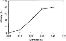
Abstract. Culture conditions for sweet potato callus were manipulated to enhance starch content. Tissue cultures were frozen in liquid nitrogen, pulverized in a ceramic mortar, and ground with a buffer and sea sand. Fractional centrifugation in a discontinuous Percoll gradient enabled separation of amyloplasts in a 40% Percoll layer sandwiched between 20% and 50% layers. A latency test of marker enzymes for amyloplast, and a preliminary feeding experiment with radio-labeled substrates for starch synthesis confirmed that the preparation is suitable for use in the study of starch biosynthesis.

Keywords: Amyloplast; Callus; Ipomoea batatas; Latency test; Starch metabolism; Sweet potato; Tissue culture.

Introduction

Starch synthesis in sink organs occurs in the amyloplasts. The precursor sucrose from the photosynthetic organ must be transformed into appropriate transportable forms to enter the organelle through the amyloplast membrane. Isolated amyloplasts are a desirable research system in which to relate sucrose metabolism in the cytosol to starch synthesis in the amyloplast. To that end, we applied many known methods (Gaynor et al., 1983; McDonald and ap Rees, 1983; Sack et al., 1983; Journet and Dounce, 1985; Macherel et al., 1985; Echeverria et al., 1985; Entwistel and ap Rees, 1988; Mohabir and Hohn, 1988) to tuberous sweet potato root, but were unsuccessful. We noticed that the large size of starch granules in root amyloplasts caused breakdown of the plastid membrane during isolation. To overcome this, we manipulated the culture conditions of sweet potato callus to induce starch synthesis and formation of amyloplasts. We now report our findings.

Materials and Methods

Tissue Culture

Leaves, stems, and young tuberous roots (ranging in size from 1 to 2 cm in diameter) of sweet potato (Ipomoea batatas cv. Tainong 57) were obtained from the University farm just prior to use. The tissue sections for callus initiation were sterilized by immersing in 70% ethanol for 30 sec and in 2.5% NaOCl (containing 1_2 drops of Tween 20 or Triton X-100 wetting agent per 100 ml) for 20 min. Tissue sections (about 5 × 8 × 7 mm) were inoculated on 0.8% agar plates containing 3% sucrose, 2 ppm NAA (1-naphthaleneacetic acid), 0.2 ppm Ki (kinetin), and MS

(Murashige-Skoog) salts and vitamins. Cultures were kept in the dark at 25°C. Callus formation was evident after one week. Subcultures employed MS medium or B5 medium (Gamborg et al., 1982) containing varying concentrations of sucrose, NAA, and Ki. The starch content of tissues was analyzed at three-week intervals. Growth in a culture medium was estimated by the change in dry weight of tissues.

Enzyme Extraction

The callus tissue was mixed with sea sand, frozen in liquid nitrogen, and ground to a powder with a mortar and pestle. One to one-and-a-half times its weight of a 20 mM HEPES buffer (pH 7.8) containing 2 mM KCl, 2 mM CaCl2, 1 mM EDTA, and 1% polyvinylpolypyrrolidone (PVPP) was added and the mixture was homogenized at 2°C for one min. After centrifugation at 10,000 g the supernatant liquid was used for assays of proteins (Bradford, 1976), SP (starch phosphorylase) (DeFekete and Vieweg, 1974), ADPGPPase(ADPG pyrophosphorylase) (Nakamura et al., 1989), PPase (inorganic pyrophosphatase) (Gross and ap Rees, 1986), SS (sucrose synthase) (Su et al., 1977), and amylase (Street, 1974).

Amyloplast Isolation

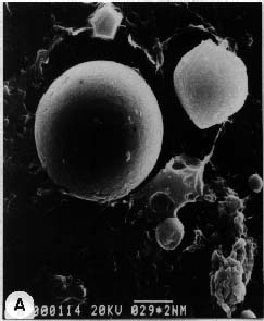
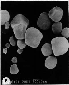
About 50 g of callus was rapidly frozen in liquid nitrogen and pulverized with a mortar and pestle. The powder was slowly added (with gentle agitation, avoiding freezing) to 500 ml of 50 mM HEPES (pH 7.8) buffer containing 5 mM MgCl2, 2 mM EDTA, 0.1% bovine serum albumin, 5 mM DTT (dithiothreitol), 1 mM CaCl2, and 0.3 M mannitol (buffer A). Subsequent operations were performed at 4°C. The suspension was filtered in succession through 300 µm and 64 µm nylon mesh. The filtrate was centrifuged at 190 g for 6 min and the supernatant liquid was discarded. The precipitate was suspended in buffer A, then Percoll was added to 30%. Three milliliters of

3Corresponding author.


Botanical Bulletin of Academia Sinica, Vol. 36, 1995

the sample was then incorporated as the 30% layer in a discontinuous Percoll gradient of 200, 50, 40, 30, 20 and 10%. The volumes of the layers were 0.5, 2, 2, 3, 2, and 2 ml, respectively. After a 20 min centrifugation at 2,270 g, intact amyloplasts in the 40% Percoll layer were collected as the amyloplast fraction (F40). The F40 fraction was washed with buffer A and sedimented by centrifuging at 250 g for 10 min. The pelleted plastids were gently resuspended in a small volume of buffer A, stored in an ice bath, and used on the day of preparation.

Analysis of Marker Enzymes

To determine the integrity and purity of isolated amyloplasts, marker enzymes specific for certain subcellular fractions were analyzed. They were StS (soluble starch synthase), G6PDH (glucose 6-phosphate dehydrogenase), 6PGDH (6-phosphogluconate dehydrogenase) (Fowler and ap Rees, 1970), PGM (phosphoglucomutase), PGI (phosphoglucoisomerase), FBPase (fructose bisphosphatase), ADH (alcohol dehydrogenase), G3PDH (glyceraldehyde 3-phosphate dehydrogenase), aldolase, TA (transaldolase), NiR (nitrite reductase) (Bergmeyer, 1974), GluS (glutamate synthase) (Hecht et al., 1988), fumarase (Stitt, 1989), ADPGPPase, SS, SP, and PPase (loc. cit.). The wholesomeness of amyloplasts was measured by the latency of marker enzymes. Latency (L) is the difference in enzyme activities in ruptured (by adding Triton X-100 to a final concentration of 0.1%, v/v) and intact (I) amyloplasts, relative to that of the ruptured (R) and expressed as percentage—L = [(R-I)/R]×100.

Fluorescence Microscopy

To observe the amyloplast membrane by fluorescent microscopy, we used the method described by Echeverria et al. (1985), using FITC (fluorescein isothiocyanate).

Scanning Electron Microscopy

Purified amyloplasts were fixed in buffer A containing 3% (v/v) glutaraldehyde, for 3 h in the cold and dark. The

fixed amyloplasts were thoroughly washed with 50 mM phosphate buffer and spread over several cover-glasses. When dry, two drops of acetone were added. The acetone-fixed amyloplasts were dehydrated through a 50, 60, 70, 80, 90, 95, and 100% ethanol series (15 min per step), and incubated in isoamyl acetate overnight. After critical point drying and gold casting, samples were examined in a Hitachi S520.

Reagents

All chemicals were reagent grade, obtained from Merck, Wako, or Sigma. Enzymes required for coupled-enzyme assays were from Sigma. Percoll from Sigma (designated 100%) was concentrated by evaporation in a 120°C oven until its volume was reduced to one-half; this product was designated 200% Percoll. Percoll concentration was calculated on the basis of percentile content of 100% Percoll by volume.

Results and Discussion

Optimization of Tissue Culture Conditions

The growth rate and starch content of calli grown under different conditions are shown in Table 1. Since callus amyloplasts could be isolated regardless of how much starch they accumulated, the conditions that gave the maximum yield of starch during a fixed period of culture was deemed optimum—25°C for 3 weeks in MS medium containing 0.15 ppm NAA, 3% sucrose, and 0.8% agar. In the absence of Ki, the starch content of calli diminished during the third week. The addition of 2 ppm Ki sustained starch accumulation, although at a much slower rate (data not shown). Because culturing for an additional week in the presence of 2 ppm Ki did not offer a significant advantage, Ki was excluded from subsequent cultures. It should be noted that under approximately equal growth conditions, a higher ratio of NAA to Ki (2 ppm/0.2 ppm) resulted in a smaller accumulation of starch than did a lower ratio (1 ppm/1 ppm) (data not shown).

Table 1. Effects of culture medium compositions on callus growth and starch accumulation.

Growth ratee Starche H2Oe Mediumc

W4/W1a mg/g fresh wt. %

N 0.15, B5, 3% Su 2.53 0.42 91.3

N 0.15, MS, 3% Su 6.57b 0.66 95.3

Ki 0.2, MS, 3% Su 2.27 0.99 91.7

Ki 2.0, MS, 3% Su 3.51 0.80 94.4

Ki 2, N 0.15, MS, 3% Su 3.31 0.71 94.8

Ki 4, N 0.15, MS, 3% Su 3.26 0.92 92.0

Ki 4, N 0.15, MS, 6% Su 3.28 0.93 92.6

Ki 4, N 0.15, B5, 6% Su 1.57 1.61 89.6

Ki 0.2 , N 2, B5, 3% Su 7.03 N.D.d 94.3

aCallus weight at the 4th week/callus weights at the time of transfer.

bCallus weight at the 3th week/callus weights at the time of transfer.

cN, 1-naphthaleneacetic acid; Ki, kinetin; Su, sucrose. The unit of concentration is ppm.

dNot Detected.

eValues are means of two experiments.


Lu et al. — Amyloplast preparation from sweet potato callus

Table 2. Effects of culture conditions on the compositions of callus cultures.

Medium* Proteinc Starchc H2Oc

mg/g fresh weight mg/g fresh weight %

N 0.15 (D)a 1.26 0.66 95.3

Ki 2.0, N 0.15 (D)a 2.47 0.71 94.8

N 0.15 (L)b 2.20 0.74 94.1

Ki 2, N 0.15 (L)b 2.13 0.70 94.6

aD, in the dark. bL, in the light.

cValues are means of two experiments.

*Calli were grown in MS medium containing 3% sucrose.

Table 3. Effects of callus culture conditions on enzyme activities of carbohydrate metabolism.

Medium* Activityab

SP Amylase ADPGPPase PPase SS

N 0.15 (D) 5.8 0.26 18.7 15.5 16.4

Ki 2.0, N 0.15 (D) 9.4 0.16 8.7 7.7 5.8

N 0.15 (L) 10.5 0.18 13.8 9.0 16.2

Ki 2, N 0.15 (L) 7.6 0.18 12.9 10.1 18.4

aValues are means of two experiments.

bSpecific activities expressed as nmol product produced / min-mg protein.

*Calli were grown in MS medium containing 3% sucrose.

Influence of Light on Callus Culture

When grown under light, the tissue turned green after three weeks and became dark green after three to four subcultures. The greening was not advantageous for starch accumulation (Table 2).

Enzymes of Starch Metabolism in the Starch Accumulating Calli

Crude extracts of the calli were analyzed for enzymes related to starch metabolism. The addition of Ki to the dark-grown callus caused a dramatic decrease of enzyme activities, except for starch phosphorylase (Table 3). The addition of Ki to light-grown calli did not produce this effect. Cultures grown in media without Ki were better suited to our purpose.

Optimum Conditions for Amyloplast Isolation

The investigation disclosed that the tissue homogenization procedure, operational temperature, Percoll-gradient-centrifugation conditions, and buffer solutions were satisfactory. We used light microscopy to estimate the yield of amyloplasts and saw that lower temperature and g values during centrifugation favored a higher yield, but that overly-low values of either or both hindered the sedimentation of amyloplasts. To maintain the integrity of amyloplasts, mannitol was added to control the osmotic conditions. The optimum concentration of mannitol was determined by the latency of PPase. The results (Figure 1) indicated that 0.3 M mannitol in the buffer solution is sufficient to prevent the plastid bodies from bursting.

Properties of Isolated Amyloplasts

Figure 2 shows a scanning electron micrograph of intact amyloplasts (2A) and one of starch granules (2B). The iodine staining for starch and FITC staining for the membrane of an amyloplast preparation are shown in Figures 3A and 3B, respectively. These images show that the majority of visible particles are spherical bodies surrounded by intact membranes. Results of the analysis of some typical marker enzymes and of enzymes of carbohydrate metabolism of the purified amyloplast are presented in Tables 4 and 5, respectively. The preparations were virtually free of contamination by cytosolic and mitochondrial components and they contained enzymes related to various facets of carbohydrate metabolism. The high latency values for these enzymes are consistent with the intact plastid structures seen by light and electron microscopy. We had previously made rather uniform and smooth 8 µm sections of amyloplasts in sweet potato tuberous roots—the organelle diameter was usually greater than 10 µm. We found

Figure 1. Latency of inorganic pyrophosphatase in the sweet potato amyloplasts prepared under different concentration of mannitol. Buffer A was used.


Botanical Bulletin of Academia Sinica, Vol. 36, 1995

Figure 2. Electron micrograph of amyloplasts (A) and starch granules (B) isolated from sweet potato callus.

Figure 3. A, Light micrograph of iodine-staining of purified amyloplasts from sweet potato callus. B, Fluorescence micrograph of purified amyloplasts. Intact amyloplasts show a FITC-FC fluorescence, but starch granules do not.


Lu et al. — Amyloplast preparation from sweet potato callus

Table 4. Enzyme activities of marker enzymes in purified amyloplasts.

Marker Associated cell Activityab Latency

enzyme compartment Intact Ruptured %

6PGDH Amyloplast 1.5 6.2 76

PPase Amyloplast 12.2 33.4 64

ADH Cytosol 0.12 0.20 _

SS Cytosol 0.23 0.21 0

Fumarase Mitochondria N.D. N.D. _

aSpecific activity expressed as nmol products/min-mg protein.

bValues are means of duplicate samples.

N.D., not detected.

*Calli were grown in MS medium containing NAA 0.15 ppm and 3% sucrose.

Chang, T.-C., S.-C. Lee, and J.-C. Su. 1987. Sweet potato starch phosphorylase—Purification and characterization. Agric. Biol. Chem. 51: 187_195.

De Fekete, M. A. R. and G. H. Vieweg. 1974. Über die Aktivität der Synthetase und Phosphorylase im Maisblatt bei verschiedenen Stärkegehalten. Planta (Berlin) 117: 83_91.

Echeverria, E. D., C. Boyer, D. C. Liu, and J. Shannon. 1985. Isolation of amyloplasts from developing maize endosperm. Plant Physiol. 77: 512_519.

Entwistle, G. and T. ap Rees. 1988. Enzymic capacities of amyloplasts from wheat (Triticum aestivum) endosperm. Biochem. J. 255: 391_396.

Fowler, M. W. and T. ap Rees. 1970. Carbohydrate oxidation during differentiation in roots of Pisum sativum. Biochim. Biophys. Acta. 201: 33_44.

Gamborg, K. Z. 1982. Regulation of cell division by auxin in isolated cultures. In P. F. Wareing (ed.), Plant Growth Substances. Academic Press, London, New York, pp. 59_67.

Gaynor, J. J. and A. W. Galston. 1983. Purification and characterization of amyloplasts from etiolated epicotyls of Pisum sativum. Plant Cell Physiol. 24: 411_421.

Gross, P. and T. ap Rees. 1986. Alkaline inorganic pyrophosphatase and starch synthesis in amyloplasts. Planta 167: 140_145.

Hawker, J. S., J. L. Osbund, and J. Preiss. 1972. Unprimed starch synthesis by soluble ADP-Glc-starch glucosyltransferase from potato tubers. Phytochemistry 11: 1287_1293.

Journet, E. P. and R. Douce. 1985. Enzymic capacities of purified cauliflower bud plastids for lipid synthesis and carbohydrate metabolism. Plant Physiol. 79: 458_467.

MacDonald, F. D. and T. ap Rees. 1983. Enzymic properties of amyloplasts from suspension cultures of soybean. Biochim. Biophys. Acta. 755: 81_89.

Macherel, D., H. Kobayashi, T. Akazawa, S. Kawano, and T. Kuroiwa. 1985. Amyloplasts nucleoids in sycamore cells and presence in amyloplast DNA of homologous sequences to chloroplast genes. Biochem. Biophys. Res. Commun. 133: 140_146.

Mohabir, G. and P. John. 1988. Effect of Temperature on starch synthesis in potato tuber tissue and in amyloplasts. Plant Physiol. 88: 1222_1228.

Nakamura, Y., K. Yuki, S. Y. Park, and T. Ohya. 1989. Carbohydrate metabolism in the developing endosperm of rice grains. Plant Cell Physiol. 30: 833_839.

Sack, F. D., D. A. Priestly, and A. C. Leopold. 1983. Surface charge on isolated maize- coleoptile amyloplasts. Planta 157: 511_517.

Stitt, M., R. M. Lilley, T. Gerhardt, and H. W. Heldt. 1989. Metabolite levels in specific cells and subcellular compartments of plant leaves. In S. Fleischer and B. Fleischer (eds.), Methods in Enzymology. Academic Press, New York, 174: 519_564.

Street, H. V. 1974. Amylase. In H. U. Bergmeyer (ed.), Methods of Enzymatic Analysis, pp. 854_858.

Su, J.-C., J.-L. Wu, and C.-L. Yang. 1977. Purification and properties of sucrose synthase from the shoot of bamboo Leleba oldhami. Plant Physiol. 60: 17_21.

Table 5. Enzyme activities of carbohydrate metabolism in purified amyloplasts from sweet potato callus*.

Enzyme Activityab Latency

Intact Rupturedc %

Starch synthesis St.S 1.3 8.2 83 SP 0.5 1.3 62 ADPGPPase 0.6 1.9 68

Glycolysis PGM 30.7 48.3 36 PGI 6.5 13.5 52 Aldolase 2.6 10.1 74 GAPDH 11.5 24.6 59

Pentose-P pathway G6PDH 0.3 2.6 88 6PGDH 1.5 6.2 76 TA 2.6 1.3 -

aSpecific activity expressed as nmol products/min-mg protein.

bValues are means of two experiments.

cRupturing of amyloplasts was done by adding 0.1% (v/v) Triton X-100.

*Calli were grown in MS medium containing NAA 0.15 ppm and 3% sucrose.

that 1_3 starch granules filled an organelle, which indicates that the diameter of starch granules was close to 10 µm (Chang et al., 1986). The largest-diameter starch granule shown in Figure 2B is 6 µm. We conclude that the manipulation of tissue culture conditions to obtain amyloplasts containing small starch granules is the key to preparing sweet potato amyloplasts suitable for experiments. How the plastid body so induced in the tissue culture can withstand the drastic treatment of freezing in liquid nitrogen followed by pulverizing in a mortar is an intriguing question.

Acknowledgment. The authors thank the National Science Council of the Republic of China for their financial support.

Literature Cited

Bradford, M. M. 1976. A rapid and sensitive method for the quantitation of microgram quantities of protein utilizing the principle of protein-dye binding. Anal. Biochem. 72: 248_254.


Botanical Bulletin of Academia Sinica, Vol. 36, 1995