Bot. Bull. Acad. Sin. (1995) 36: 235-241

Chen and Chien — Some chytrids of Taiwan

Some chytrids of Taiwan (I)

Shu-Fen Chen and Chiu-Yuan Chien

Institute of Biological Sciences, National Taiwan Normal University, Taipei, Taiwan 117, Republic of China

(Received April 28, 1995; Accepted July 18, 1995)

Abstract. This paper describes ten species of monocentric chytrids that were isolated and purified. They are Rhizophydium chaetiferum, R. haynaldii, R. laterale, Rhizophlyctis variabilis, R. mastigotrichis, Phlyctochytrium planicorne, Chytriomyces hyalinus, Catenochytridium carolinianum, Allochytridium expandens, and Entophlyctis confervae-glomeratae. Except for Phlyctochytrium planicorne, all species are new to Taiwan.

Keywords: Chytridiales; Chytrids; Taiwan.

Introduction

There have been few studies of the fungal flora of Chytridiales in Taiwan (Sawada, 1919, 1922, 1943; Volz et al., 1976; Konno, 1984). Recently, Hsu (1992) described 7 species from pure cultures. Wang and Chien (1992) recorded 4 species. The present investigation has been in progress since July 1992. Ten species, including 3 species of Rhizophydium, 2 species of Rhizophlyctis, and 1 species each of the following genera: Phlyctochytrium, Chytriomyces, Catenochytridium, Allochytridium and Entophlyctis, were isolated and purified. They are described in this paper.

Materials and Methods

A baiting technique (Fuller and Jaworski, 1987; Sparrow, 1960) was used for isolation. Samples of fresh water collected from ponds, rivers, and lakes were brought to the laboratory. Each sample was divided into three subsamples, which were placed in separate petri dishes. To each subsample was added one of the following baits: pine pollen, grass leaves, and snake skin. All subsamples were incubated at 20°C. Baits were examined every day for about three weeks. The 1/4 Emerson's YpSs agar (containing 250 ppm penicillin G and 250 ppm streptomycin sulfate) was used to isolate the organisms. All isolates were subcultured until completely purified. Axenic cultures were kept on agar slants in screw-cap tubes. Cultures were stored at 10°C and transferred every three months. All specimens have been deposited at the mycology laboratory of the Institute of Biological Sciences, National Taiwan Normal University, Taipei, Taiwan, ROC.

Sparrow's `Aquatic Phycomycetes' (1960) and Karling's `Chytridiomycetarum Iconographia' (1977) were used as references for identification.

Species Descriptions

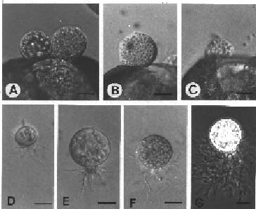
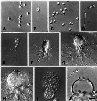
Rhizophydium chaetiferum Sparrow, Occ. Papers Boston Soc. Nat. Hist. 8: 295, 1937. (Figure 1)

On pine pollen: Sporangium epibiotic, spherical, 15_17 µm in diameter, the upper two-thirds of the wall with long, branched or unbranched hairs. After discharged, sporangium formed widening aperture.

On 1/4 YpSs agar: Young sporangium without hairs, mature sporangium with long, branched or unbranched hairs. Rhizoidal system delicate, arising from one thread-like main axis. Zoospores spherical 3_4 µm in diameter, escaping upon the deliquescence of the vesicle, amoeboid movement when water was limited. Resting spore spherical or subspherical. Color of colony : buff to brown.

Specimens examined. TAIPEI HSIEN: Pinghsi, water from stream, 16 Jul 1992, NTNU 101d; Pitan, water from stream, 31 Jul 1993, NTNU 1401; Peishih, water from creek, 10 Aug 1993, NTNU 1501b; Hsiaowulai, water from stream, 3 Jan 1995, NTNU 2401c; ILAN HSIEN: Shuanglienpyi, water from pond, 3 Oct 1992, NTNU

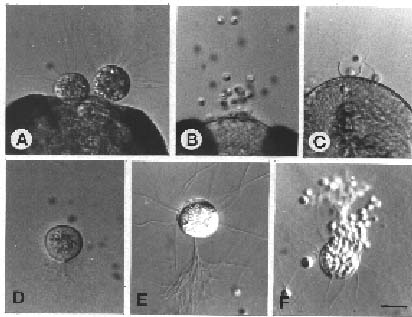
Figure 1. Rhizophydium chaetiferum. A, on pine pollen, sporangia with long hairs; B, on pine pollen, discharging sporangium; C, bowl-shaped empty sporangium; D, young sporangium; E, sporangium with hairs and thread-like rhizoids; F, discharging zoospores. (Bar = 10 µm)


Botanical Bulletin of Academia Sinica, Vol. 36, 1995

601b; Hapen, water from pond, 27 Mar 1993, NTNU 809c; Hapen, water from creek, 27 Mar 1993, NTNU 810; Fushan botanical garden, water from stream, 8 Dec 1993, NTNU 2001b, NTNU 2002a; KAOHSIUNG HSIEN: Chokouhsi, water from stream, 28 Jan1993, NTNU 902b. Isolated on pine pollen from all water samples. This species is newly recorded in Taiwan.

Note. Sporangium with long hairs is the main character. Ubiquitous.

Rhizophydium haynaldii (Schaarschmidt) Fischer, Rabenhorst. Kryptogamen-Fl. 1(4): 92, 1892. (Figure 2)

On pine pollen: Sporangium epibiotic, spherical, 15_40 µm in diameter, or irregularly shaped, with 5_8 papillae or discharge tubes, tubular diameter about 2.5 µm, up to 10 µm long; the wall of tubes remained intact after discharge. Zoospores 2.5_3.7 µm in diameter, individually released over 2 to 5 min period and becoming motile about 30 s after release, movement hopping. Resting spore spherical, 6_15 µm diameter, thick-walled.

On 1/4YpSs agar: Small sporangia varied in shape, whereas large sporangia from spherical to subspherical, papillae protruding. Rhizoids fine, arising from one to three axes on sporangium base or from knob-like extension of sporangium base. Color of colony: buff to off-white.

Figure 3. Rhizophydium laterale. A and B, on pine pollen, mature sporangia; C, on pine pollen, discharging zoospores; D and E, young sporangium; F, sporangium with 3 papillae; G, peg-like structure beneath the sporangium. (Bar = 10 µm)

Specimen examined. NANTOU HSIEN: Puli, waterfall, 20 July 1992, NTNU 201a. Isolated on pine pollen from water. This species is newly recorded in Taiwan.

Note. Papillae protruding is the main character. Sporangial base with axes or knob-like extension. The characters are similar to Barr's (1973) description.

Rhizophydium laterale (Braun) Rabenhorst, Flora Europaea Algarum 3: 281, 1868. (Figure 3)

On pine pollen: Sporangium epibiotic, occasionally interbiotic, may be spherical, slightly flattened, and oval, with one to three exit papillae. The spherical ones vary from 15_25 µm in diameter. Zoospores oval and tapering as they are emerging from the sporangia; after they have been swimming for some time, they become spherical, 2.5_4 µm in diameter, with a 15_25 µm long flagellum.

On 1/4 YpSs agar: Sporangium spherical, papillae one to three; the knob and peg-like structure beneath the sporangium; extremely fine, delicate thread-like branched rhizoids at the tip of the haustorium. Color of colony: distinctly yellow.

Specimen examined. TAIPEI CITY: Botanical garden, lake, 7 Aug 1992, NTNU 401a. Isolated on pine pollen from water. This species is newly recorded in Taiwan.

Note. The characters are similar to Karling's (1938) and Barr's (1973) descriptions.

Rhizophlyctis variabilis Karling, Sydowia 20: 104_105, 1968. (Figure 4)

On pine pollen: Sporangium epibiotic, subspherical, 30_70 µm in diameter, with 1_5 discharge papillae, papilla broad to 15 µm, gelatinous material within papilla,

Figure 2. Rhizophydium haynaldii. A, on pine pollen, sporangia with long discharge tubes; B and C, on pine pollen, empty sporangia with numerous tubes; D, on pine pollen, a resting spore; E, F, and G, young sporangium with large vacuole and 1 to 3 rhizoidal axes; H and I, large sporangium with several papillae or tubes; J, mature sporangium; K, individual zoospore escapes from pore (arrow). (Bar = 10 µm)


Chen and Chien — Some chytrids of Taiwan

22.5 µm in diameter. Rhizoids arising from three to several parts of the sporangium base. Zoospores spherical, 4_5 µm in diameter, with a 15_25 µm long flagellum; some zoospores larger—7.5_12.5 µm, with 2_4 flagella; with a large refractive globule; emerging in a spherical vesicle, soon become active and swim away or display amoeboid action. Resting spore not observed. Color of colony: white to buff.

Specimen examined. TAIPEI CITY: Chingnien garden, lake, 18 Apr 1993, NTNU 1201a. Isolated on pine pollen from water. This species is newly recorded in Taiwan.

Note. Shape of sporangia on 1/4YpSs agar is different from that in 1/4YpSs slush and on pine pollen. Size of sporangia is variable. Resting spores occasionally observed in our isolates on pine pollen.

Phlyctochytrium planicorne Atkinson, Bot. Gaz. 48: 337, 1909. (Figure 6)

On pine pollen: Sporangium epibiotic, ovoid or pyriform, variable in size, 20_50 µm high, 15_58 µm in diameter, bearing a collarette of four (rarely 6) plain teeth around the discharge papilla.

On 1/4YpSs agar: Sporangium subspherical or flask-shaped 40 × 45_70 × 85 µm, wall smooth, bearing four plain teeth at the apex. Considerable morphological varia

Figure 4. Rhizophlyctis variabilis. A, on pine pollen, sporangium with apical papillae; B, on pine pollen, sporangium with lateral papillae; C, on pine pollen, thick-walled resting spore; D, young sporangium with large vacuole; E, gradually maturing sporangium, with a subapical papilla; F, an empty sporangium with one discharge pore. (Bar = 10 µm)

rhizoids extending between pollen grains. Zoospores spherical, 4_5 µm in diameter, with a golden globule, posterior flagellum 24_25 µm long. Resting spore epibiotic or interbiotic, spherical, thick-walled, non-smooth, 12.5_20 µm in diameter, with a large eccentric oil globule.

On 1/4 YpSs agar: Immature sporangium with a large vacuole, zoospore cleavage begins from center and base; mature sporangium bears a single rhizoidal trunk at base, or massive, multibranched trunks arising from several points from its base, main axis 3.5_5 µm in diameter, branches extending for distance up to 150 µm. Zoospores swarm in a vesicale before dispersing. Color of colony: yellow brown to khaki.

Specimen examined. TAIPEI HSIEN: Pinghsi, stream, 16 Jul 1992, NTNU 101b. Isolated on pine pollen from water. This species is newly recorded in Taiwan.

Note. Sporangial papilla hemiglobose, broad to 15 µm, is the main character. Our isolate differs from Karling's (1968) by the non-smooth, thick-walled resting spore.

Rhizophlyctis mastigotrichis (Nowak.) Fischer, Rabenhorst. Kryptogamen-Fl. 1(4): 121, 1892. (Figure 5)

On pine pollen: Sporangium epibiotic or interbiotic, spherical 25_45 µm, fusiform, or ovoid, 25_35 × 37.5_45 µm, the apex prolonged into a short beak or a long tube, with gelatinous plug in the apical region. Rhizoids frequently from one to two—sometimes several, arising from the lower part of the sporangium, branched or unbranched, extending to several pollen grains. Resting spore spherical.

On 1/4YpSs agar: Sporangium spherical or subspherical, 20_150 µm, with 1_5 exit papillae, 12.5_

Figure 5. Rhizophlyctis mastigotrichis. A and B, interbiotic and epibiotic sporangia among pine pollen; C, interbiotic resting spore; D, young sporangium; E, in 1/4YpSs slush, ovoid sporangium with two rhizoidal axes and apical exit papilla; F, discharging zoospores; G, on 1/4YpSs agar, spherical sporangium with an exit papilla; H and I, zoospores discharge; J, an empty sporangium with two discharge pores. (A_F. Bar = 10 µm, G_J. Bar = 30 µm)


Botanical Bulletin of Academia Sinica, Vol. 36, 1995

ing spores spherical, 10_17.5 µm in diameter, with a smooth, thick, brownish wall, containing a large refractive globule surrounded by several smooth ones.

On 1/4YpSs agar: Sporangium usually spherical, 25_60 µm, operculum apical or subapical, with globular hyaline area below apex, 12_20 µm in diameter. Mature sporangial wall is not smooth. Rhizoids well-developed, main axes 5_17.5 µm in diameter at base, extending for distances in excess of 300 µm. Resting spore develops from the fusion body formed at the point of rhizoidal anastomosis. Color of colony: salmon-colored.

In 1/4YpSs slush: Sporangium ovate, obpyriform 55_62 × 70_80 µm, or spherical 35_65 µm in diameter. Rhizoids much-branched and arising from globular or tubular apophysis, 3_7 µm , or non-apophysate. Empty sporangium exhibits a flap operculum.

Specimens examined. ILAN HSIEN: Shuanglienpyi, water from pond, 23 Feb 1994, NTNU 604b; Hapen, water from pond, 11 Apr 1993, NTNU 811a; Fushan botanical garden, water from stream, 8 Dec 1993, NTNU 2001a; TAIPEI CITY: Chingnien garden, water from lake, 18 Apr 1993, NTNU 1201c; TAIPEI HSIEN: Pinglin, water from stream, 16 Dec 1993, NTNU 2101c; Hsiaowulai, water from stream, 3 Jan 1995, NTNU 2401b. Isolated on pine pollen from all water samples, but NTNU 811a has also been isolated on grass leaves and snail skin. This species is newly recorded in Taiwan.

Figure 6. Phlyctochytrium planicorne. A and B, uniflagellate zoospores; C and D, germination and development of a planospore; E, F, G, and H, successive stages of development of the sporangium with an apophysis or a massive rhizoids; I, zoospores emerge from a vesicle; J, four teeth at the apex of an empty sporangium. (Bar = 10 µm)

tion is shown in the apophysis and the rhizoidal system. Thalli mostly had an apophysis, from fusiform to spherical; from the apophysis the main rhizoidal axis (or axes) diverges. Zoospores spherical 4_5 µm in diameter, with a single refractive globule. On dehiscence a portion of the apex of the sporangium dissolves; zoospores emerge in a vesicle for 3_5 min, then actively swim away or display amoeboid action. Color of colony: buff to off-white.

Specimen examined. TAICHUNG HSIEN: Tungshih wooded land, lake, 6 Sep 1994, NTNU 2303c. Isolated on pine pollen from water. This species had been reported in the north of Taiwan (Wang and Chien, 1992).

Note. Sporangial apical collarette of four plain teeth is a constant feature of this chytrid. This species has been recorded on a variety of filamentous green algae and planktonic algae (Canter, 1961), but our isolate was baited with pine pollen.

Chytriomyces hyalinus Karling, Amer. J. Bot. 32: 363, 1945. (Figure 7)

On pine pollen: Sporangium epibiotic or interbiotic, spherical, 17_28 µm in diameter, or ovoid, operculum apical or subapical, 7.5 µm in diameter. Main axis of rhizoid 5 µm in diameter at base. Zoospores spherical 5_6 µm, contains a small hyaline refractive globule, emerging slowly and swarming in a vesicle one minute before dispersing; soon begin to move and jerk about slightly, and within a few seconds active swimming begins. Rest

Figure 7. Chytriomyces hyalinus. A, uniflagellate zoospores; B and C, young sporangium; D, mature operculate and apophysate sporangium; E, discharging zoospores; F, an empty sporangium with a flap operculum (arrow); G, resting spores on snake skin; H, incipient resting spore (arrow) resulting from rhizoidal anastomosis of two thalli; I, resting spore between two empty contributing thalli. (B, C, and E. Bar = 20 µm; A, D, F_I. Bar = 10 µm)


Chen and Chien — Some chytrids of Taiwan

late. Thallus saprophytic in cellulose substrate and growth is slow. The development of thallus is similar to Berdan's (1941) description. Our isolate differs from Berdan's (1939) by the waved thick-wall of the resting spore.

Allochytridium expandens Salkin, Amer. J. Bot. 57: 649, 1970. (Figure 9)

On 1/4YpSs agar: Sporangium generally irregularly globose, 55_85 µm in diameter, ellipsoidal to sac-like, 50 × 65 µm, with one to three rhizoid axes and a discharge tube. The tip of the tube appears gelatinous and has a wall that is destined to become the operculum. Zoospores are discharged at the end of the discharge tube in a vesicle, and 1_2 min later the zoospores become active and swim away. Actively swimming zoospores are globose, 5_7 µm in diameter, with a lipid globule; soon become amoeboid while still actively swimming. The operculum is conspicuous as a saucer-shaped lid to one side of the discharge tube. The zoospore cyst is spherical; contents of cyst move through the germ tube into the expanding sporangium; the empty cyst remains attached to the wall of the mature sporangium. Rhizoids arise from one or several points on the sporangium, extensively branched and irregularly tapered, often with tightly constricted points to fine ends. Resting spore never observed on agar. Color of colony: yellowish brown.

Specimen examined. ILAN HSIEN: Shunglienpyi, pond, 3 Oct 1992, NTNU601d. Isolated on grass leaves from water. This species is newly recorded in Taiwan.

Note. The thallus development is similar to the Entophlyctis type, saprophytic on a cellulose substrate, similar to Salkin's (1970) and Barr's (1986) descriptions. Resting spores are formed in older liquid 1/4YpSs culture, about 30 µm in diameter, with a smooth, thick wall.

Figure 8. Catenochytridium carolinianum. A, B, and C, successive stages in the development of a compound apophysis; D, elliptical-shaped sporangium; E, operculum (arrow) hinged to the empty sporangium; F and G, empty operculate sporangium. (Bar = 20 µm)

Note. Counter to Karlings report (1945) that this species is chitinophyllic, we collected it from a variety of baits and easily isolated and cultured it. Sporangium is operculate, but as the zoospores emerged, the low inconspicuous operculum was pushed off to one side of the sporangium and usually disappeared. Sexual reproduction is by anastomosis of rhizoids of two contributing thalli.

Catenochytridium carolinianum Berdan, Amer. J. Bot. 26: 461, 1939. (Figure 8)

On grass leaf: Sporangium extramatrical, eucarpic, operculate, spherical, ovoid, elliptical-shaped. Apophysis intramatrical, compound, consisting of linear series of constricted, catenulate segments. Rhizoidal system extensive, richly branched. Zoospores emerge in a globular mass and lie quiescent for a moment before swimming away. Resting spore spherical, with thick, wavy wall.

On 1/4YpSs agar: Catenulate segments of apophysis arranged in 1_4 (occasionally 6) linear series attached to the primary apophysate cell. They vary in size and shape. When the apophysis is fully formed, the cytoplasm contains many globules. In normal development, the contents of the rhizoids and apophysis pass out into the sporangium, which is always in an extramatrical position. At maturity, the sporangia are spherical, pyriform, elliptical-shaped, 45_60 µm in diameter. Zoospores spherical, 5_6 µm in diameter. Operculum hinged and persistent. Resting spore never observed in culture. Color of colony: yellowish brown.

Specimen examined. YILAN HSIEN: Shunglienpyi, pond, 3 Oct 1992, NTNU 601g. Isolated on grass leaf from water. This species is newly recorded in Taiwan.

Note. Apophysis catenulate segments is the main character of this species. Sporangial shape is variable, opercu

Figure 9. Allochytridium expandens. A, zoospores; B, germ tube enlarging locally to form the rudiment of the sporangium (arrow); C and D, various stages in the secondary pathway of development of the sporangium directly from the enlargement of the planospore cyst; E, mature sporangium, tip of the discharge tube appears gelatinous with operculate wall (arrow); F, vesicular zoospores releasing; G and H, various views of opercula (arrows). (A and B. Bar = 10 µm; C_H. Bar = 20 µm)


Botanical Bulletin of Academia Sinica, Vol. 36, 1995

Literature Cited

Barr, D. J. S. 1971. Morphology and taxonomy of Entophlyctis confervae-glomeratae (Chytridiales) and related species. Can. J. Bot. 49: 2215_2222.

Barr, D. J. S. 1973. Six Rhizophydium species (Chytridiales) in culture. Can. J. Bot. 51: 967_975.

Barr, D. J. S. 1986. Allochytridium expandens rediscovered : morphology, physiology and zoospore ultrastructure. Mycologia 78(3): 439_448.

Berdan, H. B. 1939. Two new genera of operculate chytrids. Amer. J. Bot. 26(7): 459_463.

Berdan, H. B. 1941. A developmental study of three saprophytic chytrids. II. Catenochytridium carolinianum Berdan. Amer. J. Bot. 28: 439_448.

Canter, H. M. 1961. Studies on British Chytrids XVII. Species occurring on planktonic desmids. Trans. Brit. mycol. Soc. 44(2):163_176.

Fuller, M. S. and A. Jaworski. 1987. Zoosporic fungi in teaching and research. Southeastern Pub. Co., Athens, Georgia, 303 pp.

Hsu, M.-L. 1992. Morphological and taxonomic studies on Chytridiales in Yangmingshan National Park. Master Thesis, Institute of Biological Science, National Taiwan Normal University, Taipei, Taiwan, 95 pp.

Karling, J. S. 1931. Studies in the Chytridiales V. A further study of species of the genus Entophlyctis. Amer. J. Bot. 18: 443_464.

Karling, J. S. 1938. Studies on Rhizophydium II. Rhizophydium laterale. Bull. Torrey. Bot. Club 65: 615_624.

Karling, J. S. 1945. Brazilian chytrids. VI. Rhopalophlyctis and Chytriomyces, two new chitinophyllic operculate genera. Amer. J. Bot. 32: 362_369.

Karling, J. S. 1968. Some zoosporic fungi of New Zealand. V. Species of Asterophlyctis, Obelidium, Rhizoclosmatium, Riphonaria and Rhizophlyctis. Sydowia 20: 96_108.

Karling, J. S. 1977. Chytridiomycetarum Iconogrophia. Luberecht J. Cramer. Monticello, New York, 414 pp.

Konno, K. 1984. Water moulds from Taiwan. Bull. Natl. Sci. Mus., Tokyo, Ser. B. 10(2): 87_99.

Salkin, I. F. 1970. Allochytridium expandens, gen. et sp. N. : Growth and morphology in continuous culture. Amer. J. Bot. 57(6): 649_658.

Sawada, K. 1919. Descriptive Catalogue of the Formosan Fungi I. Taiwan Agric. Exp. St. Spec. Bull., 695 pp.

Sawada, K. 1922. Descriptive Catalogue of Taiwan (Formosan) Fungi. Pt. II. Spec. Publ. Coll. Agric., Natl. Taiwan Univ., 173 pp.

Sawada, K. 1943. Descriptive Catalogue of Taiwan (Formosan) Fungi. Pt. IX. Spec. Publ. Coll. Agric., Natl. Taiwan Univ., 177 pp.

Sparrow, F. K. Jr. 1960. Aquatic Phycomycetes. 2nd ed. The University of Michigan Press, 1187 pp.

Volz, P. A., Y. C. Hsu, and C. H. Lin. 1976. The Thraustochytriaceae and other intertidal fungi of Taiwan. Taiwania 21(2):1_5.

Wang, P.-H. and C.-Y. Chien. 1992. Application of chytrids as a medium for mycological teaching. Biol. Bull. Natl. Taiwan Norm. Univ. 27: 35_43.

Figure 10. Entophlyctis confervae-glomeratae. A, zoospores; B and C, the tip of the germ tube beginning to enlarge; D and E, the spore and germ tube have collapsed, with or without the discharge tube; F, a mature sporangium in which cytoplasm is cleaved into zoospores; G, some zoospores remaining in the sporangia after zoospore discharge. (Bar = 10 µm)

Entophlyctis confervae-glomeratae (Cienkowski) Sparrow, Aquatic Phycomycetes, p. 258, 1943. (Figure 10)

On 1/4YpSs agar: Growth is not as good as in 1/4YpSs slush. Color of colony: white.

In 1/4YpSs slush: Sporangium formed from the expansion of the germ tube of the encysted zoospore, spherical 12.5_30 µm in diameter, somewhat oval, wall thin, smooth. Rhizoids arising from one, occasionally several, axes on the sporangium base, simple or extensively branched. Zoospores formed within the sporangium and discharged through one (or two) tube; discharge tube varying in length from a nearly sessile protuberance to 66.5 µm long. Zoospores spherical, 5 µm in diameter, with a conspicuous eccentric colorless globule, emerge singly from the discharge tube with amoeboid movement.

Specimen examined. KAOHSIUNG HSIEN: Chokouhsi, stream, 28 Jan 1993, NTNU902a. Isolated on pine pollen from water. This species is newly recorded in Taiwan.

Note. Cyst is not a functional part of the mature thallus, thin-walled and devoid of protoplasm. Rhizoids emerge typically from a main basal axis formed on the sporangium base. Karling (1931) and Barr (1971) suggested E. cienkowskiana, and E. heliomorpha (Dang.) Fischer are probably synonymous with E. confervae-glomeratae.

Acknowledgments. We thank Dr. H. S. Chang, senior specialist, Institute of Botany, Academia Sinica, Taipei, Taiwan, and Dr. Z. C. Chen, Department of Botany, National Taiwan University for revising this manuscript and for valuable criticism. This study was supported by National Science Council of the Republic of China, grant NSC84-2311-B003-006.


Chen and Chien — Some chytrids of Taiwan