Bot. Bull. Acad. Sin. (1995) 36: 243-246

Chang — Taiwan dematiaceous hyphomycetes

Notes on Taiwan dematiaceous hyphomycetes, some species of the genera Exserticlava, Craspedodidymum and Hermatomyces

H. S. Chang

Institute of Botany, Academia Sinica, Taipei, Taiwan, Republic of China

(Received May 24, 1995; Accepted August 16, 1995)

Abstract. Exserticlava uniseptata, E. globosa, Craspedodidymum proliferans, and Hermatomyces tucumanensis, all newly found in Taiwan, are described and illustrated. Exserticlava vasiformis, a fairly common dematiaceous hyphomycete is also treated.

Keywords: Craspedodidymum; Dematiaceous hyphomycetes; Exserticlava; Hermatomyces; Taiwan.

The following five dematiaceous hyphomycetes were observed on decayed twigs and wood collected from clean streams. Exserticlava unisepata, E. globosa, Craspedodidymum proliferans, and Hermatomyces tucumanensis are newly found in Taiwan. Exserticlava vasiformis, an omnipresent dematiaceous hyphomycete first recorded by Matsushima, is also included in this report on Taiwan fungal flora.

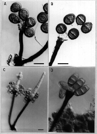
Exserticlava uniseptata Bhat and Sutton, 1985, Trans. Br. Mycol. Soc. 85: 116. Figure 1

Conidiophores macronematous, mononematous, erect, straight or flexuous, cylindrical, smooth, swollen at the apex, dark brown, unbranched, septate, up to 290 µm long, 10_15 µm wide at the base, 5_7.5 µm wide in the middle, 9_13 µm at the apex, percurrently regenerating. Conidiogenous cell formed enteroblastically by growth of the inner wall, which ruptures the outer wall; the latter remaining as tattered fragments attached to the inner conidiogenous wall, terminal, integrated, clavate, polyblastic with several indistinct, unthickened conidiogenous loci. Conidia holobastic, solitary, dry, clavate, broadly rounded at the base, 1-distoseptate, the septum in the lower half of the conidium, medium brown, 14.5_21 µm long, 9.5_15 µm wide at the widest part of upper cell.

Habitat. On unknown decaying twig at Wulai, Taipei county, collected from a stream on Sept. 5, 1994. This fungus was first found in Ethiopia (Bhat and Sutton, 1985)

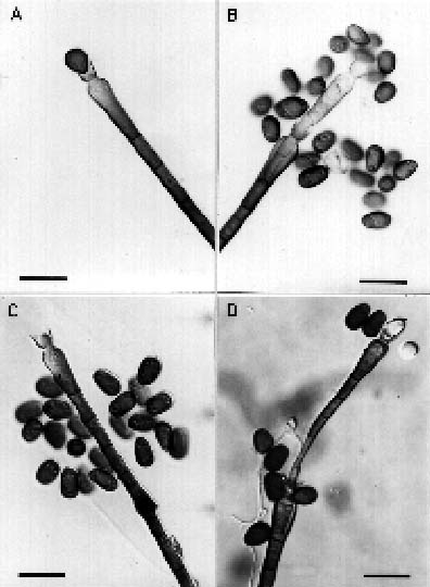
Exserticlava globosa Vasant Rao and de Hoog, 1986. Stud. Mycol. 28: 51_53. Figure 2A_B

Conidiophores macronematous, mononematous, scattered on natural substrate, erect, straight, or flexuous, cylindrical, unbranched, slightly swollen at the base, 2_6 septate, up to 270 µm long, up to 13 µm wide at the base, 9 µm wide in the middle, 20 µm wide at the apex, proliferating percurrently. Conidiogenous cells formed

enteroblastically by growth of the inner wall, which ruptures the outer wall; the latter remaining as tattered fragments attached to the inner conidiogenous wall, terminal, integrated, clavate, polyblastic with several indistinct, unthickened conidiogenous loci. Conidia holoblastic, dry, globose to subglobose, 18_25 × 17_24 µm, with thick, pale brown to brown, and mostly with rather thick median septa, regularly verruculose.

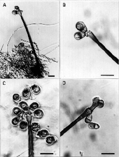
Figure 1. Exserticlava uniseptata. A_C, conidia and conidiophores; D, proliferation of conidiophore. Scale bar = 20 µm.


Botanical Bulletin of Academia Sinica, Vol. 36, 1995

septum. Conidia blastic, broadly ellipsoidal, pale golden brown smooth, thick-walled, 3-distoseptate, 27_32 µm long, 14_19 µm wide, usually produced successively in a cluster of more than 10 on the hyaline extension, which becomes subglobose.

Exserticlava vasformis, a widely distributed dematiaceous hyphomycete has been reported in Japan, New Zealand, North America, Ethiopia, Kenya, India, and Malaysia. Chen (1994) collected this fungus from an unknown dead twig, but with conidia measuring 14_20 × 9_12 µm—smaller than previously reported by Matsushima and by Hughes.

Habitat. On unknown decaying twigs collected on March 15, 1989 at Wulai, Taipei county and on Dec.25, 1991 at Puli, Nantou county. This fungus is a common one. It has been recorded in Japan (Matsushima, 1975), New Zealand (Hughes, 1978), North America (Crane and Schoknecht, 1982), and Kenya (Kirk, 1985).

Craspedodidymum proliferans Vasant Rao and de Hoog, 1986, Stud. Mycol. 28: 64_66. Figure 3

Conidiophores macronematous, mononematous, erect, straight or flexuous, unbranched, septate, mid to dark brown near the base, with the upper part pale olivaceous

Figure 2. Conidia and conidiophores. A_B, Exserticlava globosa; C_D, Exserticlava vasiformis. Scale bar = 20 µm.

Habitat. On unknown decaying twig collected from streams at Fusan Forest Branch Station of Taiwan Provincial Forest Experimental Station, Iilan and in Puli, Nantou county on Jan. 10, 1991 and Nov.1, 1994, respectively. This species were erected and described by Vasant Rao and de Hoog (1986).

Exserticlava vasiformis (Matsushima) Hughes, 1978, New Zealand J. Bot. 16: 332_334. Figure 2C_D

Cordana vasiformis Matshushima, "Icones Microfungoroum a Matsushima lectorum" 1975. Published by the author, Kobe, Japan. p. 40.

Conidiophores macronematous, mononematous, solitary or occasionally in clusters of 2 or 3 , arising from dark brown to black stromata, erect, straight or slightly curved, smooth, septate, thick-walled, up to 270 µm long, somewhat flared and up to 10 µm wide at the apex, swollen at the base up to 12 µm, and 8 µm at the middle part, dark brown to black toward the base, paler towards the apex, terminating in a conidiogenous cell. Conidiogenous cells integrated, pale brown to brown, funnel shaped, initially rounded at the apex, subsequently the pigmented outer wall of the conidiogenous cell ruptures and is left behind as a funnel-shaped structure as the hyaline inner wall layer expands outwards and finally grows upwards as a hyaline, thick-walled cone-shaped extension with more than one

Figure 3. Craspedodidymum proliferans. A_D, conidia and conidiophores, and proliferation of conidiophores. Scale bar = 20 µm.


Chang — Taiwan dematiaceous hyphomycetes

Conidiogenous cells monoblastic, integrated, terminal, determinate, 5_16 × 2.5_4 µm. Conidia solitary acrogenous, lenticular, elliptical to almost round in one plane, smooth, muriform, with pale peripheral cells surrounding central dark brown to black cells, 31_42 × 16_32 µm. Quadrilateral-form conidia (Matsushima, 1993), which Hughes (1953) described as paraphyses, rectangular, usually composed of 3_4 × 2 cells, hayline, but patched with irregularly pigmentated on upper or lower cells, 31_46 × 17_28 µm.

Hughes (1953) gave detailed account of this fungus and another species, H. sphaericum. Vasant Rao and de Hoog (1986) erected another new species, H. dimorphus, which is actually very similar to H. tucumanensis.

Habitat. On unknown decaying twig collected from a clean stream at Wuling Agricultural Farm, Taichung county on June 23, 1986 and at Puli, Nantou county on Nov.1, 1994. This fungus recorded in Argentina, Ghana, Sierra Leone (Ellis, 1971), and Peru (Matsushima, 1993)

Literature Cited

Bhat, D. J. and B.C. Sutton. 1985. New and interesting hyphomycetes from Ethiopia. Trans. Br. Mycol. Soc. 85: 107_122.

Chen, J. L. 1994. Taxonomic study of the Hyphomycetes, Deuteromycotina from Taiwan. Ph.D. thesis, National Taiwan University.

Crane, J. L. and J. D. Schoknecht. 1982. Hyphomycetes from freshwater swamps and hammocks. Can. J. Bot. 60: 369_378.

Ellis, M. B. 1971. Dematiaceous Hyphomycetes. Commonwealth Mycological Institute, Kew, Surrey, England.

Hughes, S. J. 1953. Fungi from the Gold Coast II. Mycol. Pap. 50: 1_104.

Hughes, S. J. 1978. New Zealand Fungi 25. Miscellaneous species. New Zealand J. Bot. 16: 311_370.

Kirk, P. M. 1985. New or interesting microfungi XIV. Dematiaceous Hyphomycetes from Mt. Kenya. Mycotaxon 23: 305_352.

Matsushima, T. 1975. "Icones microfungorum a Matsushima lectorum". Published by the author. Kobe, Japan. 209 pp., 415 plates.

Matsushima, T. 1993. Matsushima Mycological Memoirs, No. 7, Mastushima Fungus Collection, Kobe, Japan.

Rao, V. and G. S. de Hoog. 1986. New or Crictical Hyphomycetes from India. Studies in Mycology, No. 28, 84 pp.

Figure 4. Hermatomyces tucumanensis - Dimorphic form conidia. A_B, Hermatomyces form; C_D, quadrilateral form. Scale bar = 20 µm.

brown, smooth. up to 390 µm × 8_10 µm. Conidiogenous cells enteroblastic, monophialidic, percurrent, integrated, terminal, clavate, with a very large and distinct funnel-shaped collarette, 7.5_10 µm high, and 8_12 µm wide at the open end. Conidia ellipsoid to slightly angular and trapezium-shaped, 13_15 × 8 µm, with inconspicuous scar, usually aggregating in slimy heads, smooth and relatively thin-walled, pale brown when young and brown when mature. Our isolates frequently showed percurrent growth of conidiophore through the collarette; usually another phialide opening is formed on new conidiogenous cells. As in the original description, a phialide opening is formed by percurrent growth through the collarette.

Habitat. On unknown decaying twig collected from a stream at Wulai, Taipei county on July 4, 1989 and Feb. 13, 1990. This fungus was first recorded in India by Vasant Rao and de Hoog (1986).

Hermatomyces tucumanensis Spegazzini, 1911, An. Mus. Nac. Hist. Nat. B. Aires, Ser. 3, 13: 446. Figure 4

Conidiophores closely packed together, macronematous, mononematous, unbranched or occasionally forked, short, straight or flexuous, pale brown, smooth.


Botanical Bulletin of Academia Sinica, Vol. 36, 1995