Bot. Bull. Acad. Sin. (1996) 37: 99-105

Chen et al. — Nucleotide sequence of the nifHDK operon

Nucleotide sequence of the nifHDK operon in the aerobic

nitrogen-fixing unicellular Synechococcus RF-1

Horng-Ming Chen1,2, Tan-Chi Huang2,3, and Chiu-Yuan Chien1

1Department of Biology, National Taiwan Normal University, Taipei, Taiwan 117, Republic of China

2Institute of Botany, Academia Sinica, Nankang, Taipei, Taiwan 115, Republic of China

(Received February 5, 1996; Accepted March 19, 1996)

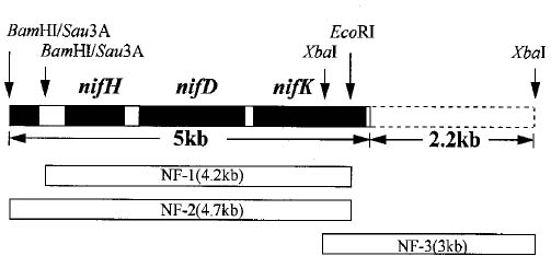
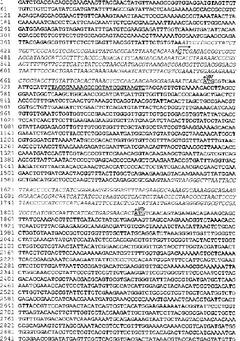
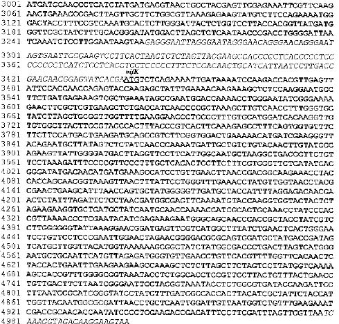
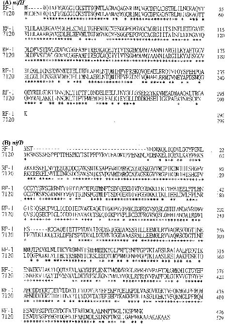
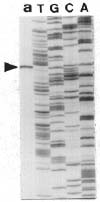
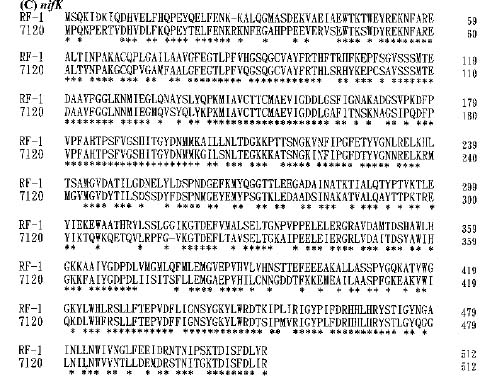
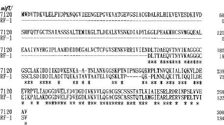
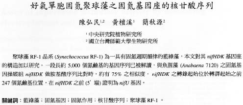
Abstract. A 5 kb nucleotide sequence which included nifHDK operon was determined. The amino acid sequences deduced from the nifH, nifD, and nifK of Synechococcus RF-1 and from Anabaena 7120 were compared. The start site of transcription was located by primer extension analysis. A region of 405 bp just upstream from the 5' end of nifH showed considerable sequence similarity to the nifU sequences of Anabaena 7120.

Keywords: Cyanobacteria; nifHDK operon; Nitrogen fixation; Nucleotide sequence; Synechococcus RF-1.

Introduction

The nitrogenase which catalyzes the reduction of atmospheric nitrogen to ammonia is a complex consisting of three different kinds of polypeptide encoded by nifH, nifD, and nifK, respectively. The molecular structure of nifHDK operon has been analyzed in eubacteria, photobacteria, and cyanobacteria. The nucleotide sequence for the coding region of nif genes among different microorganisms is highly conserved. However, the arrangement of nifH, nifD, and nifK is either in a contiguous or noncontiguous array. The nifH, nifD, and nifK in the eubacteria system such as in Klebsiella pneumoniae (Roberts and Brill, 1981), or Rhizobium meliloti (Corbin et al., 1982), and in the non-oxygenic photosynthetic bacteria, such as in Rhodopseudomonas capsulata (currently, Rhodobacter capsulatus), are linked in a single nifHDK operon (Avtges et al., 1983). In the cyanobacteria, two types of arrangement are found. Those in the nonheterocystous anaerobic nitrogen-fixing cyanobacteria such as in the Synechococcus 7335, Synechococcus 7425, or Pseudoanabaena 7409 are linked in a contiguous array (Kallas et al., 1985). However, those in heterocystous cyanobacteria such as in Anabaena 7120, nifH is linked to nifD, whereas nifK is separated from nifD by about 11 kb in the vegetative cells (Mazur et al., 1980). A rearrangement of the nif genes takes place during the initiation of heterocyst development (Golden et al., 1985).

Among the unicellular cyanobacteria, a few isolates are able to fix nitrogen aerobically. They can be morphologically grouped into sheathed and sheathless forms (Waterbury and Rippka, 1989). These aerobic nitrogen-fixing cyanobacteria fix nitrogen almost exclusively during the dark periods when grown in a diurnal 12 h/12 h light-dark regimen. The properties of the nitrogenase activity in Synechococcus RF-1, one of the sheathless aerobic nitro

gen-fixing unicellular cyanobacteria have been well characterized. When the culture was exposed to a diurnal 12 h light/12 h dark (L/D) regimen, the nitrogenase activity exhibited a rhythmic pattern with the nitrogenase activity peak occurring in the dark phase. This rhythmic pattern persisted for several days after the culture was transferred to continuous light (L/L) (Grobbelaar et al., 1986). The nitrogenase activity rhythm was also found to be temperature compensated when the temperature was changed within the physiological range. These results indicate that the nitrogenase activity in Synechococcus RF-1 is controlled by a "circadian clock" (Huang et al., 1990), a phenomenon which does not occur with other nitrogen-fixing microorganisms.

The nucleotide sequence of the nifHDK operon in heterocystous cyanobacteria has been well characterized. However, there have been no reports on the molecular structure of the nif genes in unicellular cyanobacteria. In this report, the nucleotide sequence of nifHDK operon in Synechococcus RF-1 is examined.

Materials and Methods

Isolation of DNA from Synechococcus RF-1

Axenic Synechococcus RF-1 were grown in BG-110 according to the conditions described previously (Chou et al., 1989). Since the surface of Synechococcus RF-1 contained an unknown compound which will interact with lysozyme, the cells collected by centrifugation were washed twice in distilled H2O by vibration with Vortex (Model K-550-G) at the maximum speed for 1 min at room temperature. The cells were transferred to 5 ml 6 M guanidine-HCl and vibrated for 1 min (Votex) at room temperature. The guanidine-HCl treatment was repeated once. The cells were then washed with distilled H2O three times to remove the guanidine-HCl and then suspended in 10 mM Tris-EDTA buffer (TE), pH 8.0. Lysozyme was added to have a final concentration of 10 mg/ml. The cells were then in

3Corresponding author. Fax: 886-2-7827954.


Botanical Bulletin of Academia Sinica, Vol. 37, 1996

cubated at 37°C with gentle shaking (100 rpm/min) for 1 h. The cells were collected by centrifugation and then resuspended in solution containing 2% SDS, 0.05 M EDTA, and 20 µg/ml RNase A. The cell suspension was incubated at 37°C for 1 h. Proteinase K was added after the incubation to a final concentration of 100 µg/ml followed by re-incubation at 60°C for 1 h. The final suspension was extracted gently with phenol:chloroform (1:1 v/v). The phenol contaminated in the DNA containing aqueous phase was removed by chloroform: isoamylalcohol (24:1 v/v). DNA in the liquid phase was precipitated with 2 times the volume of ethanol (-20°C) in the presence of 0.2 M NaCl. The precipitated DNA was collected with a glass stirring rod and then solubilized in TE buffer.

Construction of a Genomic Library

A genomic DNA library was prepared using DNA from Synechococcus RF-1. Genomic DNA was partially digested with Sau3A, and the DNA fragments with sizes from 10 to 23 kb were isolated from low melting-point agarose (Sambrook et al., 1989). The size-fractionated fragments were ligated into BamH1-digested EMBL4 according to the protocol provided by Stratagene Company, La Jolla, California, USA.

Cloning of the nif Genes

The genomic library of Synechococcus RF-1 prepared in l EMBL4 from a partial Sau3A digest was used for the nif gene cloning. Plasmids pBR322 containing nifH or nifK cloned from Anabaena (Rice et al., 1982) were used as probes for cloning the nif genes. The nifH or nifK probes were labeled with the digoxigenin using the DIG luminescent detection kit according to the instructions of the manufacturer Boehringer Mannheim. About 3,000 recombinant plaques of the genomic library were immobilized on nylon membranes (Micron Separations Inc.) and pretreated according to the procedures described by Sambrook et al. (1989). The membranes were hybridized with the digoxigenin (DIG)-labeled nifH or nifK probe according to the instructions of the manufacturer Boehringer Mannheim. Positive plaques were purified and amplified for DNA extraction.

Nucleotide Sequencing

Restriction fragments containing the nif gene were subcloned into pGEM7(-). The nucleotide sequence of both strands of the plasmid DNA was determined by dideoxy chain termination on an A.L.F. Pharmacia automated DNA sequencer according to the manufacturer's protocols. Sequencing of the double-stranded DNA was first carried out with the universal sequencing primer using the Auto Read Sequencing Kit (Pharmacia-LKB) following the supplier's instructions. Oligonucleotide primers were utilized to complete sequencing of both strands. The extent of identity of the derived amino acid sequence of Synechococcus nif genes to those encoded by other nif genes was searched for in the PC Gene, Intelli Genetics.

Isolation of Total RNA

Exponentially growing cells (about 2×10⁷ cells/ml) of Synechococcus RF-1 were collected by centrifugation, washed with 1 ml of distilled water, and then collected in a 1.5 ml screw-top vial (about 6×10⁸ cells per vial). Teflon-coated sea-sand (0.1 g; 0.1~0.3 mm diameter; Merck), 0.5 ml solution D (4 M guanidine thiocyanate, 25 mM sodium citrate, 0.5% sodium lauryl sarcosinate, and 0.72% mercaptoethanol), 0.5 ml water saturated hot phenol solution (80°C), 0.1 ml 2 M sodium acetate (pH 4.0), and 0.1 ml Chloroform-isoamyl alcohol (24:1) were added. The cells were broken by vibration with a Mini-Beadbeater (Biospec, USA) set at high speed for 50 sec followed by 300 sec at low speed. The broken cells were cooled in ice water for 5 min and then centrifuged at 13,000 g for 5 min. The supernatant was collected and an equal volume of absolute ethanol was added. The total RNA was collected after cooling at -70°C for more than 1 h, and then suspended in 0.3 ml diethyl pyrocarbonate (DEPC)-pretreated water in the presence of 10% (v/v) 3 M sodium acetate (pH 4.8). The RNA solution was repeatedly extracted with an equal volume of phenol-chloroform (1:1). The RNA containing aqueous phase was collected and maintained at -70°C after adding absolute ethanol (-20°C) in an amount 2.5 times its volume.

Primer Extension Analysis

The RNA prepared according to the procedures described under "Isolation of Total RNA" above was used as template. An oligonucleotide with the sequence 5'GACTTACCGATACCGCCTTTTCCGTAA3', which is complementary to the coding sequence from positions 44 to 18 of the nifH gene (Figure 1), was used as primer. About 5 to 20 µg RNA dissolved in DEPC-pretreated water was mixed with 2 µl primer (10 µg/ml) in a 1.5 ml Eppendorf tube. The mixture was kept in a 65°C water bath for 10 min and then reset at 42°C. After the temperature had decreased from 65°C to 42°C naturally (about 30 min), 1.5 µl of 10xRT buffer (reverse transcription buffer, Stratagene), 2 µl of diluted dNTP mix (Sequenase Version 2.0 DNA sequencing kit, United States Biochemical), 2

Figure 1. Schematic representation of a 7.2 kb locus of the Synechococcus RF-1 chromosome containing the nifHDK operon. The locations of the DNA fragment from the subcloned recombinants (NF-1, NF-2, and NF-3) used for the sequence analysis are shown by open bars. The 5 kb region was sequenced (Figure 2). The unsequenced downstream region (2.2 kb) is indicated by the dashed line.


Chen et al. — Nucleotide sequence of the nifHDK operon


Botanical Bulletin of Academia Sinica, Vol. 37, 1996

Figure 2. The 5 kb nucleatide sequence containing the nifHDK operon of the Synechococcus RF-1 Chromosome (Gen Bank U 22146). The open reading frame is represented by bold characters, and the non-coding region is shown in italics. The start point (underlinded G) of transcription determined by primer extension analysis is marked by an arrow. The start codon for each open reading frame is underlined. The segment from position 729 to 755 selected for primer extension analysis is also underlined.

µl [a-35S]-dATP (10 µCi/µl), 1 µl RNase inhibitor (RNase Block, 25,000_40,000 U/ml, Stratagene), and 1 µl RNase H_ reverse transcriptase (MMLV-RT, 50,000 U/ml, Stratagene) were added. Autoclaved water was added up to a final volume of 14 µl. After 10 min of incubation at 42°C, 1 µl of 25 mM dNTP was added and incubated at 42°C for 1 to 2 h. The reaction was terminated by adding an equal volume of sequencing loading dye (United States Biochemical). For the control experiment, the same synthetic oligonucleotide was used as a primer in dideoxy chain termination DNA sequencing reactions with the nifH ORF as a template. The final reaction mixtures were analysed by 6% DNA sequencing gel.

Results and Discussion

Among the nif gene positive recombinants subcloned from the l EMBL4 genomic library, recombinant NF-1, NF-2, and NF-3 were selected for DNA analysis (Figure 1). The NF-1 and NF-2, both hybridized with probe nifH and nifK, contained a 4.2 kb and 4.7 kb EcoRI fragment respectively. The NF-3 contained a 3 kb Xba1 fragment, which hybridized only with probe nifK. Results of DNA sequencing revealed that the 4.7 kb fragment overlaps with the 4.2 kb fragment, and the 4.7 kb fragment has 555 base pairs longer than the 4.2 kb at the 5'-end (Figure 1). The 5'-end of the 3 kb Xba1 fragment has 507 base pairs that


Chen et al. — Nucleotide sequence of the nifHDK operon

overlap with the 3'-end of the 4.7 kb EcoRI fragment. Based on the nucleotide sequence for fragment 4.2 kb, 4.7 kb, and 3.0 kb, and the sequence overlapping between them, a 5 kb segment containing nif gene was constructed (Figure 1).

The nucleotide sequence of the nif genes containing fragment along with its 5'-flanking sequence is given in Figure 2. Putative open reading frame (ORF) in the sequenced region was searched in all three possible translation frames with ATG as the initial codon. Three complete ORFs are present in the constructed nif gene locus. As shown in Figure 2, the first ORF beginning from the position 712 consists of 891 base pairs (bp). It encodes a polypeptide consisting of 296 amino acids (Figure 3A). The predicted amino acid sequence exhibits a high degree of similarity (76.7%) to that of the polypeptide encoded by the nifH from Anabaena 7120 (Mevarech et al., 1980). Therefore, we concluded that the ORF is the nifH from Synechococcus RF-1.

The second ORF is present 230 bp downstream from the termination codon of the nifH (from position 1833 to 3263). The ORF encodes a polypeptide of 476 amino acids (Figure 3B). As shown in Figure 3B, the predicted

Figure 4. Primer extension analysis of the 5' end of nifH gene. Lane a: A 27 oligonucleotide was annealed with total RNA and extended as described under "Materials and Methods". Lanes T,G,C,A: Electrophoretic separation of the products of dideoxynucleotide sequencing.

Figure 3. Comparison of the deduced amino acid sequences of the nifH genes (A), the nifD genes (B), and the nifK genes (C), from Synechococcus RF-1 and Anabaena 7120. The character to show that two aligned residues are identical is "*". Gaps (shown as dashes) have been introduced in the sequences where necessary to give better alignment.

Figure 5. Alignment of amino acid sequences deduced from the nifU gene of Anabaena 7120 and the partial sequence of the nifU of Synechococcus RF-1.


Botanical Bulletin of Academia Sinica, Vol. 37, 1996

amino acid sequence exhibits a high degree of similarity (77.3%) to that of the polypeptide encoded by the nifD from Anabaerna 7120 (Lammers and Haselkorn, 1983; Golden et al., 1985). The third ORF is present 176 bp downstream from the termination codon of the nifD (from position 3440 to 4978). As shown in Figure 3C, the amino acid sequence deduced from the third ORF has a 69% similarity with the polypeptide encoded by the nifK from Anabaena 7120 (Mazur and Chui, 1982). Therefore, we concluded the second and the third ORF correspond to the nifD and nifK of Synechococcus RF-1, respectively.

Transcription of the nifHDK operon of Synechococcus RF-1 was examined previously by Northern analysis (Huang and Chow, 1990). When mRNA from Synechococcus RF-1 was hybridized with nifK probe, a transcript of about 4.5 kb was detected. The size of the transcript corresponds to the size that includes the nifH, nifD, and nifK sequences. When the mRNA was probed with nifH, three bands of the sizes 1.3, 2.8, and 4.5 kb were detected, with the 1.3 kb as the most abundant band. These results indicate that nifH is the first gene of the operon. Primer extension analysis was used to locate the start site of transcription. As shown in Figure 4, a single transcript was detected. The transcription start site lies 247 bp upstream from the start codon of nifH gene (Figure 2).

A nifU gene was shown to be located upstream from the 5' end of the nifH gene from Nostoc muscorum (DeFrancesco and Potts, 1988), Anabaena 7120 (Mulligan and Haselkorn, 1989), and Plectonema boryanum (Fujita et al., 1991). The nifU gene has been suggested to be required for maturation of the MoFe protein of nitrogenase (Orme-Johnson, 1985). A partial ORF (405 bp) located 5' to the nifH gene of Synechococcus RF-1 (Figure 2) was sequenced. As shown in Figure 5, the deduced amino acid sequence (294 residues) of the partial ORF has considerable homology (higher than 64%) to the C-terminal region of the nifU product from Anabaena 7120 (Mulligan and Haselkorn, 1989). The results indicate that the same nifU-nifH order probably occurs in Synechococcus RF-1.

Acknowledgements. We thank Professor R. Haselkorn, University of Chicago, for providing the pAn207.8 and pAn154.3 plasmids. This work was funded by the Academia Sinica and the National Science Council of the Republic of China.

Literature Cited

Avtges, P., P. A. Scolnik, and R. Haselkorn. 1983. Genetic and physical map of the structural genes (nifH, D, K) coding for the nitrogenase complex of Rhodopseudomonas capsulata. J. Bacteriol. 156: 251_256.

Chou, H. M., T. J. Chow, T. Jenn, H. R. Wang, H. C. Chou, and T.C. Huang. 1989. Rhythmic nitrogenase activity of Synechococcus RF-1 established under various light-dark cycles. Bot. Bull. Acad. Sin. 30: 291_296.

Corbin, D., D. Ditta, and D. R. Helinski. 1982. Clustering of nitrogen fixation (nif) genes in Rhizobium meliloti. J. Bacteriol. 149: 221_228.

DeFrancesco, N. and M. Potts. 1988. Cloning of nifHD from Nostoc muscorum UTEX584 and a flanking region homologous to part of the Azotobacter vinelandii nifU gene. J. Bacteriol. 170: 3297_3300.

Fujita, T., Y. Takahashi, F. Shonai, Y. Ogura, and H. Matsubara. 1991. Cloning, nucleotide sequences and differential expression of the nifH and nifH-like (frxC) genes from the filamentous nitrogen-fixing cyanobacterium Plectonema boryanum. Plant Cell Physiol. 32: 1093_1106.

Golden, J. W., S. J. Robinson, and R. Haselkorn. 1985. Rearrangement of nitrogen fixation genes during hetrocyst differentiation in the cyanobacterium Anabaena. Nature 314: 419_423.

Grobbelaar, N., T. C. Huang, H. Y. Lin, and T. J. Chow. 1986. Dinitrogen-fixing endogenous rhythm in Synechococcus RF-1. FEMS Microbiol. Letts. 37: 173_177.

Huang, T. C. and T. J. Chow. 1990. Characterization of the rhythmic nitrogne-fixing activity of Synechococcus sp. RF-1 at the transcription level. Curr. Microbiol. 20: 23_26.

Huang, T. C., J. Tu, T. J. Chow, and T. H. Chen. 1990. Circadian rhythm of the prokaryote Synechococcus sp. RF-1. Plant Physiol. 92: 531_533.

Kallas, T., T. Coursin, and R. Rippka. 1985. Different organization of nif genes in nonheterocystous and heterocystous cyanobacteria. Plant Mol. Biol. 5: 321_329.

Lammers, P. J. and R. Haselkorn. 1983. Sequence of the nifD gene coding for the a subunit of dinitrogenase from the cyanobacterium Anabaena. Proc. Natl. Acad. Sci. USA 80: 4723_4727.

Mazur, B. J. and C. F. Chui. 1982. Sequence of the gene coding for the b-subunit of dinitrogenase from the blue-green alga Anabaena. Proc. Natl. Acad. Sci. USA 79: 6782_6786.

Mazur, B. J., D. Rice, and R. Haselkorn. 1980. Identification of blue-green algal nitrogen fixation genes by using heterologous DNA hybridization probe. Proc. Natl. Acad. Sci. USA 77: 186_190.

Mevarech, M., D. Rice, and R. Haselkorn. 1980. Nucleotide sequence of a cyanobacterial nifH gene conding for nitrogenase reductase. Proc. Natl. Acad. Sci. USA 77: 6476_6480.

Mulligan, M. E. and R. Haselkorn. 1989. Nitrogen fixation (nif) genes of the cyanobacterium Anabaena species strain pcc 7120. J. Biol. Chem. 264: 19200_19207.

Orme-Johnson, W.H. 1985. Molecular basis of biological nitrogen fixation. Annu. Rev. Biophys. Chem. 14: 419_459.

Rice, D., B. J. Mazur, and R. Haselkorn. 1982. Isolation and physical mapping of nitrogen fixation genes from the cyanobacterium Anabaena 7120. J. Biol. Chem. 257: 13157_13163.

Roberts, G. P. and W. J. Brill. 1981. Genetics and regulation of nitrogen fixation. Ann. Rev. Microbiol. 35: 207_235.

Sambrook, J., E. F. Fritseh, and T. Maniatis. 1989. Molecular Clonong: A Laboratory Manual. Cold Spring Harbor Laboratory, New York.

Waterbury, J.R. and R. Rippka. 1989. Order Chroococcales Wettstein 1924, emend, Rippka et al., 1979, In J. T. Staley, M. P. Bryant, N. Pfenning, and J. G. Holt (eds.), Bergey's Manual of Systematic Bacteriology, vol 3. Williams & Wilkins, Baltimore, pp. 1728_1746.


Chen et al. — Nucleotide sequence of the nifHDK operon