Bot. Bull. Acad. Sin. (1996) 37: 219-227

Chen and Hsieh — Taiwan ascomycetes

Two new species and some new records of ascomycetes from Taiwan

C. Y. Chen and W. H. Hsieh1

Department of Plant Pathology, National Chung Hsing University, Taichung, Taiwan, Republic of China

(Received March 8, 1996; Accepted June 17, 1996)

Abstract. Two new species of ascomycetes, Phyllachora schimae sp. nov. on Schima and Schiffnerula villebruneae sp. nov. on Villebrunea are described and illustrated. Additionally nine new records of ascomycetes are reported. They are Coleroa chaetomium, Dimerium meliolicola, Gloniopsis praelonga, Gnomonia setacea, Glyphium elatum, Herpotrichia macrotricha, Melomastia mastoidea, Rhytidhysteron rufulum, and Thyronectria pseudotrichia.

Keywords: New species; New records; Phyllachora schimae; Schiffnerula villebruneae; Taiwan ascomycetes.

Introduction

There are several recent reports of ascomycetes from Taiwan by Sivanesan and Hsieh (1989), Li and Hsieh (1991), Chen and Hsieh (1994a, b). Many further collections have now been examined, some of which were found to be new species and new records in Taiwan.

Materials and Methods

Specimens were collected during a continuous survey of ascomycetes from Taiwan. Thin microtome sections were mounted in lactophenol for detailed morphological observation of the fruit bodies. Asci and ascospores were also mounted in lactophenol. For some ascomycetes fluorescent staining in calcofluor (Rohringer et al., 1977) was used to obtain clear pictures of their morphology, including septation in the ascospores. Literature reference to the species of new records is given under the heading-descriptions. NCHUPP is an acronym for the herbarium in National Chung Hsing University Plant Pathology.

New Species

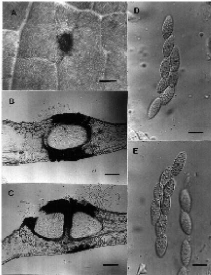
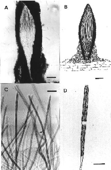
Phyllachora schimae C.Y. Chen & W.H. Hsieh, sp. nov.

Figure 1

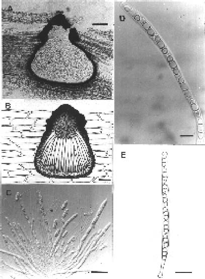
Maculae 1_2 mm lata, circulares, flavidae brunneae, cum maculis piceis, circularibus, nigris et nitidis. Stromata usque 1 mm lata, plusminusve circularis, nigra, nitida. Ascomata 240_360 mm lata, 280_380 alta, subglobosa, immersa, solitaria vel aggregata, ostiolata, clypeata. Ostiolum centrale, periphysatum. Peridium 8_16 mm crassum. Asci 65_110 × 12_13 mm, cylindrici, pedicellati, unitunicati, octospori. Ascosporae 16_20 × 7_9 mm, ovales, aseptatae, hyalinae, guttulatae, uniseriatae

vel imbricate biseriatae.TYPE: Taiwan. Taoyuan Hsien; Loloshan, In foliis Schimae superbae Gard. et Champ. (Theaccae), leg. C.Y. Chen, 20 Oct 1994, NCHUPP 2319.

Leaf spots 1_2 mm wide, mostly rounded, solitary or sometimes coalescing, discolouring the host tissue to yellow brown. Stromata up to 1 mm wide, roughly circular, black, shiny, dome-shaped tar spots scattered within the leaf spot, visible on both sides but more prominent on the upper surface. Ascomata 280_380 mm high, 240_360 mm wide, subglobose, immersed in the host mesophyll with a small conical apex forming a periphysate neck, solitary or gregarious. Clypeus black brown amorphous layer 80_120 mm wide and 360_400 mm long composed of melanized host palisade and epidermal cells and fungal hyphae beneath the cuticle. Peridium 8_16 mm, composed of several layers of thin-walled, hyaline to pale brown, elongated, compressed cells. Asci 65_110 × 12_13 mm, cylindrical, pedicellate, unitunicate, 8-spored. Ascospores 16_20 × 7_9 mm, hyaline, smooth, guttulate, aseptate, ovoid, uniseriate to overlapping biseriate inside the ascus.

Notes. Four species of Phyllachora have been reported on the genera of Theaceae (Arx and Müller, 1954; Hosagoudar, 1985; Kamat et al., 1978). These are P. cymbispora T. S. & K. Ramkr., P. euryae (Rac.) Arx & Müller, P. transiens Syd. & Butl. and P. gordoniae Hos. The first three species occur on Eurya and the last species on Gordonia. Phyllachora schimae is distinguished from them by size and shape of ascospores and asci. No Phyllachora have been reported on Schima, and Phyllachora species are based on host, so this fungus is described as a new species.

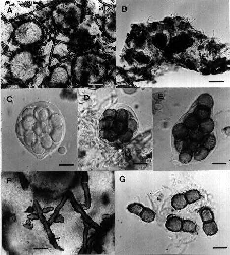
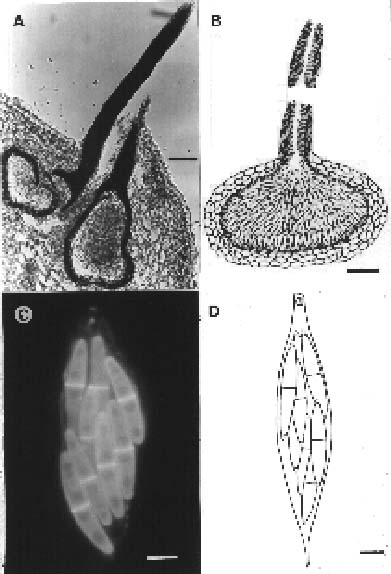
Schiffnerula villebruneae C.Y. Chen & W.H. Hsieh, sp. nov. Figure 2

1Corresponding author. Fax: (04) 2859009.


Botanical Bulletin of Academia Sinica, Vol. 37, 1996

Mycelium superficiale, hypophyllum, ex hyphis repentibus, atrobrunneis, septatis, laevibus, ramosus, 3_4 mm latis. Hyphopodia 4_8 mm lata, usque 10 mm alta, integra vel 2_3-lobata, continua, alternata, raro opposita, brunnea, laevia. Ascomata 70_90 mm lata et 35_60 mm alta, globosa, superficialia. Paries ascomati gelatinosum diffluentes. Asci 30_45 mm lati, globosi, pauci, bitunicati, crassitunicati, octospori. Ascosporae 17.5_22 × 8_11 mm, ellipsoideae vel oblongo ovoideae, atrobrunneae, mediane 1-septatae, constrictae, echinulatae.—TYPE: Taiwan, Nantou Hsien; Tungpu, In foliis Villebruneae pedunculatae Shirai (Urticaceae), leg. C.Y. Chen, 7 Apr 1995, NCHUPP 2356.

Superficial mycelium loosely reticulate, hypophyllous, dark bown, septate, smooth, branched, hyphopodiate, 3_4 mm wide. Hyphopodia 4_8 mm wide, up to 10 mm high, entire or 2_3-lobed, alternate, rarely opposite, brown. Ascomata 70_90 mm wide, 35_60 mm high, superficial on mycelia, globose. Peridium mucose diffluent early. Asci 30_45 mm wide, globose, thick-walled, few, bitunicate, 8-spored. Ascospores 17.5_22 × 8_11 mm, ellipsoid or oblong ovoid, dark brown, 1-septate in the middle, constricted at the septum, distinctly echinulate, conglobate inside the ascus. No anamorphs found associated on the material.

Notes. Many species have been described in this genus (Hughes, 1984, 1987; Sivanesan, 1984). No species have been reported either on Villebrunea (Urticaceae), or on other host genera belonging to Urticaceae.

New Records

Coleroa chaetomium (Kunze : Fr.) Rabenh., Herb. Mycol. no. 1456, 1859 Figure 3

For synonyms, see Barr (1968).

Mycelium epiphyllous, occasionally forming hypostroma of one or two layers of palisade-like cells, hyaline to pale brown in colour, not forming leaf spots. Ascomata developing originally from the subcuticular mycelium or hypostroma, erumpent and then becoming superficial, subglobose, 126_180 mm in diam., connate with a flattened base, ostiolate at the rounded apex, scattered to aggregated in small groups, often confluent, setose over the upper half, setae few to numerous, continuous or with few septa, dark brown, erect, stiff, up to 60 mm long, swollen at the base, slightly attenuate upwards. Peridium 10_14 mm thick around the upper and lateral sides, composed of 3_4 layered, dark brown, polygonal, laterally compressed cells, the basal portion 20_32 mm thick, applanate, composed of many layers of hyaline to light brown, polygonal cells, peridial wall of cells of textura angularis, up to 16 mm wide. Asci bitunicate, basal, saccate to oblong, 39_57 × 11_14 mm, sessile, apex rounded and thick-walled; pseudoparaphyses sparse, deliquescent. Ascospores biseriate, broadly clavate, 11_16 × 5_6 mm, widest at the upper third, septate below the middle, yel

Figure 1. Phyllachora schimae. A, leaf spot with ascostroma on host; B_C, vertical sections of ascostromata; D_E, asci with ascospores. Scale bars, A=1 mm, B_C=100 mm, D_E=10 mm.

Figure 2. Schiffnerula villebruneae. A, ascomata and mycelium on surface of leaf; B, vertical section of ascoma; C, immature ascus; D, mature ascus; E, ascus with protruded endotunica; F, hyphae with hyphopodia; G, ascospores. Scale bars, A=40 mm, B=20 mm, C_G=10 mm.


Chen and Hsieh — Taiwan ascomycetes

Figure 3. Coleroa chaetomium. A_B, vertical sections of ascomata; C_D, asci with ascospores. Scale bars, A_B=30 mm, C_D=10 mm.

Figure 4. Dimerium meliolicola. A_B, vertical sections of ascomata; C, ascospores; D, asci. Scale bars, A_B=30 mm, C_D= 10 mm.

lowish to olivaceous brown, smooth or minutely verruculose.

Habitat. On living leaves of Rubus incanus Liu and Yang (Rosaceae).

Specimen examined. NANTOU HSIEN: Meifeng, leg. C. Y. Chen, 23 Mar 1995, NCHUPP-2336.

Distribution. Asia, Europe, North America.

Descriptions. Barr (1968), Müller and Arx (1962).

Notes. Another specimen collected from Meifeng, Nantou Hsien (NCHUPP-2382) on the same host had hemispherical to flattened globose ascomata which were mostly gregarious, often confluent forming circular spots limited by the underlying subcuticular hypostroma. It is apparent that this Meifeng collection is a stromatic form of this species (Barr, 1968).

Dimerium meliolicola (Petr.) Hansf., Mycol. Pap. 15: 77, 1946. Figure 4

Pseudodimerium meliolicolum Petr., Ann. Mycol. 22: 21, 1924.

Hyperparasitic on Schiffnerula parasitic on living leaves of Rubus. Mycelium superficial, composed of thin-walled, roughened, interwoven, olivaceous to pale brown hyphae, 1.5_4 mm wide, forming a mycelial mat. The mycelium of Schiffnerula easily distinguished by the thick-walled, hyphopodiate, dark brown hyphae, up to 9 mm wide. Ascomata globose, 80_130 mm in diam., smooth,

scattered, with a blunt 23_40 mm wide apex, superficial on mycelia. Ascomatal wall 6_10 wide, composed of cells forming a textura angularis, the outer region composed of 1_2 layers of polygonal, brown cells, and the inner of several layers of compressed, hyaline cells. Asci bitunicate, basal, 8-spored, cylindrical-clavate to saccate, 44_67 × 10_16 mm, sessile to shortly stipitate, pseudoparaphysate. Pseudoparaphyses up to 3 mm wide, few in number. Ascospores overlapping uniseriate to biseriate, oblong to broadly clavate, straight to slightly curved, 10_13 × 4_6 mm, 1-septate slightly above the middle, rounded at apex and tapering below, brown.

Habitat. Hyperparasitic on Schiffnerula sp. on living leaves of Rubus sp. (Rosaceae).

Specimen examined. NANTOU HSIEN: Piluchi, leg. C. Y. Chen, 28 Apr 1994, NCHUPP-2368.

Distribution. Africa, Europe, Taiwan.

Description. Hansford (1946).

Notes. Hughes (1993) has discussed the taxonomic problems regarding the placement of Dimerium. He suggested that the mycoparasitic species of Dimerium may be disposed into one of the generic names listed as synonyms of Dimerium auct. (Müller and Arx, 1962) and that Phaeostigme H. Sydow & Sydow could be a suitable genus. We do not propose to transfer D. meliolicola to Phaeostigme at present as we have not been able to examine the type specimen of this fungus. Moreover, many other species in Dimerium have to be examined to ascer


Botanical Bulletin of Academia Sinica, Vol. 37, 1996

tain whether Phaeostigme or other genera are the correct choice for the transfer.

D. meliolicola is originally reported on Meliola. Although Schiffnerula is a different host genus, it is not unusual as many other fungi have been reported to parasitize more than one fungal host genus.

Gloniopsis praelonga (Schwein.) Zogg, Beitr. Kryptogamenflora Schweiz 11(3): 50, 1962. Figure 5

For synonyms, see Zogg (1962).

Hysterothecia gregarious, elongated, straight or curved up to 3 mm in length, flattened at the base, with a longitudinal and furrowed slit, 220_310 mm high, 360_470 mm wide, hyphae from basal wall penetrate into the epidermis and the host tissues below, slightly discolouring them. Peridium externally composed of blackened, pseudoparenchymatous cells, 30_70 mm wide and internally of a hyaline, 20_40 mm wide cells forming a textura intricata. Asci bitunicate, clavate, 74_100 × 18_27 mm, 8-spored, sessile or short stalked, pseudoparaphysate. Ascospores hyaline, oblong and broader in the middle, 18_25 × 6_8 mm, muriform up to six transverse septa (A-transsepta), strongly constricted at the middle septum, most segments divided by 1_2 longisepta, occasionally with B-transsepta or angular septa laid within segments, with a conspicuous mucilaginous sheath.

Habitat. On indet. wood.

Specimen examined. NANTOU HSIEN: Meifeng, leg. A. Sivanesan, 28 Jan 1992, NCHUPP-2282.

Figure 6. Glyphium elatum. A_B, vertical sections of ascomata; C_D, asci with ascospores. Scale bars, A_B=1,000 mm, C=40 mm, D=20 mm.

Distribution. Cosmopolitan.

Descriptions. Dennis (1978), Zogg (1962).

Glyphium elatum (Grev.: Fr.) Zogg, Beitr. Krypto-gamenflora Schweiz 11(3): 99, 1962. Figure 6

For synonyms, see Goree (1974).

Ascomata hysteriaceous, erect to slanted, vertically elongated and compressed, ligulate with the middle always broader and thicker, 0.8_1 mm high, 0.3_0.5 mm wide, 0.2_0.4 mm thick (the latter two measurements are made at the wider middle portion), transversely striate at the surface, blunt at apex, carbonaceous, scattered to aggregated, superficial on subiculum, with a bivalve shell-like fissuring along the lateral edges, and the two halves of shell separating to expose the lateral slits except at the apex and base, appearing lenticular in longitudinal section. Peridium 40_60 mm thick, composed of many layered, dark brown, polygonal cells, with the basal portion forming tufts of thick-walled, brown hyphae, growing downward to the underlying subiculum. Pseudoparaphyses trabeculate. Asci bitunicate, long cylindrical, rounded at apex, 270_420 × 6_8 mm, basal, 8-spored, stipitate, the

Figure 5. Gloniopsis praelonga. A_B, vertical sections of ascomata; C, asci, ascospores and pseudoparaphyses; D, asci with ascospores; E_H, ascospores. Scale bars, A_B=50 mm, C= 30 mm, D=10 mm, E_H=10 mm.


Chen and Hsieh — Taiwan ascomycetes

stipe 40_55 mm long. Ascospores filiform, almost as long as the ascus, light brown, multiseptate, septa 6_14 mm apart, fasciculate, parallel or partly twisted, obtuse at both ends.

Habitat. On indet. wood.

Specimen examined. ILAN HSIEN: Tuling, leg. C. Y. Chen, 29 Apr 1994, NCHUPP-2375.

Distribution. Europe, North America, Taiwan.

Descriptions. Barr (1990), Bisby & Ellis (1952), Goree (1974), Zogg (1962).

Gnomonia setacea (Pers.: Fr.) Ces. & De Not., Sfer. Ital. 1: 58, 1863. Figure 7

For synonyms, see Barr (1978).

Ascomata on fallen leaves, hypophyllous, subglobose and occasionally irregular in venter, 140_240 wide, 120_190 high, immersed, with a protruding long neck, scattered to aggregated, mostly on midrib and lateral veins, not forming any leaf spots. Peridium 10_30 mm thick,

Figure 8. Herpotrichia macrotrichia. A_B, vertical sections of ascomata; C, ascospores in calcofluor; D, asci and acospores. Scale bars, A=100 mm, B=50 mm, C_D=10 mm.

externally composed of polygonal to compressed, 2_4 layered, brown cells, internally of 2_3 layered hyaline cells; surface of peridium of textura angularis, each cell up to 20 mm in diam. Necks long, up to 1 mm, and 30_40 mm wide, pointed at the apex, periphysate, surface composed of parallel rows of elongated cells of textura porrecta. Asci numerous, loosening in the centrum, 8-spored, aparaphysate, cylindrical to fusiform, 23_48 × 4_8 mm, including the base which tapers to 4_14 mm long, slender stipe. Ascospores fusiform, 10_13 × 1.5_2 mm, overlapping biseriate to 4-seriate, hyaline, 1-septate in the middle, with a 1_1.5 mm long mucilaginous appendage at each end.

Habitat. On fallen leaves of Quercus variabilis Blume (Fagaceae).

Specimen examined. NANTOU HSIEN: Wushia, leg. C. Y. Chen, 10 Mar 1994, NCHUPP-2335.

Distribution. Asia, Europe, North America.

Descriptions. Barr (1978), Kobayashi (1970), Müller and Arx (1962).

Herpotrichia macrotricha (Berk. & Br.) Sacc., Sylloge Fung. 2: 213, 1883. Figure 8

For synonyms, see Barr (1984).

Mycelium superficial to partly innate in substrate, composed of thick-walled, dark brown hyphae, 5_8 mm wide

Figure 7. Gnomonia setacea. A_B, vertical sections of ascomata with one in A showing the whole neck; C, ascus in calcofluor; D, ascus with ascospores. Scale bars, A=50 mm, B=40 mm, C_D=3 mm.


Botanical Bulletin of Academia Sinica, Vol. 37, 1996

forming a dense subiculum in which the ascomata are embedded. Ascomata globose to subglobose, 290_390 mm high, 250_330 mm wide, gregarious, slightly rugose, covered with numerous hyphae with a broadly conical ostiolar region, 90_100 mm high, lined on the inside with periphyses. Peridium of two regions, the outer 30_44 mm thick, composed of brown, thick-walled polygonal cells, and the inner of hyaline, thin-walled cells, forming a textura angularis to epidermoidea, inner layer of peridium variable in thickness, widest at the basal portion, 8_20 mm wide, and becoming narrower towards the lateral sides. Asci bitunicate, basal and peripheral, cylindrical, 110_140 × 10_14 mm, shortly stipitate, 8-spored. Ascospores 30_43 × 5_7 mm, fusiform, overlapping biseriate, 3_5 septate, slightly constricted at septa, at first hyaline and then becoming brown, with mucilaginous sheath.

Habitat. On indet. wood.

Specimen examined. ILAN HSIEN: Tuling, leg. C. Y. Chen, 29 Apr 1994, NCHUPP-2364.

Distribution. America, Europe, Taiwan.

Descriptions. Barr (1984), Sivanesan (1971).

Melomastia mastoidea (Fr.) Schroet., Kryptogamenflora von Schlesien 3(2): 320, 1896. Figure 9

For synonyms, see Barr (1994).

Ascomata obpyriform with a flattened base, 450_580 mm wide, 480_600 mm high, separate and widely scattered, immersed beneath the epidermis which is pierced by a conical papillate ostiole, with the central canal up to 100 mm wide. Peridium of two regions, the outer region of variable thickness with the papillate portion wider and carbonaceous, 60_100 mm wide, while at the sides and base 14_30 mm wide composed of thick-walled, brown, compressed cells, and the inner region of hyaline, elongated, thin-walled, pseudoparenchymous cells. Asci numerous, unitunicate, cylindrical, 130_160 × 5_7 mm, 8-spored, apex rounded, stalked, with a nonamyloid apical ring. Paraphyses present. Ascospores, uniseriate, oblong with obtuse ends, hyaline, smooth, 14_17 × 4_6 mm, 2-septate, with a slightly longer middle cell and shorter end cells with mucilaginous sheath visible when mounted in water. Ascospores may separate into individual cells when slight pressure is applied on them.

Habitat. On indet. wood.

Specimen examined. NANTOU HSIEN: Piluchi, leg. C. Y. Chen, 5 Nov 1994, NCHUPP-2381.

Distribution. Europe, Taiwan.

Description. Barr (1994), Dennis (1978).

Notes. Barr (1994) described the ascospores as becoming verruculose with age. This character has not been observed in the specimen examined.

Figure 9. Melomastia mastoidea. A_B, vertical sections of ascomata, C, asci, ascospores and paraphyses; D_E, asci with ascospores. Scale bars, A_B=100 mm, C=30 mm, D=10 mm, E= 20 mm.

Figure 10. Rhytidhysteron rufulum. A_B, vertical sections of ascomata; C, asci, ascospores and pseudoparaphyses; D, ascus and ascospores, E, ascospores. Scale bars, A=300 mm, B=200 mm, C_D= 20 mm, E=10 mm.


Chen and Hsieh — Taiwan ascomycetes

Rhytidhysteron rufulum (Spreng.: Fr.) Speg., An. Soc. Cient. Argent. 90: 177, 1920. Figure 10

For synonyms see Samuels and Müller (1979).

Ascomata apothecioid, up to 1.5 mm in diam. with a brick-red hymenium, when dry the sides of apothecium rolling inwards and becoming hysterothecioid and 1_3 × 0.5_1 mm, rarely branched and triradiate, coriaceous, scattered to aggregated, erumpent-superficial, subsessile, the lips (apex of ectal excipulum) involuted, with distinct or indistinct transverse striae when viewed from the surface. The wall of two regions, the outer (ectal excipulum) 80_100 mm, composed of polygonal, thick-walled, dark brown cells, and the inner (medullary excipulum) composed of light brown, thin-walled cells which are arranged longitudinally toward the base. Asci bitunicate, cylindrical, 130_160 × 11_14 mm, with a short stipe up to 24 mm in length, 8-spored, interspersed with paraphysoidal hyphae which branch and swell at the tip to form an dark reddish epithecium. Ascospores uniseriate, broadly fusiform, straight to inequilateral, 23_30 × 7_10 mm, reddish brown, 3-septate, slightly constricted at septa.

Habitat. On indet. wood.

Specimen examined. NANTOU HSIEN: Sun-Moon-Lake, leg. A. Sivanesan, 20 Feb 1992, NCHUPP-2306.

Distribution. Common in the tropics.

Descriptions. Samuels and Müller (1979), Shear (1933), Voorhees (1939).

Thyronectria pseudotrichia (Berk. & M. A. Curtis) Seeler, J. Arnold Arbor. 21: 438, 1940. Figure 11

Nectria pseudotrichia Berk. & M. A. Curtis, J. Acad. nat. Sci. Philad., N.S. 2, 2: 289, 1853.

For other synonyms see Seifert (1985).

Anamorph: Tubercularia lateritia (Berk.) Seifert (1985).

For anamorph synonyms see Seifert (1985).

Ascomata globose to turbinate, 350_470 mm in diam., ostiole periphysate with a flattened apex, yellow to red, rough, collabent when dry, gregarious and occasionally laterally confluent on an underlying erumpent to superficial, pulvinate stroma of the same colour and texture as the ascomata. The anamorph, Tubercularia lateritia, of a characteristic capitate shape and always associated with the teleomorph. Peridium of three regions, continuous and intergrating into the underlying stroma, 60_100 mm wide, the outer region of globose to angular, yellow, thick-walled cells, the middle of elongated, yellow, thick-walled cells, the inner of elongated, hyaline, thin-walled cells, which in some cases are strongly compressed and agglutinated at the upper half, delimitation between middle and inner wall ambiguous. Region between outer and middle wall around the ostiole sometimes showing separation and gelatinization. Apical paraphyses extending from the sides below the ostiole and growing downwards, evanescent when mature and visible as gelatinous strands. Asci unitunicate, elliptical to ovoid, widest in the middle, 70_

Figure 11. Thyronectria pseudotrichia. A_B, vertical sections of ascomata; C_D, asci; E, ascospores. Scale bars, A_B=100 mm, C_E=10 mm.

90 × 15_20 mm, 8-spored, with a sessile to short stipe up to 12 mm long. Ascospores oblong to broadly clavate, variable in length, 20_40 × 8_11 mm, hyaline, partly biseriate, muriform, 6_9 transversely septate, with one or rarely two longitudinal septum in most of the cells.

Habitat. On stems of Villebrunea pedunculata Shirai (Urticaceae).

Specimen examined. NANTOU HSIEN: Puli, leg. W. H. Hsieh, 20 Apr 1995, NCHUPP-2359.

Descriptions. Booth (1959), Seaver (1909), Seifert (1985 as N. pseudotrichia).

Distribution. Cosmopolitan.

Acknowledgements. This work was supported by the National Science Council, ROC, grant number NSC 84-2321-B-005-072. The authors are grateful to Dr. A. Sivanesan for confirming the identifications and reviewing the manuscript.

Literature Cited

Arx, J. A. and E. Müller. 1954. Die Gattungen der amerosporen Pyrenomyceten. Beitr. Kryptogamenflora Schweiz 11(1): 1_434.

Barr, M. E. 1968. The Venturiaceae in North America. Can. J. Bot. 46: 799_864.

Barr, M. E. 1978. The Diaporthales in North America with emphasis on Gnomonia and its segregates. Mycol. Mem. 7: 31_62.

Barr, M. E. 1984. Herpotrichia and its segregates. Mycotaxon 20: 1_38.


Botanical Bulletin of Academia Sinica, Vol. 37, 1996

Barr, M. 1990. North American Flora: Melanommatales (Loculoascomycetes). The New York Botanical Garden, Bronx, New York, 129 pp.

Barr, M. 1994. Notes on the Amphisphaeriaceae and related families. Mycotaxon 51: 191_224.

Bisby, G. R. and M. B. Ellis. 1952. Lophium elatum Grev. Trans. Br. Mycol. Soc. 35: 299_303.

Booth, C. 1959. Studies of Pyrenomycetes: IV. Nectria (part I). Mycol. Pap. 73: 1_115.

Chen, C. Y. and W. H. Hsieh. 1994a. Some new ascomycetous fungi for Taiwan. Trans. Mycol. Soc. ROC 9(1): 1_20.

Chen, C. Y. and W. H. Hsieh. 1994b. Some new ascomycetous fungi for Taiwan. Trans. Mycol. Soc. ROC 9(1): 39_57.

Dennis, R. W. G. 1978. British Ascomycetes. J. Cramer, Vaduz, Germany, 585 pp.

Goree, H. 1974. Glyphium in western Canada and United States. Can. J. Bot. 52: 1266_1269.

Hansford, C. G. 1946. The foliicolous Ascomycetes, their parasites and associated fungi. Mycol. Pap. 15: 1_240.

Hosagoudar, V. B. 1985. New and noteworthy species of Phyllachora from South Indian. Indian Phytopathol. 38(3): 447_451.

Hughes, S. J. 1984. Schiffnerulaceous fungi on Compositae from the Americas. Can. J. Bot. 62: 2213_2222.

Hughes, S. J. 1987. Pleomorphy in some hyphopodiate fungi. In J. Sugiyama (ed.), Pleomorphic Fungi: The Diversity and Taxonomic Implications. Kodansha. Tokyo, pp. 103_139.

Hughes, S. J. 1993. Meliolina and its excluded species. Mycol. Pap. 166: 1_255.

Kamat, M. N., V. S. Seshadri, and A. A. Pande. 1978. A monographic study of Indian species of Phyllachora. University of Agricultural Sciences, Hebbal, Bangalore, 100 pp.

Kobayashi, T. 1970. Taxonomic studies of Japanese Diaporthaceae with special reference to their life-histories. Bull. Gov. Forest Exp. Sta. 226: 162_167.

Li, L.T. and W. H. Hsieh. 1991. Two new species and 13 new records of ascomycetes from Taiwan. Trans. Mycol. Soc. ROC 6(1): 57_71.

Müller, E. and J. A. von Arx. 1962. Die Gattungen der didymosporen Pyrenomyceten. Beitr. Kryptogamenflora Schweiz 11(2): 1_922.

Rohringer, R., W. K. Kim, D. J. Samborski, and N. K. Howes. 1977. Calcofluor: an optical brightener for fluorescent microscopy of fungal plant parasites. Phytopathology 67: 808_810.

Samuels, G. J. and E. Müller. 1979. Life-history studies of Brazilian Ascomycetes. 7. Rhytidhysteron rufulum and the genus Eutryblidiella. Sydowia 32: 277_293.

Seaver, J. F. 1909. The Hypocreales of North America. Mycologia 1: 177_207.

Seifert, K. A. 1985. A monograph of Stilbella and some allied Hyphomycetes. Stud. Mycol. 27: 1_235.

Shear, C. L. 1933. Life histories of Tryblidiella species. Mycologia 25: 274_285.

Sivanesan, A. 1971. The genus Herpotrichia Fuckel. Mycol. Pap. 127: 1_37.

Sivanesan, A. 1984. The Bitunicate Ascomycetes and Their Anamorph. J. Cramer, Vaduz, Germany, 701 pp.

Sivanesan, A. and W. H. Hsieh. 1989. New species and new records of ascomycetes from Taiwan. Mycol. Res. 93(3): 340_351.

Voorhees, R. K. 1939. The validity and morphology of two Tryblidiella species. Mycologia 31: 113_123.

Zogg, H. 1962. Die Hysteriaceae s. str. und Lophiaceae. Beitr. Kryptogamenflora Schweiz 11(3): 1_190.