Bot. Bull. Acad. Sin. (1997) 38: 63_72

Wu — New Hyphoderma from Taiwan

New species and new records of Hyphoderma (Basidiomycotina) from Taiwan

Sheng-Hua Wu1

Division of Collection and Research, National Museum of Natural Science, Taichung, Taiwan, Republic of China

(Received September 9, 1996; Accepted November 27, 1996)

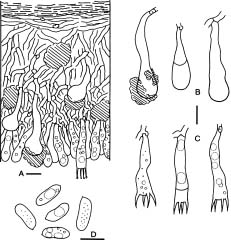
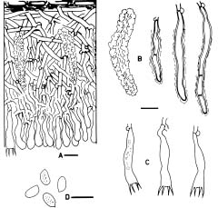
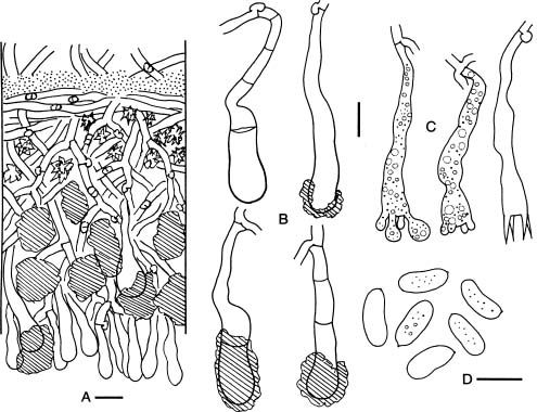
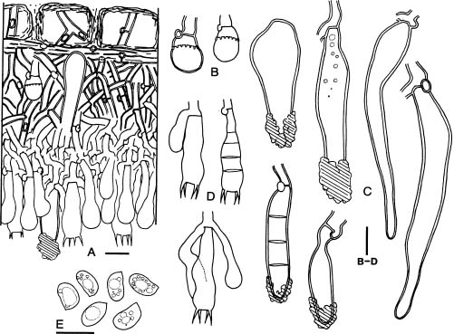
Abstract. Four new species of Hyphoderma collected from Taiwan are presented, viz. H. clavatum, H. rimulosum, H. subclavatum, and H. subpraetermissum. Two species are additionally reported from Taiwan for the first time, viz. H. definitum and H. variolosum. A key to known species of Taiwanese Hyphoderma is given. Description, microscopic line drawings, sexuality, cultural characters, and nuclear behavior are presented for the four new species.

Keywords: Basidiomycotina; Cultural studies; Hyphoderma; Taiwan; Taxonomy.

Introduction

Hyphoderma Wallr. is usually treated by mycologists under the heterogeneous Corticiaceae Herter (Donk, 1964; Eriksson and Ryvarden, 1975; Tellería, 1990; Wu, 1990). More than ninety species of Hyphoderma are known (e.g., Hjortstam, 1987; Hjortstam and Larsson, 1994; Wu, 1997), representing the largest genus among the Corticiaceae s.l.

Basidiomata of Hyphoderma are resupinate and effuse. Most species have smooth hymenial surfaces, but tuberculate, grandinioid, or odontioid cases are present in some species. Hyphoderma is microscopically characterized by the subclavate basidia with a median constriction, and typically monomitic hyphal system and nodose-septate generative hyphae, as well as nonamyloid and acyanophilic basidiospores, which are basically thin-walled. Leptocystidia, encrusted cystidia, or both are present in most species. Sexually, most species of Hyphoderma have been reported as heterothallic bipolar. Hyphoderma is lignicolous, saprobic, and causes a uniform white rot in wood. Tzean and Liou (1993) have shown that Hyphoderma is the only one among several tested genera of resupinate basidiomycetes that can capture or poison and consume nematodes.

Lin and Chen (1990) first reported Hyphoderma species from Taiwan, viz. H. praetermissum (P. Karst.) J. Erikss. & Strid, H. rude (Bres.) Hjortstam & Ryvarden (as Hyphodontia mucronata (Furukawa) S.H. Lin & Z.C. Chen), and H. setigerum (Fr.) Donk. Nine Hyphoderma species were further presented by Wu (1990) as new to Taiwan: H. allantosporum Sheng H. Wu, H. argillaceum (Bres.) Donk, H. ayresii (Berk. ex Cooke) Hjortstam (as H. macrosporum Sheng H. Wu), H. hjortstamii Sheng H. Wu, H. litschaueri (Burt) J. Erikss. & Strid, H. malenconii (Manjón & Moreno) Manjón et al., H. microcystidium

Sheng H. Wu, H. neopuberum Sheng H. Wu and, H. puberum (Fr.:Fr.) Wallr. Recently, Wu (1997) reported four additional new species of Hyphoderma from Taiwan: H. acystidiatum Sheng H. Wu, H. cremeum Sheng H. Wu, H. densum Sheng H. Wu, and H. subsetigerum Sheng H. Wu. However, the survey of Hyphoderma in Taiwan is still incomplete. In this study, four new species and two new records of Hyphoderma are proposed from Taiwan. Cultural studies including sexuality and nuclear behavior are also provided for the four new species.

Materials and Methods

Materials for this study were collected from Taiwan by the author in recent years. All studied specimens and cultures are deposited at the herbarium of National Museum of Natural Science of ROC (TNM).

Free-hand thin sections of basidiomata were prepared for microscopic studies. For observations and measurements of microscopic characters, 5% KOH was used as mounting medium to ensure rehydration. Melzer's reagent (IKI) was employed to detect amyloidity and dextrinoidity. Cotton blue (CB) was used as a mounting medium to determine cyanophily. Cultural description and species code system are basically from Nobles (1965) with amendments by Boidin and Lanquetin (1983). Minor modifications to Nobles' code system have been presented by many mycologists. The Nobles' code detailed by Nakasone (1990) is comprehensive and adopted in this study. Following Nakasone (1990) the mycelia were grown on 1.5% MEA instead of 1.25% MEA. In this study, plates were inverted to avoid accumulation of water produced by the mycelia. Inverted plates permit formation of a hymenium oriented as in nature. Nuclear staining of mycelia were made with Giemsa according to Boidin (1958). DAPI (4'-6'-diamidino-2-phenylindole) was used at a concentration of 0.25 µm/ml as fluorescent stain for nuclei of basidiospores.

1Fax: +886-4-323-5367; Email: shwu@nmns1.nmns.edu.tw


Botanical Bulletin of Academia Sinica, Vol. 38, 1997

Terminology for nuclear behavior is based on Boidin and Lanquetin (1984). The methods of cultural studies and determination of sexuality have been detailed by Wu (1996).

Taxonomy

Key to Known Species of Hyphoderma from Taiwan

1. Cystidia present 3

1. Cystidia lacking 2

2. Hymenial surface grandinioid. Subicular texture fairly loose H. acystidiatum

2. Hymenial surface smooth. Subicular texture dense H. densum

3. Septocystidia present 21

3. Septocystidia absent 4

4. Encrusted cystidia present 15

4. Encrusted cystidia absent 5

5. Hymenial surface grandinioid H. rude

5. Hymenial surface smooth 6

6. Cystidia moniliform 14

6. Cystidia not moniliform 7

7. Cystidia basally swollen, distinctly emergent H. argillaceum

7. Cystidia if basally swollen, not distinctly emergent 8

8. Cystidia distinctly clavate 13

8. Cystidia not, or only slightly clavate 9

9. Basidiospores ellipsoid 12

9. Basidiospores allantoid or suballantoid 10

10. Cystidia distinctly narrow towards apices H. allantosporum

10. Cystidia not distinctly narrow towards apices 11

11. Cystidia > 7 µm wide. Basidiospores > 3.5 µm wide H. hjortstamii

11. Cystidia < 7 µm wide. Basidiospores usually < 3.5 µm wide H. definitum

12. Basidiospores > 8 µm long H. praetermissum

12. Basidiospores < 8 µm long H. subpraetermissum

13. Heterothallic. Basidia < 40 µm long. Basidiospores colorless H. clavatum

13. Homothallic. Basidia > 40 µm long. Basidiospores brownish H. subclavatum

14. Basidiospores > 10 µm long H. malenconii

14. Basidiospores < 10 µm long H. litschaueri

15. Basidiospores > 6 µm wide H. ayresii

15. Basidiospores < 6 µm wide 16

16. Leptocystidia present 18

16. Leptocystidia absent 17

17. Basidiospores > 10 µm long H. cremeum

17. Basidiospores < 10 µm long H. rimulosum

18. Subicular hyphae brown H. variolosum

18. Subicular hyphae colorless 19

19. Lamprocystidia < 40 µm long H. microcystidium

19. Lamprocystidia > 40 µm long 20

20. Basidiospores > 11 µm long H. neopuberum

20. Basidiospores < 11 µm long H. puberum

21. Basidiospores > 3.5 µm wide H. setigerum

21. Basidiospores < 3.5 µm wide H. subsetigerum

Hyphoderma clavatum Sheng H. Wu, sp. nov. Figures 1, 5A

Holotypus. TAIWAN. MIAOLI HSIEN: Takeshan, alt. 550 m, on branch of Euphorbia pulcherrima, 25-III-1994, Wu 9403-4 (TNM).

Etymology: From clavatus (= clavate), referring to the shape of the cystidia.

Basidiocarpum effusum, membranaceum, 70_150 µm crassum; superficies hymenialis plana vel exigue tuberculata. Systema hypharum monomiticum; hyphae fibulatae. Cystidia clavata. Basidia subclavata, 30_40 × 6.5_8 µm , 4 sterigmatibus. Basidiosporae cylindricae, laeves, tenuitunicatae, 10_13 × 4.2_5.2 µm , IKI-, CB-.

Basidioma resupinate, effuse, adnate, 70_150 µm thick in section, membranaceous. Hymenial surface grayish clay in color, with numerous tiny brownish dots under the lens, smooth or slightly tuberculate, occasionally cracked; margin thinning, concolorous, slightly byssoid. Hyphal system monomitic; hyphae nodose-septate. Subiculum composed of basal layer and medullary layer. Basal layer fairly thin, with dense texture, with horizontal hyphae. Medullary layer with fairly loose texture, with variously oriented hyphae. Subicular hyphae colorless, 2_3.5 µm diam, thin-walled. Subhymenium ± thickening, with fairly dense texture; hyphae mainly vertical, colorless. Cystidia usually immersed, clavate or apically globose, colorless, 30_60 µm long, 10_15 µm diam for the globose parts, thin-walled or apically slightly thick-walled, sometimes apically covered with brownish yellow resinous material. Basidia subclavate with a median constriction, 30_40 × 6.5_8 µm , 4-sterigmate. Basidiospores cylindrical, adaxially slightly concave, smooth, thin-walled, usually guttulate, 10_13 × 4.2_5.2 µm (holotype spore-print: X = 11.11 ± 0.83 × 4.86 ± 0.25 µm , n = 30), IKI-, CB-.

Specimen examined. TAIWAN (see type).

Distribution. Known only from Taiwan.

Cultural description (polysporous mycelium of Wu 9403-4). 1 wk growth: Colony radius 15_20 mm. Advancing zone fairly even. Mats white. Aerial mycelium pellicular. 2 wk growth: Colony radius 50_60 mm. Advancing zone fairly even. Mats white. Aerial mycelium almost absent. Advancing hyphae colorless, nodose-septate, 3_6 µm diam, thin-walled. 3 wk growth: Plates covered. 6 wk growth: Mats white. Aerial mycelium absent. Hyphal system monomitic. Hyphae colorless, moderately


Wu — New Hyphoderma from Taiwan

Figure 1. Hyphoderma clavatum (holotype). A, Basidioma section; B, Cystidia; C, Basidia; D, Basidiospores. Scale bars = 10 µm.

ramified, nodose-sepate, occasionally irregularly swollen, 2_8 µm diam, thin-walled, usually guttulate, occasionally with secondary septa. Crystals present. No distinct odor. Not fruiting.

Oxidase reactions. TAA: -, 0; 0. GAA: +, 0; 0. TYA: -, 12; 25.

Species code. 2a, 3c, 26, 32, 36, 38, 43, 54, 58, 61.

Sexuality. Heterothallic, but type of incompatibility unknown. Two among eight monosporous mycelia of the holotype were paired in all combination, but all crossings were incompatible.

Nuclear behavior. Normal. Spores uninucleate, monosporous mycelium uninucleate, polysporous mycelium dikaryotic.

Remarks. Hyphoderma clavatum is allied to H. clavigerum (Bres.) Donk by the clavate cystidia and the brownish resinous material on apical parts of cystidia, but differs from it in having longer basidiospores. This new species also resembles H. subclavigerum K.H. Larss. & Hjortstam in having clavate cystidia and similar-sized basidiospores, but is distinct from the latter by the lack of projecting cylindrical cystidia.


Botanical Bulletin of Academia Sinica, Vol. 38, 1997

Holotypus. TAIWAN. NANTOU HSIEN: Entrance of Shalihsienhsi Forest Road near Tungpu, alt. 1,350 m, on culm of Miscanthus floridulus, 24-XI-1993, Wu 9311-62 (TNM).

Etymology: From rimulosus (= minutely cracked), referring to the appearance of the hymenial surface.

Basidiocarpum effusum, membranaceum, 70_130 µm crassum; superficies hymenialis plana. Systema hypharum monomiticum; hyphae fibulatae. Cystidia incrustata, cylindrica. Basidia subclavata, 22_32 × 4.5_6 µm, 4 sterigmatibus. Basidiosporae ellipsoideae, laeves, tenuitunicatae, 6_7 × 3.9_4.1 µm, IKI-, CB-.

Basidioma resupinate, effuse, adnate, 70_130 µm thick in section, membranaceous. Hymenial surface white to

Hyphoderma definitum (Jacks.) Donk, Fungus 27: 15. 1957.

A good description and illustration of this species have been given by Eriksson and Ryvarden (1975).

Specimen examined. TAIWAN. NANTOU HSIEN: Yushan National Park, Shenmu Forest Road, alt. 1,800 m, on branch of angiosperm, 7-X-1992, Wu 9210-78 (TNM).

Distribution. North America (Ginns and Lefebvre, 1993), North Europe (Eriksson and Ryvarden, 1975), Romania (Hallenberg and Toma, 1987), Taiwan.

Hyphoderma rimulosum Sheng H. Wu, sp. nov. Figures 2, 5B

Figure 2. Hyphoderma rimulosum (holotype). A, Basidioma section; B, Cystidia (left: with encrustation, right: almost without encrustation); C, Basidia; D, Basidiospores. Scale bars = 10 µm.


Wu — New Hyphoderma from Taiwan

ivory yellow in color, smooth, minutely cracked throughout; margin rather determinate, concolorous, slightly pruinose-filamentose. Hyphal system monomitic; hyphae nodose-septate. Subiculum composed of medullary layer, with loose to dense texture; hyphae variously oriented, colorless, 2_4 µm diam, thin-walled. Basal hyphae sometimes present. Subhymenium thickening, with dense texture; hyphae mainly vertical, colorless, 2_3 µm diam, thin-walled. Cystidia usually immersed, cylindrical, colorless, usually encrusted throughout, 30_55 µm long, 4_6 µm diam (without encrustation), slightly thick-walled. Basidia subclavate with a median constriction, 22_32 × 4.5_6 µm, 4-sterigmate. Basidiospores ellipsoid, adaxially slightly concave, smooth, thin-walled, 6_7 × 3.9_4.1 µm (holotype spore-print: X = 6.47 ± 0.38 × 4.03 ± 0.06 µm, n = 30), IKI-, CB-.

Specimen examined. TAIWAN (see type).

Distribution. Known only from Taiwan.

Cultural description (secondary mycelium of Wu 9311-62). 1 wk growth: Colony radius 53_56 mm. Advancing zone even. Mats white. Aerial mycelium slightly pellicular or almost absent. Advancing hyphae nodose-septate, colorless, 2.5_5 µm diam, thin-walled. 2 wk growth: Plates covered. Mats whitish. Aerial mycelium slightly pellicular or almost absent. 6 wk growth: Mats whitish-sulfur yellow. Aerial mycelium almost absent. Hyphal system monomitic. Hyphae colorless, sparsely ramified, nodose-septate, sometimes covered with minute yellowish granules, 2_5 µm diam, with thin walls to almost solid thick walls, the thick-walled hyphae usually distantly septate; vesicles occasionally present on thin-walled hyphae, 8_15 µm diam, thin-walled. Cubical crystals present. No distinct odor. Not fruiting.

Oxidase reactions. TAA: -, 0; 0. GAA: ++, 17_20; 32_36. TYA: - (brownish yellow), (+90).

Species code. 2a, 3c, 13, 21, 25, 27, 32, 36, (37), 38, 42, 54, 59, 61.

Sexuality. Bipolar (A1: 1, 3, 5, 6, 7, 8; A2: 2, 4).

Nuclear behavior. Normal. Spores uninucleate, monosporous mycelium uninucleate, secondary mycelium dikaryotic.

Remarks. This new species is closely related to Hyphoderma fouquieriae Nakasone & Gilb. by the similarity of encrusted cystidia, and similar-sized basidiospores. Holotype of H. fouquieriae (K.K. Nakasone 207, deposited in BPI) has been borrowed for this study, and its basidiospores were measured as 5.5_6.7 × 3.5_4.2 µm. The main feature that can be used to separate the two species is the difference of subicular hyphae. Hyphoderma rimulosum has subicular hyphae with 2_4 µm diam and thin walls, while those in H. fouquieriae are 3_7 µm diam, with 0.7_1.7 µm thick walls (measured from the holotype). Moreover, hymenium of H. fouquieriae turns purple in KOH, but this reaction does not occur in H. rimulosum. Incompatibility between the two species was obtained in


this study. Two monosporous mycelia sourced from holotype of H. rimulosum were respectively paired with polysporous mycelium of a H. fouquieriae specimen (K.K. Nakasone 121). Incompatibility between them was shown by the failure of dikaryotization of the monosporous mycelia after contact for four weeks. It is noted that recently Nakasone et al. (1994) transferred H. fouquieriae to the genus Ceraceomyces Jülich, i.e. C. fouquieriae (Nakasone & Gilb.) Nakasone et al.

Hyphoderma subclavatum Sheng H. Wu Figures 3, 5C

Holotypus. TAIWAN. PINTUNG HSIEN: Kenting National Park, Lanjenshan, alt. 300 m, on branch of angiosperm, leg. S.H. Wu, 1-IX-1994, Wu 9409-4 (TNM).

Etymology. From sub- (= somewhat, not completely) + clavatum, referring to the resemblance of this new species to Hyphoderma clavatum.

Basidiocarpum effusum, membranaceum, 60_130 µm crassum; superficies hymenialis plana. Systema hypharum monomiticum; hyphae fibulatae. Cystidia clavata. Basidia subclavata, 40_55 × 7_8 µm, 4 sterigmatibus. Basidiosporae cylindricae, laeves, tenuitunicatae, brunneolae, 10_12 × 4.2_5.3 µm, IKI-, CB-.

Basidioma resupinate, effuse, adnate, 60_130 µm thick in section, membranaceous. Hymenial surface clay yellow, usually with numerous tiny brownish dots under the lens, smooth, occasionally cracked; margin thinning, concolorous, pruinose. Hyphal system monomitic; hyphae nodose-septate. Subiculum composed of basal layer and medullary layer. Basal layer fairly thin, with dense texture, with horizontal hyphae. Medullary layer with fairly loose texture, with variously oriented hyphae. Crystals scattered or crowded in deep subiculum. Subicular hyphae colorless, 2.5_5 µm diam, thin- or slightly thick-walled. Subhymenium slightly thickening. Brownish resinous material scattered in subiculum and subhymenium. Cystidia clavate, colorless or with brownish contents, 40_70 µm long, 8_11 µm diam for apical parts, usually with one or more secondary septa, thin-walled, slightly thick-walled towards apices, usually covered with brownish resinous material. Basidia subclavate with a median constriction, colorless or occasionally with brownish contents, 40_55 × 7_8 µm, 4-sterigmate. Basidiospores usually with brownish contents, cylindrical, adaxially slightly concave, smooth, thin-walled, 10_12 × 4.2_5.3 µm, IKI-, CB-.

Additional specimen examined. TAIWAN. PINTUNG HSIEN: Kenting National Park, Lanjenshan, alt. 300 m, on branch of angiosperm, leg. S.H. Wu, 1-IX-1994, Wu 9409-11 (TNM).

Distribution. Known only from Taiwan.

Cultural description (polysporous mycelium of Wu 9409-4). 1 wk growth: Colony radius 12_15 mm. Advancing zone fairly even. Mats white. Aerial mycelium downy. Advancing hyphae nodose-septate, colorless, 2_5


Botanical Bulletin of Academia Sinica, Vol. 38, 1997

Figure 3. Hyphoderma subclavatum (holotype). A, Basidioma section; B, Cystidia; C, Basidia; D, Basidiospores. Scale bars = 10 µm.

µm diam, thin-walled. 2 wk growth: Colony radius 30_35 mm diam. 3 wk growth: Colony radius 55_60 mm diam. Advancing zone fairly even, slightly fimbriate. Mats white. Aerial mycelium pellicular near inoculum, downy elsewhere. 4 wk growth: Plates partly covered. 6 wk growth: Mats white or whitish. Aerial mycelium almost absent, slightly zonate. Hyphal system monomitic. Generative hyphae nodose-septate, moderately or sparsely ramified, colorless, sometimes guttulate, 2_6 µm diam, thin-walled, secondary septa occasionally present. Cubical crystals present. No distinct odor. Not fruiting.

Oxidase reactions. TAA: -, 0; 0. GAA: +++, 0; 0. TYA: -, 14_17; 27_30.

Species code. 2a, 3c, 7, 32, 36, 38, (44), 54, 57, 68.

Sexuality. Homothallic (indicated by the presence of clamped hyphae of all monosporous mycelium).

Nuclear behavior. Holodikaryotic. Spores binucleate, monosporous mycelium dikaryotic, polysporous mycelium dikaryotic.

Remarks. This species is very close to H. clavatum in characters, such as the clavate cystidia and the almost

same-sized basidiospores. However, it differs from the latter in having homothallic sexuality and brownish basidiospores.

Hyphoderma subpraetermissum Sheng H. Wu, sp. nov. Figures 4, 5D

Holotypus. TAIWAN. TAICHUNG HSIEN: Anmashan, alt. 2250, on branch of angiosperm, 7-VI-1995, leg. S.H. Wu & J.Y. Tseng, Wu 9506-27 (TNM).

Etymology. From sub- (= somewhat, not completely) + praetermissum, referring to the resemblance of this new species to Hyphoderma praetermissum.

Basidiocarpum effusum, membranaceo-subceraceum, 50_120 µm crassum; superficies hymenialis plana. Systema hypharum monomiticum; hyphae fibulatae. Stephanocystae praesentes. Cystidia cylindrica vel ventricosa, 30_100 × 7_15 µm. Basidia subclavata, 20_35 × 6_8 µm, 4 sterigmatibus. Basidiosporae ellipsoideae vel late ellipsoideae, laeves, tenuitunicatae, 6.2_7.5 × 4_5 µm, IKI-, CB-.

Basidioma resupinate, effuse, ± adnate, 50_120 µm thick in section, membranaceous-subceraceous. Hymenial


Wu — New Hyphoderma from Taiwan

Figure 4. Hyphoderma subpraetermissum (holotype). A, Basidioma section; B, Stephanocysts; C, Cystidia; D, Basidia; E, Basidiospores. Scale bars = 10 µm.

surface whitish, cream-colored when old, slightly purplish tinted due to the small excreted dots, smooth, not cracked; margin thinning, concolorous or paler, pruinose-filamentose. Hyphal system monomitic; hyphae nodose-septate. Subiculum composed of a thin basal layer and a medullary layer, the former sometimes indistinct, medullary layer with fairly loose texture; hyphae colorless, 3_6 µm diam, with 0.5_1 µm thick walls. Subhymenium ± thickening. Stephanocysts located in deep subiculum, 8_11 µm diam for global apices, Cystidia abundant, emergent, or immersed, colorless, usually apically covered with brownish resinous material, cylindrical or ventricose, sometimes tapering towards apices, 30_100 × 7_15 µm, with 0.5_1.2 µm thick walls, occasionally with secondary septa. Basidia subclavate with a median constriction, 20_35 × 6_8 µm, 4-sterigmate, occasionally with secondary septa. Basidiospores ellipsoid or broadly ellipsoid, adaxially slightly concave, smooth, thin-walled, 6.2_7.5 × 4_5 µm (holotype spore-print: X = 6.94 ± 0.31 × 4.44 ± 0.37 µm, n = 30), IKI-, CB-.

Specimen examined. TAIWAN (see holotype).

Distribution. Known only from Taiwan.


Cultural description (secondary mycelium of Wu 9506-27). 1 wk growth: Colony radius 3_6 mm. Mats white. 2 wk growth: Colony radius 16_18 mm. Mats white. Aerial mycelium almost absent. Advancing zone fairly even. Advancing hyphae colorless, nodose-septate, 2.5_5 µm diam, thin-walled. 3 wk growth: Colony radius 27_30 mm. 4 wk growth: Colony radius 38_42 mm. 5 wk growth: Colony radius 48_53 mm. 6 wk growth: Colony radius 58_63 mm. Mats white. Aerial mycelium absent. Advancing zone fairly even. Hyphal system monomitic. Hyphae colorless, moderately ramified, nodose-sepate, occasionally irregularly swollen, 2_6 (-9) µm diam, thin-walled, usually guttulate, occasionally with secondary septa. Cystidia present, colorless, irregularly cylindrical, guttulate, 15_40 × 4_8 µm, thin-walled. Stephanocysts present. Cubical crystals present. No distinct odor. Not fruiting.

Oxidase reactions. TAA: -, 0; -, 0. GAA: +++, 0; +++, 0. TYA: +, 7_10; ++, 25_30.

Species code. 2a, 3c, 15b, 30, 32, 36, 38, 47, 54, 60, 61.


Botanical Bulletin of Academia Sinica, Vol. 38, 1997

Figure 5. Cultures after 6 wk of growth on 1.5% MEA at 25°C. A, Hyphoderma clavatum (polysporous mycelium of holotype); B, Hyphoderma rimulosum (secondary mycelium of holotype); C, Hyphoderma subclavatum (polysporous mycelium of holotype); D, Hyphoderma subpraetermissum (secondary mycelium of holotype).

Sexuality. Tetrapolar (A1B1: 1, 3, 5; A1B2: 2; A2B1: 6, 8: A2B2: 4, 7).

Nuclear behavior. Normal. Spores uninucleate, monosporous mycelium uninucleate, secondary mycelium dikaryotic.

Remarks. This new speies can be regarded as belonging to Hyphoderma praetermissum (P. Karst.) J. Erikss. & Strid group due to the presence of similar cystidia and stephanocysts. Hallenberg et al. (1994) have detected five incompatible sibling species of H. praetermissum complex, based on collections from various countries of Europe, Turkey, and Canada. Nevertheless, basidiospore lengths

measured from all of their studied specimens are longer than this new species. Wu (1990) measured basidiospores of Taiwanese H. praetermissum as 8.8_12 × 4.2_5, which is also distinctly longer than this proposed new taxon. Both bipolar heterothallism and homothallism within H. praetermissum complex have been detected by Boidin (1958). Tetrapolar sexuality of H. subpraetermissum as detected in this study is the only case known for a species of Hyphoderma.

Hyphoderma variolosum Boidin, Lanquetin & Gilles, Bull. Soc. Mycol. France 107: 143. 1991.


Wu — New Hyphoderma from Taiwan

Description, drawn figure, and cultural studies of this species have been adequately provided in the protologue (Boidin et al., 1991).

Specimen examined. TAIWAN. KAOHSIUNG HSIEN: Liukuei, Shanping, on branch of angiosperm, 14-VII-1989, Wu 890714-14 (TNM).

Distribution. Central African Republic, Gabon (Boidin et al., 1991), Taiwan.

Remarks. Boidin et al. (1991) have noted that the brown subicular hyphae in this species are reminiscent of the genus Peniophora Cooke. According to their study, this species is homothallic. Sexually, Hyphoderma is typically bipolar and Peniophora is typically tetrapolar. However, the subclavate basidia with a median constriction clearly indicates the placement of this species in the genus Hyphoderma. Species of Peniophora have clavate basidia.

Acknowledgments. The author is indebted to curators of BPI and CFMR for arranging the loan of specimens and providing cultures. Dr. K.K. Nakasone has provided valuable suggestions on the manuscript. This study was supported by the National Science Council of the ROC (No. NSC 84-2311-B-178-002) in surveying Hyphoderma of Taiwan.

Literature Cited

Boidin, J. 1958. Essai biotaxonomique sur les Hydnés résupinés et les Corticiés. Rev. Mycol. Mém. Hors-Sér. 6: 1_388.

Boidin, J. and P. Lanquetin. 1965. Hétérobasidiomycètes saprophytes et Homobasidiomycètes résupinés X. Nouvelles données sur la polarité dite sexuelle. Rev. Mycol. 30: 3_16.

Boidin, J. and P. Lanquetin. 1983. Basidiomycètes Aphyllophoralés épithéloïdes étalés. Mycotaxon 16: 461_499.

Boidin, J. and P. Lanquetin. 1984. Répertoire des données utiles pour effectuer les tests d'intercompatibilité chez les Basidiomycètes. I. Introduction. Crypt. Mycol. 5: 33_45.

Boidin, J., P. Lanquetin, and G. Gilles. 1991. Les Peniophoraceae de la zone intertropicale (Basidiomycetes, Aphyllophorales). A. Especes Paleotropicales. Bull. Soc. Mycol. France 107: 91_147.

Donk, M.A. 1964. A conspectus of the families of Aphyllophorales. Persoonia 3: 199_324.

Eriksson, J. and L. Ryvarden. 1975. The Corticiaceae of North Europe volume 3. Coronicium-Hyphoderma. Fungiflora, Oslo, pp. 285_546.

Ginns, J. and M.N.L. Lefebvre. 1993. Lignicolous corticioid fungi (Basidiomycota) of North America. Mycol. Mem. 19: 1_247.

Hallenberg, N., K.-H. Larsson, and E. Larsson. 1994. On the Hyphoderma praetermissum complex. Mycol. Res. 98: 1012_1018.

Hallenberg, N. and M. Toma. 1987. Species of Corticiaceae (Basidiomycetes) new to the mycoflora of Romania. Rev. Roumaine Bio. 32: 1_8.

Hjortstam, K. 1987. A check-list to genera and species of corticioid fungi (Hymenomycetes). Windahlia 17: 55_85.

Hjortstam, K. and K.-H. Larsson. 1994. Annotated check-list to genera and species of corticioid fungi (Aphyllophorales, Basidiomycotina) with special regards to tropical and subtropical areas. Windahlia 21: 1_75.

Lin, S.H. and Z.C. Chen. 1990. The Corticiaceae and the resupinate Hydnaceae of Taiwan. Taiwania 35: 69_111.

Nakasone, K.K. 1990. Cultural studies and identification of wood-inhabiting Corticiaceae and selected Hymenomycetes from North America. Mycol. Mem. 15: 1_412.

Nakasone, K.K., C.R. Bergman, and H.H. Burdsall, Jr. 1994. Phanerochaete filamentosa-Corticium radicatum species complex in North America. Sydowia 46: 44_62.

Nobles, M.K. 1965. Identification of cultures of wood-inhabiting Hymenomycetes. Canad. J. Bot. 43: 1097_1139.

Tellería, M.T. 1990. Annotated list of the Corticiaceae, sensu lato (Aphyllophorales, Basidiomycotina), for Peninsular Spain and Balearic Islands. Biblioth. Mycol. 135: 1_152.

Tzean, S.S. and J.Y. Liou. 1993. Nematophagous resupinate basidiomycetous fungi. Phytopathology 83: 1015_1020.

Wu, S.H. 1990. The Corticiaceae (Basidiomycetes) subfamilies Phlebioideae, Phanerochaetoideae and Hyphodermoideae in Taiwan. Acta Bot. Fennica 142: 1_123.

Wu, S.H. 1996. Studies on Gloeocystidiellum sensu lato (Basidiomycotina) in Taiwan. Mycotaxon 58: 1_68.

Wu, S.H. 1997. New species of Hyphoderma from Taiwan. Mycologia 89: 132_140.