Bot. Bull. Acad. Sin. (1997) 38: 191-195

Wu and Huang — Studies of Hyphodontia

Cultural and ultrastructural studies of Hyphodontia mollis and H. subglobosa

Sheng-Hua Wu1 and Yu-Ying Huang

Division of Collection and Research, National Museum of Natural Science, Taichung, Taiwan, Republic of China

(Received December 10, 1996; Accepted March 8, 1997)

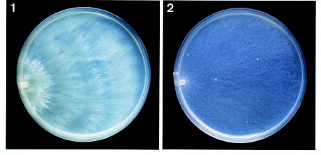
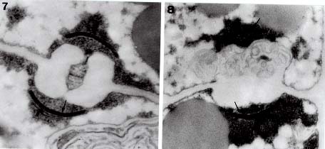
Abstract. Cultural and ultrastructural studies are reported for Hyphodontia mollis and H. subglobosa, which are known only from Taiwan. These two species have bipolar sexuality, normal nuclear behavior, and dolipores with continuous parenthesomes. Cultural characters are also provided for both species.

Keywords: Cultural studies; Hyphodontia mollis; H. subglobosa; Taiwan; Ultrastructure.

Introduction

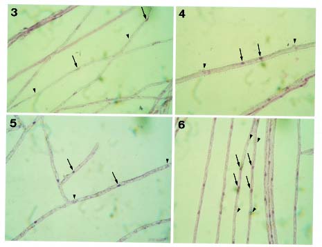
Hyphodontia J. Erikss. is generally regarded by mycologists as a member of the heterogeneous family Corticiaceae Herter, encompassing about 50 species (Langer, 1994). Species of Hyphodontia basically have resupinate basidiomata. Odontioid or grandinioid hymenial surfaces are commonly found in Hyphodontia, but smooth or poroid surfaces are present in some species. The Hyphal system of Hyphodontia is basically monomitic, with nodose-septate generative hyphae present in most species. The Basidia of Hyphodontia are uniformly suburniform and bear fairly small basidiospores, which usually contain one to a few distinct oily drops. The Mycelia of Hyphodontia species generally grow fairly slowly on MEA and usually develop malocysts and/or drepanocysts in cultures (Hassan Kasim and David, 1983). These structures are consistently present in a few species, but typically appear only in some cultures of a species (Nakasone, 1990). Most studied species of Hyphodontia show tetrapolar sexuality and have normal nuclear behavior. Studies of ultrastructure by Langer and Oberwinkler (1993) have shown that Hyphodontia species have dolipores with continuous parenthesomes. This type of parenthesome is not common in the homobasidiomycetes, but has been extensively detected in the heterobasidiomycetes (Wells, 1994).

Most studied species of Hyphodontia show tetrapolar sexuality, with only a few exceptions. Hyphodontia arguta (Fr.) J. Erikss. was reported by Brown (1935) as homothallic. More recently tetrapolarity has been reported for this species (Boidin, 1951; Boidin, 1958; Boidin and Lanquetin, 1965). Hyphodontia efibulata J. Erikss. & Hjortstam was reported by Hassan Kasim and David (1983) as presumed haploid parthenogenetic because uni

nucleate status was indicated for basidiospores, monosporous mycelium, and polysporous mycelium. However, H. efibulata has simple-septate hyphae, whereas most Hyphodontia species bear nodose-septate hyphae. Hyphodontia gossypina (Parmasto) Hjortstam was reported by Hassan Kasim (1981) as having bipolar sexuality. This species was originally known as Fibrodontia gossypina Parmasto. Hjortstam (1990) transferred this species to Hyphodontia, believing the presence of skeletal hyphae insufficient to separate Fibrodontia Parmasto from Hyphodontia. This species (Langer and Oberwinkler, 1993) has dolipores with continuous parenthesomes.

Hyphodontia mollis and H. subglobosa were proposed by Wu (1990) as new species from Taiwan, and both are still known only from Taiwan. At an earlier stage of this study, H. mollis was determined to be bipolar in sexuality. Hyphodontia subglobosa is a species with simple-septate hyphae. As above-mentioned, bipolar sexuality and simple-septate hyphae are rarely present in Hyphodontia. This study was designed to clarify the placement of these two species in Hyphodontia, through the analysis of their sexuality, cultural characters, nuclear behavior, and septal pore ultrastructure.

Materials and Methods

Fungal Specimens and Cultures

Studied fungal specimens and cultures collected from Taiwan, are deposited at the herbarium of the National Museum of Natural Science, ROC (TNM). Monosporous mycelia and polysporous mycelia offered for this study, have been obtained from germinated basidiospores according to Wu (1996).

Cultural Studies

Cultural description and the species code system are basically from Nobles (1965) with amendments by Boidin and Lanquetin (1983). Minor modifications to Nobles'

1Corresponding author. Fax: +886-4-323-5367; Email: shwu@ nmns1.nmns.edu.tw


Botanical Bulletin of Academia Sinica, Vol. 38, 1997

code system have been presented by many other mycologists. The Nobles' code detailed by Nakasone (1990) is adopted in this study. Following Nakasone (1990), the mycelia were grown on 1.5% MEA instead of 1.25% MEA. In this study, plates were inverted to avoid accumulation of water drops on the inner surface of the lid. Inverted plates also permit formation of a hymenium oriented as in nature. Determination of sexuality of single-clamped species has been described by Wu (1996), and this method was used in this study for Hyphodontia mollis. Detection of clamped hyphae between crossings among monosporous mycelia can not be used for determining sexuality of the clampless H. subglobosa. Nuclear staining in this study has shown that the monosporous mycelium of H. subglobosa is uninucleate, and the polysporous mycelium is dikaryotic. Therefore, nuclear staining of the crossed mycelia of all combinations was used to determine the sexuality of H. subglobosa. Nuclear staining of mycelia was made with Giemsa according to Boidin (1958). DAPI (4'-6'-diamidino-2-phenylindole) was supplementally used at a concentration of 0.25 µg/ml as a rapid fluorescent stain for mycelial nuclei. The same concentration of DAPI was also used for staining nuclei of basidiospores. Terminology for nuclear behavior is based on Boidin and Lanquetin (1984). The methods of cultural studies have been detailed by Wu (1996).

Ultrastructural Studies

Polysporous mycelia have been used for studies of transmission electron microscopy of septal pore ultrastructure. Pieces of mycelia grown on 2% MEA were picked and prefixed in 2% glutaraldehyde in 0.1 M sodium cacodylate buffer (pH 7.2) at room temperature for three days. Following six transfers in 0.1 M sodium cacodylate

buffer (pH 7.2), samples were postfixed in 1% osmium tetroxide in the same buffer for 2 h in the dark, six times washed in distilled water for 10 min each, and stained in 1% aqueous uranyl acetate for 1 h in the dark. The samples were dehydrated in ethanol, using 10 min changes at 20%, 40%, 60%, 80%, 90% and 3 times at 100% ethanol. Samples were embedded in Spurr's plastic (Spurr, 1969). Serial sections 70 nm thick were cut on a Reichert Ultracut S ultramicrotone with a diamond knife. Sections were mounted on formvar-coated copper grids, poststained with lead citrate (Reynolds, 1963) at room temperature for 4 min, and washed again with distilled water. The ultrathin sections were examined with a Hitachi H-600 electron microscope at 75 kV.

Results

Hyphodontia mollis Sheng H. Wu, Acta Bot. Fennica 142: 95. 1990.

Material studied. TAIWAN. NANTOU HSIEN: Neimaopu, Tachiehshan, alt. 750 m, on branch of angiosperm, leg. S.H. Wu & J.Y. Tseng, 13-X-1994, Wu 9410-19 (TNM).

Cultural description (polysporous mycelium).

Growth at 1 wk: Colony radius 5_8 mm. Mat whitish. Aerial mycelium downy. Advancing zone slightly bayed. Growth at 2 wk: Colony radius 20_23 mm. Mat slightly yellow. Aerial mycelium slightly pellicular. Advancing zone bayed. Advancing hyphae colorless, nodose-septate, 2.5_4.5 µm diam, thin-walled. Growth at 3 and 4 wk: Colony radius 35_42 and 55_65 mm. Growth at 5 wk: Plates partly covered. Growth at 6 wk (Figure 1): Mat

Figures 1_2. Cultures after 6 wk of growth on 1.5% MEA at 25°C. 1, Hyphodontia mollis (polysporous mycelium). 2, H. subglobosa (polysporous mycelium).


Wu and Huang — Studies of Hyphodontia

Figures 3_6. Nuclei of mycelia stained with Giemsa (arrows indicate nuclei; arrowheads indicate septa). 3, Uninucleate monosporous mycelium of Hyphodontia mollis. 4, Dikaryotic polysporous mycelium of H. mollis. 5, Uninucleate monosporous mycelium of H. subglobosa. 6, Dikaryotic polysporous mycelium of H. subglobosa.

slightly yellow. Aerial mycelium almost absent, zonate. Hyphal system monomitic. Hyphae nodose-septate, moderately ramified, colorless or slightly yellow, usually guttulate, 1.5_5 µm diam, thin-walled. Malocysts rarely present. Crystals present, sometimes rod-like. No distinct odor. Not fruiting.

Oxidase reactions. TAA: +, 0; 0. GAA: + to ++, 0; +++, 0. TYA: -, 0; 0.

Species code. 2a, 3c, (31a), 32, 36, 38, (45), 54, 59, 61.

Sexuality. Bipolar (A1: 1, 2, 5, 6, 7; A2: 3, 4, 8).

Nuclear behavior. Normal. Spores uninucleate, monosporous mycelium uninucleate (Figure 3), secondary mycelium dikaryotic (Figure 4).

Ultrastructure. Dolipore parenthesomes continuous (Figure 7).

Hyphodontia subglobosa Sheng H. Wu, Acta Bot. Fennica 142: 106. 1990.

Material studied. TAIWAN. MIAOLI HSIEN: Takeshan, alt. 600 m, on rotten branch of angiosperm, leg. S.H. Wu, 17-IV-1995, Wu 9504-7 (TNM).

Cultural description (polysporous mycelium).

Growth at 1 wk: Colony radius 13_16 mm. Mat white. Aerial mycelium slightly pellicular. Advancing zone slightly uneven. Growth at 2 wk: Colony radius 33_36 mm. Mat white. Aerial mycelium almost absent. Advancing zone fairly even. Advancing hyphae colorless, simple-septate, 2_4 µm diam, thin-walled. Growth at 3 and 4 wk: Colony radius 53_56 and 72_76 mm. Growth at 5 wk: Plates covered. Growth at 6 wk (Figure 2): Mat white. Aerial mycelium absent. Hyphal system monomitic. Hyphae colorless, moderately ramified, simple-septate, sometimes guttulate, 2_5 µm diam, thin-walled. Rod-like crystals present. No distinct odor. Not fruiting.

Oxidase reactions. TAA: -, 0; -, 0. GAA: -, 0; +, 0. TYA: -, 35; - (yellowish), 60-70.

Species code. 2a, 6, 7, 32, 36, 38, 45, 54, 59, 61.


Botanical Bulletin of Academia Sinica, Vol. 38, 1997

Figures 7_8. Dolipores with continuous parenthesomes (arrows indicate parenthesomes). 7, Hyphodontia mollis. 8, H. subglobosa.

Sexuality. Bipolar (A1: 1, 2, 5, 6, 8; A2: 3, 4, 7).

Nuclear behavior. Normal. Spores uninucleate; monosporous mycelium usually uninucleate (Figure 5); polysporous mycelium dikaryotic (Figure 6).

Ultrastructure. Dolipore parenthesomes continuous (Figure 8).

Discussion

Suburniform basidia, development of malocysts and/or drepanocysts in cultures, and the dolipores with continuous parenthesomes are key features for recognizing the genus Hyphodontia. However, the malocysts and/or drepanocysts do not occur in all cultures of Hyphodontia species. In this study, Hyphodontia mollis and H. subglobosa were shown to have a bipolar mating system; a tetrapolar mating system occurs in most species of Hyphodontia. Sexuality is usually fairly fixed for a genus of the homobasidiomycetes, but exceptions have been detected in many homogeneous genera. In this study, malocysts were rarely found in the culture of H. mollis, and were not seen in the culture of H. subglobosa. The malocyts in culture and the continuous parenthesomes of septal dolipores in H. mollis has confirmed its placement in Hyphodontia. The continuous parenthesomes of septal dolipores in H. subglobosa, as observed in this study, strongly support its taxonomic position in this genus.

Acknowledgments. The authors are indebted to Dr. M.L. Lee for TEM technical guidance. Drs. J. Ginns and N. Hallenberg and an anonymous reviewer provided valuable suggestions in improving the manuscript. This study was supported by the National Science Council, ROC (No. NSC 86-2311-B-178-001).

Literature Cited

Boidin, J. 1951. Caryologie des spores, germinations et mycéliums de quelques Basidiomycètes résupinés (Hydnés et Mérulinés). C. R. Acad. Sc. Paris 233: 707_709.

Boidin, J. 1958. Essai biotaxonomique sur les Hydnés résupinés et les Corticiés. Rev. Mycol. Mém. Hors-Sér. 6: 1_388.

Boidin, J. and P. Lanquetin. 1965. Hétérobasidiomycètes saprophytes et Homobasidiomycètes résupinés X. Nouvelles données sur la polarité dite sexuelle. Rev. Mycol. 30: 3_16.

Boidin, J. and P. Lanquetin. 1983. Basidiomycètes Aphyllophoralés épithéloïdes étalés. Mycotaxon 16: 461_499.

Boidin, J. and P. Lanquetin. 1984. Répertoire des données utiles pour effectuer les tests d'intercompatibilité chez les Basidiomycètes. I. Introduction. Crypt. Mycol. 5: 33_45.

Brown, C.A. 1935. Morphology and biology of some species of Odontia. Bot. Gaz. 96: 640_675.

Hassan Kasim, F. 1981. Contribution à la Connaissance du Genre Hyphodontia Erikss. (Basidiomycètes). Étude Systématique et Culturale. Thèse Lyon, 87 pp.

Hassan Kasim, F. and A. David. 1983. Studies on the cultural characterization of 16 species of Hyphodontia Eriksson and Chaetoporellus Bond. and Sing. ex Sing. Sydowia 36: 139_149.

Hjortstam, K. 1990. Corticioid fungi described by J. Berkeley II. Species from Cuba. Mycotaxon 39: 415_423.

Langer, E. 1994. Die Gattung Hyphodontia John Eriksson. Biblioth. Mycol. 154: 1_298.

Langer, E. and F. Oberwinkler. 1993. Corticioid Basidiomycetes. I. Morphology and ultrastructure. Windahlia 20: 1_28.

Nakasone, K.K. 1990. Cultural studies and identification of wood-inhabiting Corticiaceae and selected Hymenomycetes from North America. Mycol. Mem. 15: 1_412.


Wu and Huang — Studies of Hyphodontia

Nobles, M.K. 1965. Identification of cultures of wood-inhabiting Hymenomycetes. Can. J. Bot. 43: 1097_1139.

Reynolds, E.S. 1963. The use of lead citrate at high pH as an electron-opaque stain in electron microscopy. J. Cell Biol. 17: 208_212.

Spurr, A.J. 1969. A low-viscosity epoxy resin embedding medium for electron microscopy. J. Ultrastruct. Res. 26: 31_43.

Wells, K. 1994. Jelly fungi, then and now! Mycologia 86: 18_48.

Wu, S.H. 1990. The Corticiaceae (Basidiomycetes) subfamilies Phlebioideae, Phanerochaetoideae and Hyphodermoideae in Taiwan. Acta Bot. Fennica 142: 1_123.

Wu, S.H. 1996. Studies on Gloeocystidiellum sensu lato (Basidiomycotina) in Taiwan. Mycotaxon 58: 1_68.