Bot. Bull. Acad. Sin. (1998) 39: 23_27

Rout et al. — Genetic stability of micropropagated plants of ginger

Determination of genetic stability of micropropagated plants of ginger using Random Amplified Polymorphic DNA (RAPD) markers

G.R. Rout1,3, P. Das1, S. Goel2, and S.N. Raina2

1Plant Biotechnology Division, Plant Tissue Culture Laboratory, Regional Plant Resource Centre, Bhubaneswar - 751 015, Orissa, India

2Plant Cytogenetics and Molecular Biology Laboratory, Department of Botany, Delhi University, Delhi - 110 007, India

(Received July 16, 1997; Accepted October 3, 1997)

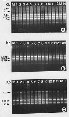
Abstract. Random amplified polymorphic DNA (RAPD) markers were used to evaluate the genetic stability of micropropagated plants of Zingiber officinales cv. V3S18. Fifteen arbitrary decamers were used to amplify DNA from in vivo and in vitro plant material to assess the genetic fidelity. All RAPD profiles from micropropagated plants were monomorphic and similar to those of field grown control plants. No variation was detected within the micropropagated plants. The utilization of RAPD markers both for the assessment of genetic stability of clonal materials and to certify genetic stability throughout the systems of micropropagation is discussed.

Keywords: Genetic markers; Genetic stability; Micropropagation; RAPD.

Introduction

Zingiber officinales Rosc. (ginger) of the family Zingiberaceae is an important tropical horticultural plant, valued all over the world as a spice in culinary preparations and for its medicinal properties. It is rich in secondary metabolite such as Oleoresin (Bhagyalakshmi and Singh, 1988). Breeding of ginger is seriously handicapped by poor flowering and seed set. Most crop improvement programs of this species are confined to evaluation and selection of naturally occurring clonal variations. In vitro culture techniques provide an alternative means of plant propagation and a tool for crop improvement (Vasil, 1988). Clonal multiplication of ginger through shoot multiplication has been reported (Hosoki and Sagawa, 1977; Wang, 1989; Balachandran et al., 1990; Rout et al., 1997). Plantlets derived from in vitro culture might exhibit somaclonal variation (Larkin and Scowcroft, 1981) which is often heritable (Breiman et al., 1987). Other reports claim that useful morphological, cytological, and molecular variations may be generated in vitro (Larkin et al., 1989). Any system which significantly reduces or eliminates tissue culture generated variations can be of much practical utility. The variations may be due to several factors (Vasil, 1987; 1988), such as genotypes used (Breiman et al., 1987), pathways of regeneration, and parameters employed for assessing the effect of in vitro culture, such as gross morphology and cytology (Swedlund and Vasil, 1985), field assessment, and molecular studies (Breiman

et al., 1989; Chawdhury et al., 1994; Shenoy and Vasil, 1992).

Several strategies can be used to assess the genetic fidelity of in vitro derived clones, but most have limitations. Karyological analysis, for example, cannot reveal alterations in specific genes or small chromosomal rearrangements (Isabel et al., 1993). Isozyme markers provide a convenient method for detecting genetic changes, but are subject to ontogenic variations. They are also limited in number, and only DNA regions coding for soluble proteins can be sampled. Using the polymerase chain reaction (PCR) in conjuction with short primers of arbitrary sequence (Williams et al., 1990), randomly amplified polymorphic DNA (RAPD) markers were recently shown to be sensitive for detecting variations among individuals between and within species (Carlson et al., 1991; Roy et al., 1992). RAPD markers have been used successfully to assess genetic stability among somatic embryos in spruce species (Isabel et al., 1993; 1996) and among micropropagated plants of poplar (Rani et al., 1995). The present study was undertaken to determine the genetic stability of the micropropagated plants of Zingiber officinales cv. V3S18 by RAPD analysis.

Materials and Methods

The standard protocol of micropropagating Zingiber officinales cv. V3S18 plants through meristem culture was developed and tested at the Regional Plant Resource Centre, Bhubaneswar (Rout et al., unpublished). The protocol was used for assessment of genetic stability. In the present

3Corresponding author. Fax: 91-0674-450274.


Botanical Bulletin of Academia Sinica, Vol. 39, 1998

study, a total of 15 micropropagated plants were randomly selected from a particular batch and analyzed.

The method of raising plants was as follows: rhizomes of ginger (Zingiber officinales cv. V3S18) were collected from the Orissa University of Agriculture and Technology, Bhubaneswar, Orissa, India, and incubated in a bed of sterile moist, sandy soil to induce sprouting. The pale-white sprouts were collected and washed with 2% (v/v) detergent solution (Teepol, Qualigen, India) and surface sterilized by immersing in 0.2% (w/v) aqueous solution of mercuric chloride for 25 min. The shoot buds were asceptically examined under a dissecting microscope, and meristems with only the apical dome or with one or two leaf primordia and ranging in size from 0.1 to 0.5 mm were removed and inoculated on MS solid medium (Murashige and Skoog, 1962) supplemented with 4.0 mg/l 6-benzylaminopurine (BAP), 1.0 mg/l indole-3-acetic acid (IAA), and 100 mg/l adenine sulfate. After 8 weeks on the multiplication medium, the propagules were transferred to half-strength MS basal salts supplemented with 1.0 mg/l indole-3-butyric acid (IBA). Well rooted plantlets were hardened under greenhouse conditions for 2 weeks before transfer to the field conditions.

DNA Extraction

DNA was extracted from fresh leaves of micropropagated and field grown plants by the CTAB method (Bousquet et al., 1990). Approximately, 200 mg of fresh leaf were ground to a powder in liquid nitrogen, using a mortar and pestle. The powder was transferred to a 25 ml sterile Falcon tube with 10 ml of CTAB buffer. The extraction buffer consisted of 2% (w/v) CTAB (cetyltrimethyl amonium bromide, Sigma), 1.4 M NaCl, 20 mM EDTA, 100 mM Tris-HCl pH 9.5, and 0.2% (v/v) b-mercaptoethanol. The homogenate was incubated at 65°C for 1 h, extracted with an equal volume of chloroform, and centrifuged at 10,000 rpm for 20 min. DNA was precipitated from the aqueous phase by mixing with 1/10 volume (ml) of 3 M sodium acetate and an equal volume of isopropanol. After centrifugation at 10,000 rpm for 10 min, the DNA pellet was washed with 70% ethanol, air dried, and resuspended in 10 mM Tris pH 8.0, 0.1 mM EDTA buffers. DNA quantity was estimated spectrophotometrically by measuring absorbance 260 nm.

PCR Amplification

Fifteen arbitrary 10-base primers (Operon Technologies Inc., Alameda, California) were used for polymerase chain reaction (PCR), following the protocol of Williams et al. (1990), with minor modifications. Amplification reactions were performed with 25 µl of 10× assay buffer (Stratagene), 2.0 µl of 1.25 mM each of dNTP's (Pharmacia), 15 ng of the primer, 1× Taq polymerase buffer, 0.5 units of Taq DNA polymerase (Genei, India), 2.5 mM MgCl2, and 30 ng of genomic DNA. The reaction mixture was overlaid with 25 µl of mineral oil (Sigma, USA). DNA amplification was performed in a Perkin Elmer Cetus 480 DNA Thermal Cycler (Perkin Elmer Ce

tus, Norwalk, Conn, USA) programmed for 45 cycles as follows: 1st cycle of 3.5 min at 92°C, 1 min at 35°C, 2 min at 72°C; followed by 44 cycles each of 1 min at 92°C, 1 min at 35°C, 2 min at 72°C followed by one final extension cycle of 7 min at 72°C. The amplification products were size separated by electrophoresis in 1.2% (w/v) agarose (Pharmacia) gels with 0.5× TBE buffer, stained with ethidium bromide, and photographed under UV light. In all cases l-DNA digested with Hind III (Genei, Bangalore, India) was used as molecular size marker. All the reactions were repeated at least twice.

Amplified DNA Marker Scoring

Amplified DNA markers were scored as present or absent in each micropropagated plant. Electrophoretic DNA bands of low visual intensity that could not be readily distinguished as present or absent were considered ambiguous markers and were not scored.

Results and Discussion

Optimum conditions for DNA extraction from ginger leaves for reproducible PCR amplification in the presence of primers were first investigated. The results were scored as patterns of bands obtained from in vitro micropropagated plants and compared with plants maintained in the field. Out of fifteen different decamers tested, three (OPC 04, OPC 19, and OPC 07) produced amplification products that were monomorphic across all micropropagated plants (Figure 1A_C). The twelve other primers did not yield good amplified product. The size of the monomorphic DNA fragments, produced by these primers ranged from 9.4 to 1.0 Kb for OPC 04, 0.972 to 0.756 Kb for OPC 19, and 1.32 to 0.434 Kb for OPC 07. Primer OPC 04 produced five DNA fragments common to all 14 micropropagated plants, but in the case of primer OPC 19 and OPC 07 only three monomorphic fragments were obtained. The variation of monomorphic bands in micropropagated plants by using different primers has been reported earlier (Potter and Jones, 1991; Rani et al., 1995; Angel et al., 1996). No polymorphisms or changes in the amplified DNAs were detected after amplification by PCR within micropropagated ginger plants. Shenoy and Vasil (1992) reported that micropropagation through meristem culture is generally associated with low risk of genetic instability because the organised meristems are generally more resistant to genetic changes that might occur during cell division or differentiation under in vitro conditions. The genetic stability of ginger (Zingiber officinales cv. V3S18) shown in our study is in agreement with Angel et al. (1996), who found no RAPD fingerprint variation when cassava (Manihot esculenta) plants derived from in vitro stored apical meristems. This study provides the first information on the molecular basis of polymorphism detected as RAPD markers in micropropagated plants of Zingiber officinales cv. V3S18. The DNA amplification products, which represent one allele per locus, could result from changes in either the sequence of the primer bind


Rout et al. — Genetic stability of micropropagated plants of ginger

sidered. Many authors using RAPD or RFLP have failed to observe intraclonal variations in various species including Picea mariana, Festuca pratensis, Saccharum, Pennisetum purpureum, or Liliodendron tulipifera (Isabel et al., 1993; Valles et al., 1993; Chowdhury and Vasil, 1993; Shenoy and Vasil, 1992; Merkle et al., 1988). Similarly, Rani et al. (1995) found RAPD variations among 23 micropropagated Populus deltoides plants originating from the same clone and morphologically similar. Bouman et al. (1992) and Bouman and Kuijpers (1994) also found intraclonal RAPD polymorphism amongst micropropagated Begonia but at a lower frequency than phenotypic variations and without any correlation with the phenotype.

In conclusion, our results demonstrate that RAPD analysis can be applied to assess the genetic fidelity of plants derived in vitro on an industrial scale as part of crop improvement programs. This method might be useful for monitoring the stability of in vitro germplasm collections and cryopreserved material.

Acknowledgement. The first author (GRR) wishes to acknowledge the Department of Science and Technology, Government of India for awarding a SERC visiting fellowship for conducting this investigation.

Literature Cited

Angel, F., V.E. Barney, J. Tohme, and W.M. Roca. 1996. Stability of cassava plants at the DNA level after retrieval from 10 years of in vitro storage. Euphytica 90: 307_313.

Balachandran, S.N., S.R. Bhat, and K.P.S. Chandel. 1990. In vitro clonal multiplication of turmeric (Curcuma spp.) and ginger (Zingiber officinales Rosc.). Plant Cell Rep. 8: 521_524.

Bhagyalakshmi, N. and N.S. Singh. 1988. Meristem culture and micropropagation of a variety of ginger (Zingiber officinales Rosc.) with a high yield of Oleoresin. J. Hortic. Sci. 63: 321_327.

Breiman, A., D. Rotem-abarbanell, A. Karp, and H. Shaskin. 1987. Heritable somaclonal variation in wild barley (Hordeum spontaneum). Theor. Appl. Genet. 71: 637_643.

Breiman, A., T. Felsenburg, and E. Galun. 1989. Is Nor region variability in wheat invariably caused by tissue culture? Theor. Appl. Genet. 77: 809_814.

Bousquet, J., L. Simon, and M. Lalonde. 1990. DNA amplification from vegetative and sexual tissues of trees using polymerase chain reaction. Can. J. For. Res. 20: 254_457.

Bouman, H., A.M. Kuijpers, and G.J. deKlerk. 1992. The influence of tissue culture methods on somaclonal variation in Begonia. Physiol. Plant. 85: A45.

Bouman, H. and A.M. Kuijpers. 1994. RAPD-patterns and somaclonal variation in Begonia. In Abst. 4th Int. Congv. Plant Mol. Biol., Amsterdam, Poster 37.

Carlson, J.E., L.K. Tulsieram, J.C. Glaubitz, V.W.K. Luk, C. Kauffeldt, and R. Rutledge. 1991. Segregation of random amplified DNA markers in F1 progeny of conifers. Theor. Appl. Genet. 83: 194_200.

Chowdhury, M.K.U. and J.K. Vasil. 1993. Molecular analysis of plants regenerated from embryogenic cultures of hybrid

Figure 1. A_C, RAPD profiles generated by primer OPC 04 (A), OPC 19 (B) and OPC 07 (C). Lane 1 shows RAPD bands from the field grown mother plant (M). Lanes 2_14 show RAPD products from micropropagated plants. Arrow indicates the size of the fragments as compared with markers.

ing site or changes which alter the size and prevent the successful amplification of target DNA. In our study, the amplified products exhibited monomorphisms among all the in vitro plants and were similar to those from control plants. The method is simple and the results are reproducible. Because only micro-amounts of material are necessary, this approach can be used to assess tissue at several stages of in vitro culture. Large sample sizes can be handled rapidly, and the technique lends itself to automation (Williams et al., 1990; Welsh and McClelland, 1990; Hedrick, 1992). Furthermore, the genome is most probably randomly sampled without the influence of ontogeny. However, only major fragments genetically characterized through segregation analysis should be used as markers. Minor fragments, which tend to be unstable in staining intensity, are unreliable and should not be con


Botanical Bulletin of Academia Sinica, Vol. 39, 1998

Rani, V., A. Parida, and S.N. Raina. 1995. Random amplified polymorphic DNA (RAPD) markers for genetic analysis in micropropagated plants of Populus deltoides Marsh. Plant Cell Rep. 14: 459_462.

Rout, G.R., S.K. Palai, and P. Das. 1997. In vitro micropropagation of ginger (Zingiber officinales Rosc): interaction of growth regulators and culture conditions. Indian J. Herbs Spices. (In press)

Roy, A., N. Frascaria, J. MacKay, and J. Bousquet. 1992. Segregating random amplified polymorphic DNAs (RAPD) in Betula alleghaniensis. Theor. Appl. Genet. 85: 173_180.

Shenoy, V.B. and I.K. Vasil. 1992. Biochemical and molecular analysis of plants derived from embryogenic tissue cultures of napiergrass (Pannisetum purpureum K. Schum). Theor. Appl. Genet. 83: 947_955.

Swedlund, B.D. and I.K. Vasil. 1985. Cytogenetic characterization of embryogenic callus and regenerated plants of Pennisetum americanum (L.) K. Schum. Theor. Appl. Genet. 69: 575_581.

Vasil, I.K. 1987. Developing cell and tissue culture systems for the improvement of cereal and grass crops. J. Plant Physiol. 128: 193_218.

Vasil, I.K. 1988. Progress in the regeneration and genetic manipulation of cereal crops. BioTechnology 6: 397_402.

Valles, M.P., Z.Y. Wang, P. Montavon, I. Potrykus, and G. Spangenberg. 1993. Analysis of genetic stability of plants regenerated from suspension cultures and protoplasts of Meadow fescue (Festuca pratensis Huds). Plant Cell Rep. 12: 101_106.

Wang, H. 1989. In vitro clonal propagation of ginger sprouts. Acta Bot. Yunnanica 11: 231_233.

Welsh, J. and M. McClelland. 1990. Fingerprinting genomes using PCR with arbitrary primers. Nucleic Acids Res. 18: 7213_7218.

Williams, J.G.K., A.R. Kubelik, K.J. Livak, J.A. Rafalski, and S.V. Tingey. 1990. DNA polymorphism amplified by arbitrary primers are useful as genetic markers. Nucleic Acids Res. 18: 6531_6535.

sugarcane cultivars (Saccharum spp.). Theor. Appl. Genet. 86: 181_188.

Chawdhury, M.K.U., V. Vasil, and I.K. Vasil. 1994. Molecular analysis of plants regenerated from embryogenic cultures of wheat (Triticum aestivum L.). Theor. Appl. Genet. 87: 821_828.

Hosoki, T. and Y. Sagawa. 1977. Clonal propagation of ginger (Zingiber officinales Rosc.) through tissue culture. Hort Sci. 12: 451_452.

Hedrick, P. 1992. Shooting the RAPD. Nature 355: 679_680.

Isabel, N., L. Tremblay, M. Michaud, F.M. Tremblay, and J. Bousquet. 1993. RAPDs as an aid to evaluate the genetic integrity of somatic embryogenesis-derived populations of Picea mariana (Mill.) B.S.P. Theor. Appl. Genet. 86: 81_87.

Isabel, N., L. Tremblay, M. Michaud, F.M. Tremblay, and J. Bousquet. 1996. Occurrence of somaclonal variation among somatic embryo-derived white spruces (Picea glauca, Pinaceae). Amer. J. Bot. 83: 1121_1130.

Larkin, P.J. and W.R. Scowcroft. 1981. Somaclonal variation- a novel source of variability from cell cultures for plant improvement. Theor. Appl. Genet. 60: 197_214.

Larkin, P.J., P.M. Banks, R. Bhati, R.S. Bretell, P.A. Davies, S.A. Rayan, W.R. Scowcroft, L.H. Spindler, and G.H. Tanner. 1989. From somatic variation to variant plants: mechanisms and applications. Genome 31: 705_711.

Merkle, S.A., P.L. Chou, and H.E. Sommer. 1988. Stability of highly repeated sequences in the DNA of embryogenic cultures of yellow poplar. In W.M. Cheliak and A.C. Yapa (eds.), Molecular Genetics of Forest Trees. Proc. 2nd IUFRO Work Party Molecular Genet., Petawawa National Forestry Institute, Chalk River, Canada, Information Report PI-x-80.

Murashige, T. and F. Skoog. 1962. A revised medium for rapid growth and bio-assays with tobacco tissue cultures. Physiol. Plant. 15: 473_497.

Potter, R. and M.G.K. Jones. 1991. An assessment of genetic stability of potato in vitro by molecular and phenotypic analysis. Plant Sci. 76: 239_248.