Bot. Bull. Acad. Sin. (1998) 39: 181_186

Chen et al. — A pectin lyase from Pythium splendens

Purification and characterization of a pectin lyase from Pythium splendens infected cucumber fruits

Wei-Chen Chen1, Huann-Ju Hsieh1, and Tsung-Che Tseng2,3

1Department of Plant Pathology and Entomology, National Taiwan University, Taipei, Taiwan, Republic of China

2Institute of Botany, Academia Sinica, Nankang, Taipei, Taiwan, Republic of China

(Received February 24, 1998; Accepted April 3, 1998)

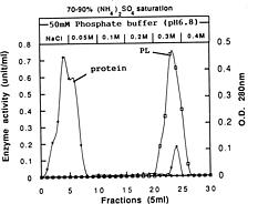
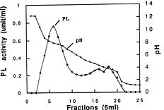
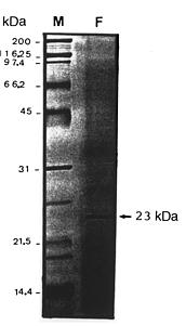
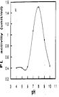
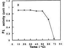
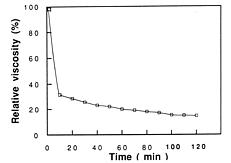
Abstract. A pectin lyase (PL, EC 4.2.2.10) was produced by Pythium splendens Braun in infected cucumber fruits. The enzyme was purified to near homogeneity by ammonium sulfate fractionation, CM - Sepharose column chromatography, and preparative isoelectric focusing. The enzyme had a molecular mass of 23 kDa as determined by SDS-PAGE. The pI of the PL was 8.0, which was also the optimum pH for the enzyme activity. The enzyme was stable at 4_50°C, but beyond 50°C, its activity decreased rapidly. The pectin lyase degraded high methyl-esterified pectin by trans-elimination in an endo-manner, and was stimulated by some divalent cations including Ca2+, Mg2+, and Sr2+. Addition of Zn2+ inhibited the enzyme activity by 36%. The purified PL caused rapid maceration of potato and cucumber discs. This is the first report of a PL produced by Pythium splendens, a plant pathogenic fungus which causes seedling damping-off and fruit rot in cucumber.

Keywords: Cucumber (Cucumis sativa L.); Pectin lyase; Pythium splendens.

Introduction

Plant pathogenic fungi produce an array of extracellular degradative enzymes that may be important in pathogenesis (Walton, 1994). Most attention has focused on enzymes that depolymerize pectin (Collmer and Keen, 1986; Cooper, 1984). Pectin lyase (PL, EC 4.2.2.10) catalyzes the trans-elimination of pectin and has been isolated from a variety of bacterial and fungal pathogens (Collmer and Keen, 1986; Linhardt et al., 1986). Its role in tissue maceration by plant pathogenic bacteria has been demonstrated using mutants that lack PL activity (Beaulieu et al., 1993). Recently, several reports have suggested that PL may be a pathogenicity factor in fungal-plant interactions (Crawford and Kolatlukudy, 1987; Wattad et al., 1994; Wattad et al., 1995; Wijesundera et al., 1984).

Pythium splendens Braun is an economically important soil borne plant pathogenic fungus which causes seedling damping-off and fruit rot in cucumber (Cucumis sativa L.). This fruit rot is apparently caused by the activity of pectolytic and/or cellulolytic enzymes, but this is still unverified. In many pythium diseases, secretion of cell wall-degrading enzymes has been reported (Cherif et al., 1991; Dube and Prabakaran, 1989). The loosening of primary walls by pectolytic enzymes may cause osmotic stress and subsequent cell death (Hall and Wood, 1973). The softening of cucumber fruit in cottony leak disease, caused by Pythium aphanidermatum, has been demostrated to be related to the activities of endo-polygalacturonase and cellulase (Zamski and Peretz, 1996).

However, neither the production of pectolytic enzymes nor cell wall-degrading enzymes are available for P. splendens infected cucumber. This is the first report on the production, purification, and characterization of a pectin lyase for P. splendens infected cucumber fruits.

Materials and Methods

Pythium splendens was isolated from the roots of a diseased cucumber seedling near Taipei and was maintained routinely on V-8 agar slants (200 ml V-8 juice, 3 g CaCO3, 15 g agar in 800 ml distilled water) at 28°C.

Inoculum

Three discs (10 mm dia) were cut from a V-8 agar plate and were grown in a 10 ml V-8 liquid medium in a 50 ml Erlenmeyer flask for 4 days at room temperature. Inocula were obtained after homogenization and centrifugation of the culture at 20,000 g for 10 min. The sporangia-containing pellets were resuspended in distilled water and used as inoculum.

Enzyme Preparation

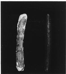
Cucumber fruits (Cucumis sativa L.) were purchased from a local supermarket. They were surface sterilized by washing in 10% (v/v) sodium hypochlorite for 10 min and then rinsed twice in distilled water. The fruits were aseptically punched with a needle before inoculation with the sporangium suspension (2×10³ sporangia/ml). Controls were inoculated with several drops of sterilized water on the fruit surface. Inoculated fruits were kept in chambers under 100% relative humidity at 25°C for 3 days. Rotted

3Corresponding author. Tel: 886-2-27823050 ext. 424; Fax: 886-2-26515042.


Botanical Bulletin of Academia Sinica, Vol. 39, 1998

tissue was collected, removed with a scalpel, weighed, and ground in a Waring blender for 2 min in 20 ml of distilled water. Ground tissue was strained through two layers of cheesecloth and centrifuged at 20,000 g for 15 min at 4°C. The supernatants were dialyzed against distilled water for at least 16 h at 4°C, lyophilized and used as crude enzyme preparations.

Enzyme Assay

The PL activity was measured by the periodate-thiobarbituric acid (TBA) method (Preiss and Ashwell, 1963). The activity was indicated by the formation of a red chromagen which had a maximum absorbance at 548 nm. The reaction mixture contained 0.25 ml of 0.5% pectin [93% degree of esterification (DE90), Sigma P-9561] in 50 mM Tris-HCl buffer (pH 8.0) plus 0.2 mM CaCl2 and 0.25 ml enzyme. The reaction mixture was incubated for 1 h at 30°C. One unit of the PL activity was defined as the amount of enzyme that increased the absorbance by 0.05 in 1 h at 548 nm.

Controls contained autoclaved enzymes instead of active enzymes. Protein concentration was determined by the method of Lowry et al. (1951). Crystalline bovine serum albumin was used as the reference protein.

Enzyme Purification

Five grams of lyophilized crude enzyme extract of P. splendens was dissolved in 100 ml of 50 mM Tris-HCl buffer (pH 8.0) and dialyzed against two liters of distilled water for 24 h at 4°C. The dialyzed solution was brought to 30% saturation with powdered ammonium sulfate, allowed to stand for 30 min at 4°C and centrifuged for 10 min at 20,000 g. The precipitate was collected and dissolved in 10 ml of cold distilled water. This procedure was repeated for ammonium sulfate saturations of 50, 70, and 90%. The precipitate of each fraction was immediately assayed for protein and PL activity. The fraction, which contained a large amount of PL was used for further purification on CM-Sepharose (Pharmacia) column chromatography. The column size was 1.7×40 cm and the CM-Sepharose was equilibrated with 50 mM phosphate buffer (pH 6.8). The column was eluted with 60 ml of 50 mM phosphate (pH 6.8), followed by a 25 ml step-wise NaCl gradient in buffer until 0.3 M NaCl was reached. Five ml fractions were collected and assayed for PL activity.

The fractions from the CM-Sepharose elution, containing the PL enzyme, were combined and dialyzed for 24 h at 4°C. The enzyme was subjected to isoelectric focusing in a LKB 8101 Ampholine electrofocusing apparatus equipped with 110 ml column (LKB Produkter AB, Bromma, Sweden) containing a pH 3.0_10 Ampholine carrier. After electrofocusing, 5-ml fractions were collected, and the pH of each fraction was immediately measured.

The endo- or exo- mode of PL was determined by measuring the decrease in relative viscosity in an Ostwald vis

cometer as described by Hancock et al. (1964). Reaction mixtures contained 5 ml of 0.5% pectin (DE 90) in 50 mM Tris-HCl buffer pH 8.0 with 0.2 mM CaCl2 plus 1 ml of distilled water and 1 ml of enzyme at 30°C.

Sodium Dodecyl Sulphate Polyacrylamide Gel Electrophoresis (SDS-PAGE)

SDS-PAGE was performed by the method of Laemmli (1970) on a 12% acylamide (Sigma) 75 mm thick gel. Proteins were analyzed electrophoretically on a Hoefer Cooled Dual Slab Unit SE-600 (Hoefer Scientific Instruments, San Francisco, USA) at 200 v for 45 min before silver staining the gels as described by Blum et al. (1987). The molecular weight of PL was estimated by comparing its mobility with that of protein standards (Bio Rad Lab. Inc., CA, USA).

Effect of Temperature and pH on Enzyme Activity

The stability of PL was performed by placing enzyme samples (5 ml) in a water bath at the tested temperatures for 30 min. The tubes were cooled in cold water (12°C) and then assayed for PL activity. The effect of pH on the enzyme activity was carried out by 50 mM citrate buffer (pH 4_5), 50 mM phosphate buffer (pH 8_9), and 50 mM borax buffer (pH 10).

Effect of Divalent Cations on PL Activity

The divalent cations (Ca2+, Ba2+, Sr2+, Mg2+, Mn2+, Zn2+ and Co2+) were added to the substrate buffer (0.5% pectin in 50 mM Tris-HCl buffer, pH 8.0) in place of EDTA to give a final concentration of 0.2 mM, and the PL activity was measured as previously described.

Tissue Maceration

Tissue maceration was evaluated and rated as described by Tseng and Mount (1974). Discs (10×1 mm) of potato tubers and cucumber fruit tissues were used as the substrate. Discs were placed in enzyme preparations buffered at pH 8.0 with 5 mM Tris-HCl buffer. Measurements of tissue maceration were made over a 100 min incubation period with the enzyme. Discs which were not macerated received a rating of 0 and those macerated completely received a score of 5.

Chemicals

All chemicals were reagent grade and were purchased from Sigma Co (St. Louis, MO. USA), except periodate-thiobarbituric acid was purchased from E. Merck (Germany).

Results

Pectin lyase was isolated from cucumber rot tissues after inoculation with P. splendens at 25°C for 72 h as shown in Figure 1. No PL enzyme was detected in either uninoculated and inoculated sterile water fruit tissues.


Chen et al. — A pectin lyase from Pythium splendens

Figure 2. Elution profile of the pectin lyase from Pythium splendens infected cucumber fruits by CM-Sepharose column chromatography. Three ml of dialyzed enzyme from the 70_90% ammonium sulfate saturation were applied to the column (1.7×40 cm) at 4°C. Fractions (5.0 ml) were collected, and the enzyme activity was immediately assayed.

Figure 1. Symptom expression in cucumber fruit after inoculation with Pythium splendens at 25°C for 72 h. R: rot tissue with the fungus; L: control, fruit inoculated with sterile water.

Purification of Pectin Lyase

Figure 2 showed the elution profile of a major PL peak in the 70_90% (NH4)2SO4 fraction from CM-Sephaose column. The enzyme within the 105_130 ml eluate (fractions 21 to 26) from the column were pooled, dialyzed, and subjected to isoelectric focusing using an Ampholine carrier which had a pH range of 3_10 (Figure 3). Fraction 6, which showed the highest level of the PL activity, was pooled and concentrated by acetone precipitation. The recovery and purification of the pectin lyase were achieved (Table 1). The purified enzyme was used for the following experiments: (a) tissue maceration and (b) enzyme characterization (i.e., molecular mass, pH activity, heat stability, and cofactor dependancy).

Characterization of the Pectin Lyase

The purified PL had a molecular mass of 23 kDa by SDS-PAGE (Figure 4) and a pI of 8.0 (Figure 3). The enzyme caused a rapid decrease in the viscosity of pectin

Figure 3. Resolution of the pectin lyase from fractions 21_26 eluted from the CM-Sepharose column described in Figure 2. Separation was accomplished by isoelectric focusing using Ampholine carriers with a pH range of 3_10 at 4°C for 48 h. Fractions (5.0 ml) were collected and immediately measured for pH values. After dialysis at 4°C for 24 h, each fraction was checked for enzyme activity.

Table 1. Purification of the pectin lyase from Pythium splendens infected cucumber fruits.

Purification steps Total protein Total activity Specific activity Purification Yeid

(mg)b (unit)a (units/mg protein) (fold) (%)

Culture filtrate 276.00 1,892.0 6.9 1.0 100.0

70_90% (NH4)2SO4 saturation 38.30 1,101.0 28.8 4.2 58.1

CM-Sepharose column 0.20 91.0 455.0 66.0 4.8

Isoelectric focusing 0.15 80.2 534.6 77.4 4.2

aOne unit of the pectin lyase activity is defined as that amount of enzyme giving an increase in absorbance of 0.05 in 1 h at 548 nm.

bProtein was determined by the method of Lowry et al. (1951).


Botanical Bulletin of Academia Sinica, Vol. 39, 1998

Figure 4. SDS-PAGE of the pectin lyase of Pythium splendens after purification by CM-Sepharose column chromatography and isoelectric fousing. Lane F, pectin lyase from infected cucumber fruits; lane M, molecular weight size standards (kDa).

Figure 6. Effects of pH (A) and temperature (B) on the purified pectin lyase activity of Pythium splendens.

(DE 90), suggesting a random (endo) cleavage of the substrate (Figure 5). The pectin lyase had maximum activity under alkaline conditions (Figure 6A). Treatment of the enzyme at temperature ranging from 4 to 50°C for 30 min did not affect its activity as compared with that of 4°C. At temperatures higher than 50°C, the enzyme activity was drastically reduced. At 70°C, the enzyme lost its activity completely (Figure 6B). The effect of Ca2+, Mg2+, Sr2+, Mn2+, Ba2+, Zn2+, and Co2+ on the PL activity was also de

termined. The enzyme activity was stimulated by the divalent cations including Ca2+, Mg2+, and Sr2+. Addition of Zn2+ inhibited the enzyme activity by 36%. EDTA did not affect enzyme activity but inhibited stimulation by Ca2+ (Table 2). As for tissue maceration, both tuber discs of potato or cucumber were macerated completely by the purified PL preparation within 60 min, while the controlled discs in 50 mM Tris-HCl buffer (pH 8.0) were unaffected (Table 3).

Table 2. Effect of divalent cations on the activity of purified pectin lyase from Pythium splendens infected cucumber fruitsa.

Cationb addition Relative activity

Control (no addition) 100

Control+EDTAc 103

Control+EDTAc+Ca2+ 101

Ca2+ 134

Mg2+ 126

Ba2+ 90

Co2+ 91

Mn2+ 89

Zn2+ 64

Sr2+ 116

aCofactors and EDTA were dissolved in 0.5% pectin substrate in 50 mM Tris-HCl buffer (pH 8.0).

bReaction mixture without adding any cofactors or EDTA.

cEDTA was added at a final concentration of 0.2 mM.

Figure 5. The rates of decrease in the viscosity of pectin (DE 90) by the purified pectin lyase of Pythium splendens.


Chen et al. — A pectin lyase from Pythium splendens

Table 3. Tissue maceration ratings of potato and cucumber discs treated with the purified pectin lyase from Pythium splendens infected cucumber fruits.

Tissue Time (min) Macerationa

Potato 20 2

40 3

60 5

80 5

100 5

Cucumber 20 2

40 4

60 5

80 5

100 5

aTissue maceration of potato or cucumber disc (10×1 mm) was measured in 3 ml of enzyme preparation buffered with 50 mM Tris-HCl buffer (pH 8.0) at 30°C. For cucumber seedling maceration tested, segments (0.5×10 mm) instead of discs were used. All experiments were performed twice with buffer (50 mM Tris-HCl at pH 8.0) as control. A value of 1 indicates no maceration, whereas a value of 5 indicates complete maceration.

0.2 mM Ca2+, which increased activity by 1.34 times (Table 2). Similar results have been reported by elsewhere (Delgado et al., 1992; Starr and Moran, 1962; Whilaker, 1972). Slight stimulation was obtained with Mg2+ and Sr2+. Addition of Zn2+ inhibited the enzyme activity. This inhibition has also been reported elsewhere (Delgado et al., 1992). Addition of EDTA did not affect enzyme activity, but did inhibit stimulation by Ca2+ (Table 2).

The current study illustrated that purified P. splendens PL was able to macerate both potato and cucumber tissues rapidly. This is a clear indication that the enzyme is involved in the disease development. Ours is the first report of PL produced by P. splendens, a plant pathogenic fungus which causes seedling damping-off and fruit rot of cucumber.

Achnowledgements. We are grateful to Dr. J. C. Tu for his constructive comments in preparation of this paper and Miss H. N. Tsai for her help in typing manuscript. This work was partially supported by Academia Sinica and the National Science Council (Grant NSC 83-0211-B-001-071), Taiwan, Republic of China.

Literature Cited

Beaulieu, C., M. Boccara, and F. Van Gijsegem. 1993. Pathogenic behavior of pectinase-defective Erwinia Chrysanthemi mutants on different Plants. Mol. Plant-Microbe Interact. 6: 197_202.

Blum, H., H. Beier, and H.J. Gross. 1987. Improved silver staining of plant proteins, RNA and DNA in polyacrylamide gels. Electrophoresis 8: 93_99.

Bugbee, W.M. 1990. Purification and characteristics of pectin lyase from Rhizoctonia solani. Physiol. Mol. Plant Path. 36: 15_25.

Cherif, M., N. Benhamou, and R.R. Belanger. 1991. Ultranstructural and cytochemical studies of fungal development and host reactions in cucumber plants infected by Pythium ultimum. Physiol. Mol. Plant Path. 39: 353_375.

Collmer, A. and N.T. Keen. 1986. The role of pectic enzymes in plant pathogenesis. Ann. Rev. Phytopath. 24: 383_409.

Cooper, R.M. 1984. The role of cell wall degrading enzymes in infection and damage. In R.K.S. Wood and G. J. Jellis (eds.), Plant diseases: Infection, Damage and Loss. Blackwell Scientific Publications, Oxford, pp. 13_27.

Crawford, M.S. and P.E. Kolatlukudy. 1987. Pectate lyase from Fusarium solani f. sp. pisi: Purification, characterization, in vitro translation of the mRNA, and involvement in pathogenicity. Arch. Biochem. Biophys. 258: 196_205.

Delgado, L., A.T. Blanca, C. Huitron, and G. Aguilar. 1992. Pectin lyase from Aspergillus sp. CH-Y-1043. Appl. Microbiol. Biotechnol. 39: 515_519.

Dube, H.C. and K. Prabakaran. 1989. Cell wall degrading enzymes of Pythium. Perspective Plant. Path. 89: 181_188.

Godfrey, V., L.J. Hyman, G.P. McMillan, D.C. Old, and M.C.M. Perombelon. 1994. Purification and characterization of mitomycin C-induced pectate lyase of Erwinia carotovora subsp. atroseptica strain SCRI 1043. J. Appl. Bacteriol. 76: 13_21.

Hall, J.A. and K.R.S. Wood. 1973. The killing of plant cells by pectolytic enzymes. In R.J.W. Byde and C.V. Cutting (eds.),

Discussion

Tissue disintegration is commonly found in a variety of rots and damping-off diseases caused by Pythium spp. The involvement of enzymes such as pectinolytic enzymes and cellulases in cell wall degradation, in tissue disintegration, and in softening of cucumber fruits by P. aphanidermatum has been demonstrated (Winstead and McCombs, 1961). Several investigations have been made on the production of pectinolytic enzymes by Pythium spp. (Dube and Prabakaran, 1989; Turner and Bateman, 1968; Zamski and Peretz, 1996). However, there are no reports on the production of the enzymes by P. splendens. In order to elucidate the role of the pectinolytic enzyme in the pathogenesis of cucumber fruit rots, a highly purified enzyme is needed.

The PL was purified to near homogeneity and was characterized. No polygalacturonase, pectate lyase, pectate methyl esterase, or cellulase activity were found. The enzyme degraded methyl-esterified pectin by trans-elimination in an endo-manner and showed similarities to other fungal and bacterial endo-PLs (Bugbee, 1990; Godfrey et al., 1994; Wijesundera et al., 1984).

Temperature and pH optima (43°C/8.0) were similar to endo PLs from Rhizoctonia solani (Bugbee, 1990) Collectotrichum lindemuthianum (Wijesundera et al., 1984) and Erwinia chrysanthemi (Tsuyumu and Funakbo, 1985). With a pI of 8.0 and molecular mass of 23 kDa, the PL closely resembled the PL from C. lindemuthianum (8.2/23 kDa) (Wijesundera et al., 1984), whereas the enzymes from R. solani, Penicillium italicum, and E. chrysanthemi had higher pIs and mol. wts. (Laemmli, 1970; Tsuyumu and Funakbo, 1985).

The effect of divalent cations on the PL activity was measured. The highest stimulation level was obtained at


Botanical Bulletin of Academia Sinica, Vol. 39, 1998

Fungal Pathogenicity and the Plant's Response. Academic Press, London, pp. 19_38.

Hancock, J.G., R.L. Millar, and J.W. Lorbeer. 1964. Pectolytic and cellulolytic enzymes produced by Botrytis allii, B. cinerea and B. squamosa in vitro and in viro. Phytopathology 54: 928_935.

Laemmli, U.K. 1970. Cleavage of structural proteins during the assembly of the head of bacteriophage T4. Nature 227: 680_685.

Linhardt, R.J, P.M. Galliher, and C.L. Cooney. 1986. Polysaccharide lyases. Appl. Biochem. Biotechnol. 12: 135_176.

Lowry, O.H., N.J. Rosebrough, A.L. Farr, and R.J. Randall. 1951. Protein measurement with the Folin phenol reagent. J. Biol. Chem. 193: 265_275.

Preiss, J. and G. Ashwell. 1963. Polygalacturonic acid metabolism in bacteria. I. Enzymatic formation of 4-deoxy-L-threo-5-hexocellulose uronic acid. J. Biol. Chem. 283: 1571_1576.

Starr, M. and F. Moran. 1962. Eliminative split of pectic substances by phytopathogenic soft-rot. Science 135: 920_921.

Tseng, T.C. and M.S. Mount. 1974. Toxicity of endopoly-galacturonate trans-eliminase, phosphatidase and protease to potato and cucumber tissue. Phytopathology 64: 229_236.

Tsuyumu, S. and T. Funakbo. 1985. Purification and partial characterization of a mitomycin C-induced pectin-lyase of Erwinia Chrysanthemi stain EC183. Physiol. Plant Path. 27: 119_130.

Turner, M.R. and D.F. Bateman. 1968. Maceration of plant tis

sues susceptible and resistant to soft-rot pathogens by enzymes from compatible host-pathogen combinations. Phytopathology 58: 1509_1515.

Walton, J.D. 1994. Deconstructing the cell wall. Plant physiol. 104: 1113_1118.

Wattad, C., A. Dinoor, and D. Prusky. 1994. Purification of pectate lyase produced by Colletotrichum gloeosporioides and its inhibition by epicatechin: a possible factor involved in the resistance of unripe avocado fruits to anthracnose. Mol. Plant-Microbe Interact. 7: 293_297.

Wattad, C., S. Freeman, A. Dinnor, and D. Prusky. 1995. A nonpathogenic mutant of Collectotrichum magna in deficient in extracellular secretion of pectate lyase. Mol. Plant-Microbe Interact. 8: 621_626.

Whilaker, I.R. 1972. Pectic substances, pectic enzymes and haze formation in fruit juices. Enz. Microbiol. Technol. 6: 341_349.

Wijesundera, R.L.C., J.A. Bailey, and R.J.W. Byrde. 1984. Production of pectin lyase by Collectrotrichum lindemuthianum in culture and in infected bean (Phaseolus vulgaris) tissue. J. Gen. Microbiol. 130: 285_290.

Winstead, N.N. and M.C. McCombs. 1961. Pectinolytic and cellulolytic enzyme production by Pythium aphanidermatum. Phytopathology 51: 270_273.

Zamski, E. and I. Peretz. 1996. Cavity spot of carrots: II Cell-wall-degrading enzymes secreated by Pythium and pathogen-related porteins produced by the root cells. Ann. Appl. Biol. 128: 195_207.