Bot. Bull. Acad. Sin. (1998) 39: 209_212

Zheng and Ho — The rediscovery of Phytophthora polygoni

The rediscovery of Phytophthora polygoni Saw.

Xiabo Zheng1 and H.H. Ho2,3

1Plant Pathogen Research Institute, Nanjing Agricultural University, Nanjing 210095, People's Republic of China

2Department of Biology, State University of New York, New Paltz, New York, 12561, USA

(Received January 14, 1998; Accepted March 16, 1998)

Abstract. Phytophthora polygoni Saw. was first described as a new species by Sawada in 1922, causing leaf spot disease of Polygonum japonicum Meisn. in Taiwan. However, there has been no report of the fungus since its first discovery. Recently, a similar disease occurred on Rumex dentatus L. in Nanjing of Jiangsu Province in mainland China and the pathogen was identified as P. polygoni based on the similar biological characteristics.

Keywords: Phytophthora polygoni; Rumex dentatus.

Introduction

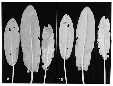
Phytophthora polygoni Saw. was first discovered and described on the leaves of Polygonum japonicum Meisn. (Family Polygonaceae) in Taipei and Taichung of Taiwan causing leaf spot disease from the late winter/early spring to early summer (Sawada, 1922). The disease first appeared as small, round yellow green spots which became dull yellow brown with a tint of purple. Subsequently, the lesions enlarged and turned brown and diffuse, resulting in the death of the lower leaves.

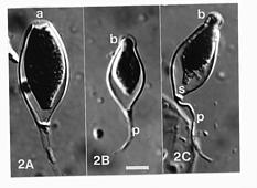
The hyphae of the pathogen were intercellular within the leaf spots, aseptate, colorless and branching with hyphal diameter 5_15 µm (usually 12_14 µm) wide. Appresoria in host cells were mostly obovate in shape. Intercellular hyphae grew through the stomata to form clusters of slender, unbranched or sympodially branched sporangiophores, 15_75 µm long and 1.5_5 µm wide (4_20 sporangiophores per stoma) each terminating in a sporangium with a conical papillum. The sporangia were mostly elongated ellipsoidal to fusiform or pyriform, 17_42 × 4_20 µm. When placed in water, a sporangium germinated either by a germ tube or by releasing motile laterally biflagellated ovoid zoospores with obtuse apex and rounded base, 11_15 × 6_9 µm, encysting to become 10_15 µm in diameter. Neither chlamydospores nor resting sporangia were observed, and no sexual structures were found. Attempts to grow the fungus on culture media failed.

Unfortunately, type specimen of P. polygoni is no longer in existence for re-evaluation, and the fungus has not been reported again since its first description. Efforts were thus made to locate Polygonum and related genera within the family Polygonaceae, with the symptoms as described by Sawada, hoping that P. polygoni could be rediscovered.

Materials and Methods

One genus of the family Polygonaceae, Rumex is a common weed in the Nanjing region of Jiangsu Province in mainland China. Field trips were made to detect and collect Rumex plants whose leaves showed leaf spot disease as described by Sawada (1922).

Freshly collected leaf specimens were prevented from dessication by keeping them in a petri dish with two pieces of moist filter paper on the bottom. They were exposed to natural daylight for 28_48 h at 18_20°C to induce sporangial formation from leaf lesions. The diseased leaf was then examined under microscope at ×150 for the presence of sporangia, which were then carefully scaped off on a glass slide and mounted in a drop of lactophenol, with or without 0.05% cotton blue.

In order to isolate the fungus, two kinds of culture media were used: Lima bean agar (Zhou et al., 1997) and V-8 agar (Ann et al., 1990). Prior to pouring agar medium into sterilized petri dishes, 50 µg/ml each of penicillin, rifampicin, and PCNB (Pentachloronitrobenzene) were added. Diseased leaf was not surface-sterilized, but washed clean with running tap water and allowed to air-dry. Six small pieces of diseased tissue (about 4 × 4 mm) were cut out with a pair of scissors and placed along the edge of an agar plate, at equal distance from one another. At least three agar plates were used for each diseased leaf. In addition, diseased leaf tissues were also surface-sterilized for 5 minutes in 2% chlorine solution prepared by dissolving 1 bleach tablet in 10 ml distilled water. The bleach tablets are produced by Xuhang Chemical Company in Shanghai and marketed by Shanghai Pharmaceutical Company. The sterilized plant material was placed on Lima bean agar and V-8 agar plates without the addition of the above antimicrobial chemicals. The inoculated plates were incubated in dark at 18°C or 25°C for at least one week.

3Corresponding author. Tel: 914-257-3780; Fax: 914-257-3791; E-mail: hoh@npvm.newpaltz.edu


Botanical Bulletin of Academia Sinica, Vol. 39, 1998

Results

Leaf spot disease was commonly seen on Rumex dentatus L. in Nanjing City in the spring (late March to Mid-April) when the outdoor temperature was about 5_20°C. Diseased specimens were collected from early spring till as late as early June when the temperature rose to 33°C.

The lesion spots on the leaf were roundish to irregular in shape with diffuse edges. Initially, the lesion appeared as blurry, pinkish brown spots on the leaf surface (Figure 1A), but the color was not altered or light pink when viewed from the leaf bottom (Figure 1B). No significant senescence of leaf cells was observed in the lesion. The leaf spots then changed to reddish brown in color and eventually turned light brown to greyish black. Under humid conditions downy mildew-like fungus could be vaguely observed on the surface of a fresh leaf specimen's lesions. Under the microscope, clusters of ten to more than thirty sporangia could be seen emerging from a single stoma on the leaf surface. Fewer sporangia were found on old lesions with mostly dead leaf cells.

The fungal hyphae (8_12 µm wide) were colorless, aseptate, branching freely between leaf cells within the lesions sending oval haustoria into host cells. The sporangia were small, mostly ellipsoidal to elongated ellipsoidal, fusiform or obpyriform, each with a tapered base and a slightly elongated beak (Figure 2A_C). Sporangia measured (18.9-) 37.1 (-28.8) × (11.3-) 17.7 (-23.8) µm with length/breadth ratio of (1.86-) 2.1 (-2.8) and were semi-papillate with the apical thickening (0.5-) 1.7 (-2.7) µm deep. Zoospores were formed within the sporangium and expelled through the sporangial apex via an exit pore (5.0-) 6.4 (-7.5) µm in width. The sporangiophores were (1.8-) 2.2 (-2.5) wide, unbranched or occasionally branched sympodially in later stages. Mature sporangia were often deciduous each with a medium pedicel 20_45 µm in length, from which it was cut off by a basal cross wall.

Sexual structures were not found in diseased tissues, cleared by immersing in lactophenol or 2% potassium hydroxide solution and then examined under microscope, stained or unstained with 0.05% cotton blue. No fungal growth was detected on Lima bean agar or V-8 juice agar medium, despite numerous attempts.

Figure 1. Leaf spot disease symptoms of Rumex dentatus caused by Phytophthora polygoni, (from left to right, early, intermediate, and final stages). (A) Upper surface of diseased leaf; (B) Lower surface of diseased leaf.


Zheng and Ho — The rediscovery of Phytophthora polygoni

spite numerous attempts. However, the pathogenicity of P. polygoni to Rumex dentatus still needs to be tested by artificial inoculation. The absence of sex organs within the host plant can be attributable to the possible heterothallism of the fungus. The inoculating of zoospores from sporangia collected from different localities on the same leaf may increase the chance of finding the sexual stage. On the other hand, the production of sex organs may be induced by manipulating the environmental conditions. The absence of sex organ description has not prevented Stamps et al. (1990) from including P. gonapodyides (Peterson) Buisman, P. japonica Waterh., P. undulata (Petersen) Dick and P. palmivora var. heterocystica Babacauh in their revised tabular key to the species of Phytophthora. Although P. botryosa Chee produces small, narrowly elongated sporangia it can be easily distinguished from P. polygoni due to its ease of growth on common agar plates (Chee, 1969). Of those few Phytophthora species which are not known in culture, and produce semi-papillate sporangia on the host plant (Stamps et al., 1990; Waterhouse, 1963), P. cyperi (Ideta) S. Ito, P. cyperi-bulbosa Seethal. & Ramakr., and P. lepironiae Saw. all produce sex organs and significantly larger sporangia, distinct from P. polygoni. Thus, P. polygoni should be accepted as an identifiable Phytophthora species. Further attempts will be made to search for the sexual stage of P. polygoni in the field and under experimental conditions in the laboratory or greenhouse, in order to elucidate the life cycle of this obscure fungus.

Dried diseased specimens of Rumex dentatus with P. polygoni sporangia have been deposited in the herbarium of the Department of Plant Protection at Nanjing Agricultural University.

Acknowledgements. The work was supported in part by a grant from the National Geographic Society to H.H. Ho.

Literature Cited

Ann, P.J., R. Kumimoto, and W.H. Ko. 1990. Phytophthora wilt of carnation in Taiwan and Hawaii. Plant Prot. Bull. 32: 145_147.

Chee, K.H. 1969. Variability of Phytophthora species from Hevea brasiliensis. Trans. Br. Mycol. Soc. 52: 425_436.

Ho, H.H. and H.S. Chang. 1992. A re-evaluation of Phytophthora species described by K. Sawada in Taiwan. Mycotaxon 43: 297_316.

Sawada, K. 1922. Descriptive Catalogue of the Formosan Fungi II. Rept. Dept. Agr. Gov. Res. Inst. Formosa 2: 1_173.

Stamps, D.J., G.M. Waterhouse, F.J. Newhook, and G.S. Hall. 1990. Revised tabular key to the species of Phytophthora. Mycol. Pap. 162. C.A.B. International, Kew, Surrey, England, 28 pp.

Waterhouse, G.M. 1963. Key to the species of Phytophthora de Bary. Mycol. Pap. 92. C.M.I., Kew, Surrey, England, 22 pp.

Zhou, Y.C., X.B. Zheng, J.Y. Lu, and W.H. Ko. 1997. Sexuality type change from self-induction to cross-induction in Phytophthora boehmeriae. Mycol. Res. 101: 1047_1050.

Figure 2. Detached sporangia of Phytophthora polygoni. (A) Shallow apical thickening (a); (B) and (C). Beaked papillum (b) with pedicel of medium length (p) and basal septum (s). Scale bar equals 10 µm. All at same magnification.

Discussion

Sawada (1922) erected the new species Phytophthora polygoni primarily based on the small, narrowly elongated to fusiform sporangia, distinct from the significantly larger, oval to obpyriform sporangia of other Phytophthora species known at that time. He did point out that Dastur (no literature cited) found an unnamed species of Phytophthora from Hevea in Burma with sporangia of similar size and shape. Waterhouse (1963) excluded P. polygoni from her key to Phytophthora species for the lack of description on its sex organs. Ho and Chang (1992) re-examined the taxonomic status of all 23 species of Phytophthora described by Sawada in Taiwan based on a study of available dried plant specimens, type/authentic cultures, and the original publications. Unfortunately, neither of the two specimens of P. polygoni, collected from Taipei and Fangyun of Taichung, as cited by Sawada (1922) could be located and they tentatively accepted the judgment of Waterhouse that this species is insufficiently described to be recognized.

However, the rediscovery of a Phytophthora species in the Nanjing area of mainland China with pathological and morphological characteristics matching closely with those described for P. polygoni permitted us to re-evaluate the taxonomy of this species. Firstly, the newly found Phytophthora species is associated with a similar leaf spot disease on related plant hosts within the same family Polygonaceae, which includes both Polygonum and Rumex. Secondly, the disease occurred under similar environmental conditions when the temperature was cool and wet. The two specimens of P. polygoni on Polygonum japonicum were collected from Taipei and Taichung in March and May, when the weather was similar to that of the Nanjing area in springtime, when the leaf spot disease on Rumex dentatus was most prevalent. In both cases, the fungus could not be cultured on common agar media de