Bot. Bull. Acad. Sin. (1998) 39: 261_267

Yang and Lin — Characteristics from Rhizobium fredii

Cytostructure, lipopolysaccharides, and cell proteins analysis from Rhizobium fredii

Fang-Lin Yang and Liang-Ping Lin1

Graduate Institute of Agricultural Chemistry, National Taiwan University, Taipei 106, Taiwan, Republic of China

(Received January 10, 1997; Accepted June 2, 1998)

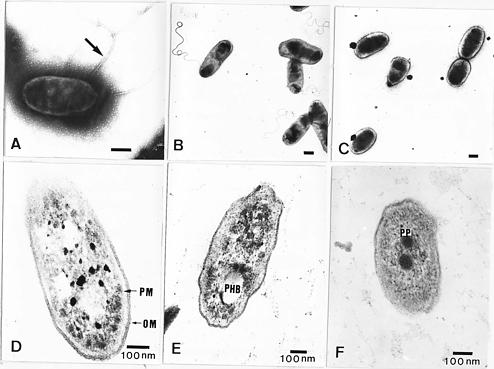
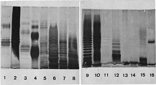
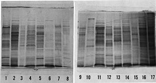
Abstract. The symbiotic roles of rhizobia with legumes have been extensively investigated for agricultural applications. We isolated several strains of rhizobia from Taiwanese soils and analyzed their cellular proteins and lipopolysaccharide (LPS) profiles as well as their ultrastructure by electron microscopy. It was found that the flagella have different forms and locations on the surface of fast-growing and slow-growing rhizobia. In addition, the results of their ultrastructural visualization confirmed that poly-b-hydroxybutyrate (PHB) and polyphosphates are present in rhizobia. The ladder profiles of LPS and cellular proteins on sodium dodecylsulfate-polyacrylamide gel electrophoresis (SDS-PAGE) indicated that different strains of rhizobia possess different profiles, but strains which belong to the same species have similar profiles. It was also shown by Western blot and immunoassay that strain specificity exists between rhizobia cellular proteins and the antisera of anti-rhizobia cells. These results confirm that indigenous R. fredii exhibits a significant difference from other species of Rhizobiaceae.

Keywords: Anti-cellular protein antibody; Anti-LPS antibody; Cell proteins; Cytostructure; Lipopolysaccharides; Rhizobium fredii; Soybean.

Introduction

The taxonomy of rhizobia, based on bacteria-plant cross inoculation groups, was at one time widely used (Elkan, 1992). However, it is now being replaced by numerical taxonomy, serology, metabolic components, and various molecular techniques (Hennecke et al., 1985; Javis et al., 1986; De Lajudie et al., 1994). All of these methods are able to recognize the fast-growing genus Rhizobium and the slow-growing genus Bradyrhizobium by their bacterial growth rate (Jordan, 1984; Elkan, 1992).

Based on our knowledge of Rhizobium-legume symbiosis, the rhizobial cell is involved in interactions with host plants during nodule development (Schmidt et al., 1989; Stacey et al., 1995). The cell surface components of Rhizobium have always been considered important factors in symbiotic effectiveness (Halverson and Stacey, 1986; Hitchcock et al., 1986; Dylan et al., 1990; Gray and Rolfe, 1990; Carlson et al., 1991; Yokota and Sakane, 1991; Niehaus et al., 1993; Ozga et al., 1994) in the specific interactions of rhizobia with legumes. Lipopolysaccharide (LPS) is one component of the bacterial outer membrane and consists of a polysaccharide and lipid A (Hitchcock et al., 1986). Various rhizobial strains differ in LPS composition and serological characters that can nodulate the legume host. Thus, LPS has taxonomic significance in Rhizobiaceae (Yokota and Sakane, 1991). In bacterial taxonomy, the diversity of cellular proteins

in different species is also considered an important factor.

In the present study, we extracted LPS and cellular proteins from several strains of Rhizobium and Bradyrhizobium. These strains were isolated from indigenous soils and purchased from the American Type Culture and Collection (Rockville, MD). The LPS and cellular proteins were analyzed by SDS-polyacrylamide gel electrophoresis and by immunoassay after Western blotting. Based on the results, we can compare the SDS-PAGE profiles of Rhizobium and Bradyrhizobium as well as different species of Rhizobium. In addition, we analyzed the ultrastructural differences between Rhizobium and Bradyrhizobium by electron microscopy, because these results also provide some information for taxonomy, especially in Rhizobiaceae. Significant differences exist in the cellular structure between that of Rhizobium and Bradyrhizobium. From these results, we can obtain more information to confirm the taxonomic significance of indigenous Rhizobium fredii.

Materials and Methods

Strains of Rhizobia and Cultural Conditions

Nine strains of R. fredii and R. loti Lu7 were isolated from Taiwanese soils. The collected legumes (soybean) nodules (ca. 0.3 cm diameter) were washed with distilled water several times. This washing and cleaning procedure was done within 4 h of nodule collection. The nodules were then soaked in 4% H2O2 for 2 min and then washed

1Corresponding author. Fax: (02) 2362-6455.


Botanical Bulletin of Academia Sinica, Vol. 39, 1998

with sterile water for 3 min. Final nodule treatment included soaking in a 0.1% HgCl2 solution for 2 min and then washing with distilled water. We visually selected the effective nodules based on their pink coloration, which indicated that they contained leghemoglobin. The nodule juice extract was streaked on YEM medium and incubated at 28°C for 4_5 days. After selecting resulting white colonies, the medium was restreaked several times to purify the culture growth. The cultural characteristics, biochemical identification, physiological characteristics and soybean-inoculation tests were compared. The control standard strains of R. fredii, R. loti ATCC 33669, R. leguminosarum bv. viceae, R. leguminosarum bv. trifolii, R. meliloti ATCC 9930, B. japonicum USDA 110, B. japonicum ATCC 103324 and B. japonicum ATCC 10927 were purchased from the American Type Culture Collection (Rockville, MD) and the United States Department of Agriculture (Beltsville, MD). All assays with seedlings were performed under microbiologically controlled conditions using nitrogen-free Fåhraeus medium (-NF) (Fåhraeus, 1957) as the plant growth medium.

Extraction of Lipopolysaccharides and Cellular Proteins

Both fast-growing and slow-growing rhizobia were grown on TY medium (tryptone 5 g, yeast extract 3 g, and CaCl2 1 g per 1 L distilled water) at 28°C for 3 days and 6 days, respectively (Beringer, 1974). They were then centrifuged, suspended in 0.1 M phosphate-buffered saline (0.9% NaCl, pH 7.0 PBS), stirred for 15 min, and recentrifuged at 8,000 rpm for 10 min. The cells were reacted with hot phenol water (0.1% NaHCO3, 95% phenol) at 65°C for 15 min, cooled at 10°C for 15 min, centrifuged, and then the water layer was dialyzed against distilled water for 24 h. After being centrifuged, the distilled water layer was reacted with DNase I (10 µg/ml), RNase A (10 µg/ml), and proteinase K (600 µg/ml) for 1 h at 37°C, then incubated at 65°C for overnight. The solution was then dialyzed against distilled water and centrifuged at 9,000 rpm for 10 min. The supernatant was collected and lyophilized as partially purified LPS (Carlson et al., 1978; Cava et al., 1989). The DNase I and RNase A were purchased from Sigma Chemical Co., St. Louis, MO. The proteinase K was purchased from Merck, Germany.

To obtain cellular proteins, rhizobia were cultured and collected as above, then washed twice with TES buffer (10 mM Tris, 1 mM EDTA, 100 mM NaCl, pH 7.0). The OD600 of the cell suspension was adjusted to 0.6. Then the rhizobia were added to 1/4 volume of 5X sample buffer (0.3 M Tris, pH 6.8, 10% [w/v] SDS, 25% [v/v] 2-mercaptoethanol, 50% glycerol, 0.01% bromophenol blue) (Lipsanen and Lindström, 1989). The mixture was boiled for 5 min, centrifuged at 8,000 rpm for 10 min, and finally analyzed with electrophoresis (12.5% SDS-PAGE) and immunoassays (Western blots and agglutination tests) (Catty, 1989).

Preparation and Purification of Anti-Lipopolysaccharide and Anti-Bacterial Cell Antibody

The partially purified LPS (500 mg) or rhizobial cells (108 cells/ml) were suspended in 0.1 M PB, pH 7.0 and emulsified with an equal volume of complete Freund's adjuvant (Difco, Detroit, MI). Antiserum against the LPS or rhizobial cells was produced in two New Zealand white rabbits by administering four injections over 1_2 ml adjuvant treated cells over a 2 month period. The antiserum was further purified with ammonium sulphate (33%, w/v) and centrifuged at 7,800 g for 20 min. The pellets were resuspended in the same buffer and then dialyzed in 0.01 M PB for 24 h. Final purification used a DEAE-Sephacel (2.4 × 24 cm, Pharmacia) ion exchange column that was eluted with 0_0.5 M NaCl gradient in 5 mM Tris-HCl (pH 8.0) (Catty, 1989). Protein concentration was measured by Lowry's method and OD280 (Lowry et al., 1951).

SDS-PAGE, Immunoblotting and Agglutination Analyses of LPS and Cell Proteins

Twenty-five µg LPS from rhizobial cells were separated by SDS-PAGE and detected by silver stain after oxidation with periodic acid (Tsai and Frasch, 1982). The LBS was then electrotransferred to nitrocellulose paper (Transphorr, 0.45 µm) and then immunoblotted with a 100 dilution of rabbit anti-LPS antisera, a 2,000 dilution of HRP [goat anti-rabbit Ig(A,G,M)]. Finally, AEC (3-amino-9-ethyl-carbazole, C14H14N2, Pierce, Rockford, IL) was used for staining. Agglutination analysis was carried out by the addition of a 25 µl resuspension of cells to a serial dilution of rabbit anti-LPS or anti-cell antisera, which was then agglutinated overnight at 4°C. The titer of this reaction above the original antigen-antibody was determined, and we defined the reaction as positive (Catty, 1989).

Electron Microscopic Observation

Rhizobia were embedded in 2% agar at 40°C, cut into small pieces (1 mm3), fixed with 2.5% glutaraldehyde in 0.1M PB (pH 7.0) for 2 h at 4°C, and then postfixed with 1% osmium tetroxide in the same buffer for 2_4 h at 4°C. The rhizobia were then dehydrated in an acetone (or ethanol) series and embedded Spurr resin (Yoshiharu and Nakamura, 1980). Ultrathin sections (80 nm, LKB Super Nova) were collected on Formvar-coated copper grids and stained with uranyl acetate, and lead citrate before examination with a JOEL 1200 EX II electron microscope at 80 kV. For negative contrast staining, rhizobia were centrifuged at low speed and washed. The cell suspensions were then dropped on Formvar-coated 300 mesh copper grids and stained with 1% phosphotungstic acid, and the type of flagella was determined with a transmission electron microscope at 80 kV.

Results and Discussion

Fast-growing rhizobia, R. fredii, have 2 to 6 peritrichous flagella (Figure 1A). Slow-growing rhizobia, B.


Yang and Lin — Characteristics from Rhizobium fredii

Figure 1. Surface structure and ultrasection of rhizobia. (A, D) R. fredii TU6, pH 6.8; (B, F) B. japonicum USDA 110; (C ) R. fredii TU6, pH 5.0; (E) R. loti Lu7. OM: outer membrane, PM: plasma membrane, PHB: poly-b-hydroxybutyrate, PP: polyphosphate. Flagella (arrow). Bar present 0.5 µm.

japonicum, are sub-polar (Figure 1B). When the medium pH was decreased from 6.8 to 5.0, the number of R. fredii flagella fell (Figure 1C). This may be caused by the pI of flagellin near this pH value (Power et al., 1992). The rhizobia cells tend to be round because the low pH incubation will induce the secretion of extracellular polysaccharides (Song and Lin, unpublished data). Acidic conditions will also affect the activity of the bacterial colonies in the rhizosphere. According to some reports (Smiley and Cook, 1983; Doherty et al., 1988; Streeter, 1988; Ozga et al., 1994) soybean nodulation and nitrogen-fixation activity are reduced in an acid environment. Such environments also influence root hair extension and curling (Lie, 1969), and/or expression of gene nodulation (Richardson et al., 1988).

The ultrastructures of R. fredii TU6, R. loti Lu7, and B. japonicum USDA 110 are shown in Figure 1D, E, F and present a double layer envelope. The dehydration step used for electron microscopy processing could make the capsule structure hard to retain because water-soluble gum components occupy 99% of exopolysaccharide (Gray and

Rolfe, 1990). Large amounts of poly-b-hydroxybutyrate (PHB) are accumulated during the late-log or stationary phases (Figure 1E). Polyphosphates could be found in any growth phase (Figure 1F). Incubation of the ultrasection of R. fredii KR23 with anti-LPS-gold complex resulted in a deposition of gold particles in the outer layer and cell surface of the rhizobia.

Electrophoretic profiles of rhizobial LPS and cell proteins are shown in Figures 2 and 3. They profiles represent the ladder pattern, except for those of LPS of R. fredii TU6 and RO. Some reports (De Maagd et al., 1988; Carrion et al., 1990) have indicated that the O-antigens of these LPSs might commonly consist of oligosaccharides of specific lengths and presented in doublet form. The profiles of the cell proteins and LPS were to be strain-specific (Carrion et al., 1990; De Lajudie et al., 1994). Serological specificity of anti-LPS or anti-cell antisera was assessed by Western blots (Tables 1 and 2). The antisera prepared by injecting rabbits strongly hybridized with original antigen but, in general, failed to react with other rhizobia. The LPS of


Botanical Bulletin of Academia Sinica, Vol. 39, 1998

Figure 2. Silver-stained SDS-PAGE (12.5%) containing 25 µg of LPS. Lane 1_8: R. fredii TU6, R. fredii RO, R. leguminosarum bv. viceae, R. loti Lu7, R. leguminosarum bv. trifolii, R. fredii SB54, R. fredii KR23, B. japonicum USDA 110; lane 9_16: R. fredii KR25, R. fredii N23, R. fredii SB138, B. japonicum ATCC 11927, R. fredii NT, R. meliloti ATCC 9930, R. lotii ATCC 33669, B. japonicum ATCC 10324. Molecular markers are indicated.

Figure 3. Electrophoretic banding pattern of total cellular proteins (12.5% SDS-PAGE). Lane 1_8: R. fredii TU6, R. fredii KR23, R. fredii KR25, R. leguminosarum bv. viceae, R. meliloti ATCC 9930, R. lotii ATCC 33669, R. fredii NT; lane 9_16: R. fredii RO, R. fredii SB138, R. leguminosarum bv. trifolii, R. fredii RO, R. fredii TuSO, R. fredii N23, B. japonicum USDA 110, B. japonicum ATCC 11927, B. japonicum ATCC 10324. Molecular markers are indicated.

Table 1. Western blots between Rhizobium LPS and rabbit anti-Rhizobium LPS antisera.

Antisera

Strain R. fredii TU6 R. fredii KR23 R. loti Lu7 B. japonicum USDA 110

R. fredii TU6 + _ _ _

KR23 _ + _ _

KR25 _ _ _ +

SB54 _ _ _ _

RO + _ _ _

N23 _ + _ _

NT _ _ _ _

TuSO _ _ _ _

SB138 _ _ _ _

R. loti Lu7 _ + + +

ATCC 33669 _ _ _ _

R. leguminosarum bv. viceae _ _ _ _

R. leguminosarum bv. trifolii _ _ _ _

R. meliloti ATCC 9930 _ _ _ _

B. japonicum USDA 110 _ _ _ +

ATCC 10324 _ _ _ _

ATCC 11927 _ _ _ _


Yang and Lin — Characteristics from Rhizobium fredii

Table 2. Western blots between Rhizobium cell proteins and rabbit anti-Rhizobium whole cell antisera.

Antisera

Strain R. fredii TU6 R. fredii KR23 R. loti Lu7 B. japonicum USDA 110

R. fredii TU6 + _ _ _

KR23 _ + _ _

KR25 _ _ _ _

SB54 _ _ _ _

RO _ _ _ _

N23 _ + _ _

NT _ _ _ _

TuSO _ _ _ _

SB138 _ _ _ _

R. loti Lu7 _ _ + _

ATCC 33669 _ _ _ _

R. leguminosarum bv. viceae _ _ _ _

R. leguminosarum bv. trifolii _ _ _ _

R. meliloti ATCC 9930 + _ _ _

B. japonicum USDA 110 _ _ _ +

ATCC 10324 _ _ _ _

ATCC 11927 _ _ _ _

R. loti Lu7 did also react with the antisera of R. fredii KR23 and B. japonicum USDA 110. Antisera of R. fredii KR23 reacted with R. fredii N23 and R. loti Lu7. As to anti-rhizobial cell antisera, those of R. fredii KR23 reacted with R. fredii KR23 and N23. Antisera of R. loti Lu7 and B. japonicum USDA 110 only interacted with the original antigen. Antisera of R. fredii TU6 cross-reacted with R. meliloti ATCC 9930. These results show that the R. fredii KR23 and N23 have the same epitopes on their surfaces, and R. loti Lu7 might have the same LPS structure, but not in cellular proteins.

The results of electrophoresis and immunoassays indicated that LPS of R. fredii was strain-specific, but not species-specific. Thus, the rhizobial LPS of our studies might not behave as a host-recognition determinant because specific relationships exist between the rhizobial species and the leguminous host. We have previously shown that LPS participates in the attachment step of rhizobia to the soybean root hairs (Yang and Lin, 1993). In Rhizobium-clover symbiosis, the preferential adsorption to clover root hairs is through a 2-deoxy-glucose sensitive receptor site (Dazzo et al., 1976). Various rhizobial strains differing in LPS composition, serological character, and gel patterns were indicated in previous reports (Carlson et al., 1978; Lipsanen and Lindström, 1989; Yokota and Sakane, 1991). Generally, R. leguminosarum strains were found to be formed by a heterogeneous group of biovar subspecies; in which individual strains could be identified by their LPS profiles (Carrion et al., 1990). Given the differences in LPS profiles among strains, separate analysis of them may be very useful for distinguishing between the individual strains of R. fredii.

Recently, taxonomic classifications based on a combination of nucleic acid hybridization, protein analysis, cross-inoculation groups, and serology have offered rhizobia as two genus, Rhizobium and Bradyrhizobium. From the results herein, it is clear that rhizobial cell ultrastructure and some cell components obtained by

electron microscopy can be used to distinguish between Rhizobium and Bradyrhizobium. The profiles of LPS and cellular proteins on SDS-PAGE can reveal the differences that exist between species and strains. Also, we are able to compare the differences of surface epitopes among different species and the relation of phylogeny through serological identification. We can therefore confirm, through the combination of the results in this and our other studies, that R. fredii isolated from Taiwanese soil exhibits significantly more taxonomic diversity than other species of Rhizobiaceae.

Acknowledgment. This work was supported by grant NSC 83-0409-B-002-016 from the National Science Council, Republic of China.

Literature Cited

Beringer, J.E. 1974. R factor transfer in Rhizobium leguminosarum. J. Gen. Microbiol. 84: 188_198.

Carlson, R.W., R. Saunders, and C. Napoli. 1978. Host-symbiont interaction. III. Purification and partial characterization of Rhizobium lipopolysaccharides. Plant Physiol. 62: 912_917.

Carlson, R.W., U.R. Bhat, and B. Reuhs. 1991. Biochemical and genetic analysis of genetic expression in plant and bacteria. Rhizobium lipopolysaccharides: Their Structures and Evidence for their Importance in the Nitrogen-Fixation Symbiotic Infection of their Legumes. Chapman & Hill, New York.

Carrion, M., U.R. Bhat, B. Reuhs, and R.W. Carlson. 1990. Isolation and characterization of the lipopolysaccharides from Bradyrhizobium japonicum. J. Bacteriol. 172: 1725_1731.

Catty, D. 1989. Antibodies. vol. I. A Practical Approach. IRL Press, Oxford, pp. 19_79; 162_167.

Cava, J.R., P.M. Elies, D.A. Truowski, and K.D. Neol. 1989. Rhizobium leguminosarum CFN 42 genetic regions encoding lipopolysaccharide structures essential for complete nodule development on bean plants. J. Bacteriol. 171: 8_15.


Botanical Bulletin of Academia Sinica, Vol. 39, 1998

Dazzo, F.B., C.A. Napoli, and D.H. Hubbell. 1976. Adsorption of bacteria to roots as related to host specificity in the Rhizobium-clover symbiosis. Appl. Environ. Microbiol. 32: 168_171.

De Lajudie, P., A. Willems, B. Pot, D. Dewettinck, G. Maestrojuan, M. Neyra, M.D. Collins, B. Dreyfus, K. Kersters, and M. Gillis. 1994. Polyphasic taxonomy of rhizobia: Emendation of the genus Sinorhizobium and description of Sinorhizobium meliloti comb. nov., Sinorhizobium saheli sp. nov., and Sinorhizobium teranga sp. nov. Int. J. Sys. Bacteriol. 44: 715_733.

De Maagd, R.A., C. van Rossum, and B.J.J. Lugtenberg. 1988. Recognition of individual strains of fast-growing rhizobia by using profiles of membrane proteins and lipopolysaccharides. J. Bacteriol. 170: 3782_3788.

Doherty, D., J.A. Leigh, J. Glazebrook, and G.C. Walker. 1988. Rhizobium meliloti mutants that overproduce the R. meliloti acidic calcofluor binding exopolysaccharide. J. Bacteriol. 170: 4249_4256.

Dylan, T., D.R. Helinski, and G.S. Ditta. 1990. Hypoosmotic adaptation in Rhizobium meliloti requires b-(1®2)-glucan. J. Bacteriol. 172: 1400_1408.

Elkan, G.H. 1992. Taxonomy of the rhizobia. Can. J. Microbiol. 38: 446_450.

Fåhraeus, G. 1957. The infection of clover root hair by nodule bacteria studies by a simple glass slide technique. J. Gen. Microbiol. 16: 1647_1664.

Gray, J.X. and B.J. Rolfe. 1990. Exopolysaccharide production in Rhizobium and its role in invasion. Mol. Microbiol. 4: 1425_1431.

Halverson, L.J. and G. Stacey. 1986. Signal exchange in plant-microbe interactions. Microbiol. Rev. 50: 193_225.

Hennecke, H., K. Kaluza, B. Thöny, M. Fuhrmann, M. Ludwig, and E. Stackebrandt. 1985. Concurrent evolution of nitrogenase genes and 16S rRNA in Rhizobium species and other nitrogen-fixing bacteria. Arch. Microbiol. 142: 342_348.

Hitchcock, P.J., L. Leive, H. Mäkelä, E.T. Rietschel, W. Strittmatter, and D.C. Morrison. 1986. Lipopolysaccharide nomenclature, past, present, and future. J. Bacteriol. 166: 699_705.

Javis, B.D.W., M. Gillis, and J. DeLey. 1986. Intra- and intergeneric similarities between the ribosomal ribonucleic acid citrons of Rhizobium and Bradyrhizobium species and some related bacteria. Int. J. Sys. Bacteriol. 36: 129_138.

Jordan, D.C. 1984. Family III. Rhizobiaceae Conn 1938, 321AL. In N.R. Krieg and J.G. Holt (eds.), Bergey's Manual of Systematic Bacteriology. Williams and Wilkins, Baltimore, MD, pp. 234_254.

Lie, T.A. 1969. The effect of low pH on different phases of nodule formation in pea plants. Plant Soil. 31: 391_405.

Lipsanen, P. and K. Lindström. 1989. Lipopolysaccharides and protein patterns of Rhizobium sp. (Galega). FEMS Microbiol. Lett. 58: 323_328.

Lowry, O.H., N.J. Rosebrough, A.L. Farr, and R.J. Randall. 1951. Protein measurement with the Folin phenol reagent. J. Biol. Chem. 193: 265_275.

Niehaus, K., D. Kapp, and A. Pühler. 1993. Plant defense and delayed infection of alfafa pseudonodules induced by an exopolysaccharide (EPS-I)-deficient Rhizobium meliloti mutant. Planta 190: 415_425.

Ozga, D.A., J.C. Lara, and J.A. Leig. 1994. The regulation of exopolysaccharide production is important at two levels of nodule development in Rhizobium meliloti. Mol. Plant-Microbe Interact. 7: 758_765.

Power, M.E., A.A. Richard, and T.J. Trust. 1992. Biochemical and antigenic properties of Campylobacter flagellar hook protein. J. Bacteriol. 174: 3874_3883.

Richardson, A.E., R.J. Simpson, M.A. Djordjevic, and B.J. Rolfe. 1988. Expression of nodulation genes in Rhizobium leguminosarum bv. trifolii is affected by low pH and by Ca and Al ions. Appl. Environ. Microbiol. 54: 2541_2548.

Schmidt, J., D.T. Cam Ha, M. Lados, B. Horvath, K. Slaska-Kiss, and J. Schell. 1989. Molecular genetic basis of Rhizobium-legume interaction. Genome 31: 350_353.

Smiley, R.W. and R.J. Cook. 1983. Relationship between take-all of wheat and rhizosphere pH in soil fertilized with ammonium vs. nitrate-nitrogen. Phytopathology 63: 882_890.

Stacey, G., J. Sanjuan, S. Luka, T. Dockendroff, and R.W. Carlson. 1995. Signal exchange in the Brdyrhizobium-soybean symbiosis. Soil Biol. Biochem. 27: 473_483.

Streeter, J.G. 1988. Inhibition of legume nodule formation and N2 fixation by nitrate. CRC Crit. Rev. Plant Sci. 7: 1_23.

Tsai, C.M. and C.E. Frasch. 1982. A sensitive silver stain for detecting lipopolysaccharide in polyacrylamide gels. Anal. Biochem. 119: 115_119.

Yang, F.L. and L.P. Lin. 1993. Role of lipolysaccharides in the initial infection process of Rhizobium-soybean symbiosis. J. Chin. Agric. Chem. Soc. 32: 75_90.

Yokota, A. and T. Sakane. 1991. Taxonomic significance of fatty acids components in whole cells and lipopolysaccharides in Rhizobiaceae. Inst. Ferment. Osaka Res. Commun. 15: 57_75.

Yoshiharu, M.G.K. and M. Nakamura. 1980. Role of bacterial polysaccharides in the adsorption process of the Rhizobium-pea symbiosis. Agric. Biol. Chem. 44: 2843_2855.