Bot. Bull. Acad. Sin. (1999) 40: 73_78

Song and Lin — Transition of Rhizobium lipopolysaccharides by root exudation

The transition of Rhizobium fredii lipopolysaccharides induced by soybean root exudation

Shuh-Chyung Song and Liang-Ping Lin1

Graduate Institute of Agricultural Chemistry, National Taiwan University, Taipei 106, Taiwan, Republic of China

(Received April 10, 1997; Accepted June 2, 1998)

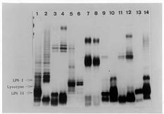
Abstract. The chemical compounds contained in leguminous plant root exudation induce structural and compositional transitions of the lipopolysaccharides of Rhizobium that occur during rhizobia-legume symbiotic infection processes. The root exudate was extracted from the seed germinated solution and seedling roots of soybean. We added the exudation to bacteria cultured medium. Rhizobium and Bradyrhizobium strains were purified through gel filtration chromatography followed by SDS-PAGE analysis. Different chromatographic and SDS-PAGE patterns among the different rhizobia strains were obtained. We determined by means of the gel filtration chromatography that soybean root exudations induced a molecular weight range change in the lipopolysaccharides of these strains. The band of this LPS-II region exhibited diffused patterns in SDS-PAGE as a result of soybean root secretion. The LPS-I region of four strains cultured in TY medium with root exudates exhibited clearer and denser bands than those of rhizobia from TY medium without added soybean root exudates. Based on the composition analysis, we found that the KDO ratio increased in R. fredii TU6, R. meliloti ATCC 9930, R. leguminosarum bv. viciae ATCC 10004 and B. japonicum USDA 110, but decreased in R. fredii KR23, R. loti Lu7, and R. loti ATCC 33669 when soybean root exudation was added to the medium. The hexouronate ratio decreased except in R. loti Lu7. This result was consistent with the SDS-PAGE profile. The LPS-I region of R. loti LU7 showed little variation. It was therefore ascertained that soybean root secretion induces a variation in the structure of rhizobial lipopolysaccharides.

Keywords: Bradyrhizobium japonicum; Lipopolysaccharide; Rhizobium fredii; Root exudation.

Abbreviations: LPS, lipopolysaccharide; Hex, hexose; HexA, hexuronic acid or hexuronate; KDO, 2-keto-3-deoxyoctanate; O-Ag, O-antigen; MWCO, molecular weight cut off; SDS-PAGE, sodium dodecyl sulfate-polyacrylamide gel electrophoresis.

Introduction

Lipopolysaccharide (LPS) is a component of the outer membrane of Gram negative bacteria and is composed of O-antigen (O-Ag), core oligosaccahride, and lipid A. In the nitrogen fixing bacteria Rhizobium, it has been suspected of playing a role in early symbiotic processes. It has been pointed out in some research results that LPS can affect the ability of rhizobia to nodulate leguminous hosts. For instance, an LPS mutant of Bradyrhizobium japonicum, defective in O-Ag, cannot infect soybean root hair (Puvanesarajah et al., 1987). LPS mutants of R. phaseoli also form a defective infectious thread, so that rhizobial cells cannot be released and form nodules (Cava et al., 1989). Thus, rhizobial LPS is known to be an essential factor in nodule forming processes.

Leguminous root exudations were previously thought to be an important factor in the infection processes and to contain a variety of different molecular weight fractions. Recently, some reports have showed that flavonoid chemicals, secreted in root exudates, can activate nod

genes and regulate the nod gene product as part of a signal compound participating rhizobia-host recognition reaction (Peter et al., 1986; Peter and Long, 1988; Lerouge et al., 1990). The signal compound of R. meliloti was identified as a tetrasaccharide which could be derived from cell wall (Lerouge et al., 1990). In rhizobial extracellular polysaccharides, some alterations in composition, structure, and molecular weight of exopolysaccharides were induced by 1 µM genistein (Dunn et al., 1992). No one has yet reported on the effect on LPS by low molecular weight root exudates although LPS plays an important role in infectious processes.

In this study, we decided to investigate the effect of a mixture, instead of any single chemical substance, because the low molecular weight fraction was always a mixture in the rhizosphere. According to the results of Gaworzeska and Carlile (1982), the low molecular fractions are secreted during 0_3 days of germinating seedling. We therefore collected the root secretion of 3-day-old soybean seedlings for our research. In this paper, we will use the results of gel filtration chromatography and SDS-PAGE to demonstrate that the transition of LPS can be induced by those small molecules.

1Corresponding author: Tel: 886-2-363-5523; Fax: 886-2-362-6455.


Botanical Bulletin of Academia Sinica, Vol. 40, 1999

Materials and Methods

Bacterial Strains

Rhizobium fredii KR23 and TU6 were isolated from indigenous soils of Taiwan and stored in yeast-mannitol agar at 4°C in our laboratory. Rhizobium loti Lu7, an isolate in Taiwan, was provided by Prof. C.C. Young, Dept. of Soil Science, National Chung Hsing University, Taichung, Taiwan. Bradyrhizobium japonicum USDA110 was donated by USDA. Rhizobium meliloti ATCC 9930, R. leguminosarum bv. viciae ATCC 10004 and R. loti ATCC 33669 were purchased from the American Type Culture Collection, Rockville, MD, USA. The rhizobial strains and their host are described in Table 1.

LPS Purification and Analysis

The LPS was extracted from bacteria by phenol-water method as modified by Carlson et al. (1978) and Carrion et al. (1990). Crude LPS was purified through Fractogel TSK HW-65 (F) (Merck, Germany) gel filtration chromatography with 0.1 M EDTA-0.3 M triethylamine buffer, pH 7.0. Fractions were collected and assayed for hexose (Hex) by the phenol-sulfuric acid method, hexuronic acid (HexA) by the m-hydroxydiphenyl method (Blumenkrantz and Asboe-Hansen, 1973), and 2-keto-3-deoxyoctanate (KDO) by the thiobarbituric acid (Karhanis et al., 1978).

The purified LPS was analyzed by 12.5% SDS-PAGE (16×16 cm), 20 mA/gel in a Hoeffer SE600 electrophoresis apparatus (Hoeffer Scientific Instruments, San Francisco, CA). The gels were stained with silver nitrate solution according to the method of Tsai and Frasch (1982).

Results

The crude rhizobial LPS was purified using a Fractogel TSK HW-65 (F) gel filtration column (Figure 1). The gel filtration chromatography was performed with an EDTA-triethylamine buffer (pH 7.0), which separates the LPSs from residual capsular polysaccharides, extracellular polysaccharides, and smaller molecular glucans (Carlson, 1984). From these results, we determined that soybean root exudates induce alterations in LPS expression. A later eluted peak corresponded to the position of R. fredii cultured in TY-soybean root exudation containing medium. As shown in Figure 1, we also found some transitional peaks in the early eluted regions.

Table 1. Strains and their symbiotic hosts.

Bacteria Host genus

Rhizobium fredii Glycine

KR23

TU6

Bradyrhizobium japonicum Glycine

USDA 110

R. loti Leucaena

Lu7

ATCC 33669

R. meliloti Medicago

ATCC 9930

R. leguminosarum bv. viciae Pisum

ATCC 10004

Soybean and Preparation of Root Exudate

Soybean seeds (Glycine max) were obtained from the Tainan Agricultural Experiment Station, Tainan, Taiwan. Surface sterilized seeds were incubated and allowed to germinate on a sterilized petri dish with a stainless grid. N-free Fahraeus' solution was used for plant incubation. Preparation of the root secretions were modified from the method of Wall and Favelukes (1991).

The seedlings were cultured in a plant growth chamber, in the dark, at 28°C for 3 days. The incubation solution was used as one source of root secretion. The roots of seedlings were cut off, and washed in distilled water. Two of these root exudations were combined and dialyzed (MWCO<3000, Spectrum Med. Ind. Inc., CA, USA) against distilled water. The dialyzable fraction was collected, concentrated, and lyophilized for further incubating preparation.

Bacteria were grown in TY medium at 28°C until the late log phase. The medium was composed of 0.5% tryptone, 0.3% yeast extract, and 10 mM CaCl2. Root exudation was added to the TY medium at a 93 µg/ml concentration. As a control, bacteria were incubated in the TY medium only. In order to avoid root-exudations from denaturation during autoclaving, a 0.22 µm membrane (Millipore Co., Bedford, MA, USA) was used for sterilization by filteration through this membrane.

Figure 1. TSK HW-65 gel filtration chromatography of LPS from Rhizobium fredii species. Column size 82×1.6 cm, 4.0 ml each fractions, eluted with EDTA-triethylamine. (l) LPS extracted from rhizobia incubated in medium without soybean root exudations, and (ˇ) extracted from rhizobia incubated in medium with soybean root exudations. (A) R. fredii KR23, (B) R. fredii TU6.


Song and Lin — Transition of Rhizobium lipopolysaccharides by root exudation

Our compositional analysis revealed that hexose, hexuronate, and KDO were ubiquitously present in each strain and that the Hex/HexA and Hex/KDO ratio of each strain had changed in the root exudates as shown in Table 2. We found that the Hex/HexA ratio always increased, except in R. loti Lu7, when the medium contained soybean root exudation. The Hex/KDO ratios were divided into two groups, one with an increased and the other with a decreased ratio. Four strains belonged to the increased Hex/KDO group—R. fredii TU6, R. meliloti ATCC 9930, and R. leguminosarum bv. viciae ATCC 10004, as well as B. japonicum USDA 110. By contrast, R. fredii KR23, R. loti Lu7 and R. loti ATCC 33669 exhibited decreased Hex/KDO ratios.

We obtained different LPS patterns by SDS-PAGE analysis (Figure 2). Based on the position of lysozyme used as the molecular weight standard, we identified two regions: LPS-I and LPS-II. The LPS-I region, with slower mobility than that of lysozyme, is derived from the complete LPS molecule with O-antigen while the faster-moving LPS-II region represents the LPS without O-chain (Bhat and Carlson, 1992; Carlson, 1984). Each strain showed quite a different electrophoretic pattern. Except for R. loti Lu7, differences were seen between the LPS structures derived from cultures with and without soybean exudates. Thus, a major difference was observed in the LPS-II region of R. fredii species. In R. fredii KR23, variations in LPS involved a decrease in molecular weight (Figure 2) and a different SDS-PAGE expression while R. fredii TU6 exhibited different LPS-II SDS-PAGE patterns. Other rhizobial strains showed different transitions in the LPS-I and LPS-II regions. There were fewer LPS-I bands in B. japonicum USDA 110 and R. meliloti ATCC 9930 after incubation with soybean root exudation medium. The number of LPS-I bands increased in R. leguminosarum bv. viciae ATCC10004 and R. loti ATCC 33669. As to the difference in band forms, we found a very prominent doublet band in the LPS-I region of R. loti ATCC 33669 not present in the TY medium without root secretion. The transition in LPS-I region seems to indicate the heterogeneity in size induced by soybean root exudates.

Figure 2. SDS-PAGE analysis of lipopolysaccharides. The LPS samples loaded are Lane 1_2, Rhizobium fredii KR23; Lane 3_4, R. fredii TU6; Lane 5_6, Bradyrhizobium japonicum USDA 110; Lane 7_8, R. loti Lu7; Lane 9_10, R. meliloti ATCC 9930; Lane 11_12, R. leguminosarum bv. viciae ATCC 10004; Lane 13_14, R. loti ATCC 33669. The odd lanes loaded with LPS were extracted from rhizobia incubated in medium with soybean root exudations. The even lanes loaded with LPS were extracted from rhizobia incubated in medium without soybean root exudations.

Discussion

Rhizobia will move to the surface of root hairs from the rhizosphere because of the chemotaxis caused by leguminous plant root exudation. They then tend to bind to the root hair surfaces (Bergman et al., 1988; Gaworzeska and Carlile, 1982). Root secretions contain many compounds that are capable of inducing nod gene expression determining infection specificity. Flavonoids have been identified to trigger nod gene expression (Puppke, 1984; Firmin et al., 1986; Peter et al., 1986; Djordjevic et al., 1987). In those studies, different Rhizobium species were affected by different types of chemical compounds. Isoflavones induced activation of B. japonicum nod genes, but not those of R. leguminosarum bv. trifolii (Kosslake et al., 1987). In addition to these effects, nodD gene encodes a regulatory

Table 2. Soybean root exudate influences the variation of rhizobial lipopolysaccharides on hexose/hexuronate and hexose/KDO.

Bacteria Without root exudate With root exudate

Hex/HexAa Hex/KDO Hex/HexAa Hex/KDO

Rhizobium fredii

KR23 57.7 27.2 111.7 10.8

TU6 15.0 9.2 30.9 21.2

Bradyrhizobium japonicum

USDA 110 19.9 10.5 34.5 34.5

R. loti

Lu7 7.3 206.9 8.1 59.3

ATCC 33669 7.4 154.8 24.1 43.3

R. meliloti

ATCC 9930 5.0 20.0 15.3 50.8

R. leguminosarum bv. viciae

ATCC 10004 10.4 16.1 20.7 31.1

aThe abbreviations: Hex, hexose; HexA, hexuronate; KDO, 2-keto-deoxy-octanic acid.


Botanical Bulletin of Academia Sinica, Vol. 40, 1999

1992). Tao et al. (1992) cultured rhizobia under a variety of different conditions including low pH, high temperature, low phosphate, and low oxygen concentration. Antigenic changes at low pH were dependent on growth of the bacteria, but independent of nitrogen and carbon sources and the rich or minimal quality of the medium. This means that these LPSs will have less or no reaction to antibodies but will have the same mobility in SDS-PAGE, except when rhizobia are incubated in a low pH. Bhat and Carlson (1992) studied the pH-dependent structural epitopes of LPS from R. leguminosarum bv. phaseoli. The results showed that the methylated glycosyl residues were either affected by growth pH or were part of the O-chain polysaccharide of LPS in the nodule when the bacteria were incubated in a pH 4.8 medium. These research results showed that the LPS changes were affected by conditions of low pH and that these changes only showed up in the LPS-I region. In addition, LPSs are involved at a later stage in the nodule development, such as forming infection threads and releasing bacteroids from infection threads. We found that the changes are expressed not only in the LPS-I region but also in the LPS-II region, and showed transitions in the SDS-PAGE pattern. We can therefore conclude that alterations in LPSs will be affected by plant exudates in the rhizosphere environment and that the changes are related to the recognition or attachment in the infection processes.

In our agronomy experiments, we observed that soybean infected by R. fredii KR23 had a 50% higher nodulation rate than R. fredii TU6. When comparing the LPS-I regions by SDS-PAGE, the band of R. fredii KR23 showed more alterations than that of R. fredii TU6, indicating that LPSs play an important role in infection processes, especially after transitions are induced through root secretions. We therefore conclude from both agronomic and biochemical results that LPSs are important determinants for successful infection between soybean and R. fredii.

Acknowledgment. This work was supported by a grant from the National Science Council, Taipei 106, Taiwan, Republic of China.

Literature Cited

Bergman, K., M. Gulash-Hoffee, R.E. Hovestadt, R.C. Larosiliere, P.G. Ronco II, and L. Su. 1988. Physiology of behavioral mutants of Rhizobium meliloti: Evidence for a dual chemotaxis pathway. J. Bacteriol. 170: 3249_3254.

Bhat, U.R. and R.W. Carlson. 1992. Chemical characterization of pH-dependent structural epitopes of lipopolysaccharides from Rhizobium leguminosarum biovar phaseoli. J. Bacteriol. 174: 2230_2235.

Blumenkrantz, N. and G. Asboe-Hansen. 1973. New method for quantitative determination of uronic acids. Anal. Biochem. 54: 484_489.

Carlson, R.W. 1984. Heterogeneity of Rhizobium lipopolysaccharides. J. Bacteriol. 158: 1012_1017.

Carlson, R.W., R.E. Sanders, C. Napoli, and P. Albersheim. 1978. Host-symbiont interaction. III. purification and partial char

protein which not only controls the expression of other nod genes but also autoregulates nodD itself (Pinero et al., 1988; Rossen et al., 1985; Sherman et al., 1986). These data strongly suggest that infection recognition is a multi-step process.

Once rhizobial nod genes are activated, rhizobia produce a signal compound, which correlates with a specific recognition reaction. The nodH and nodPQ genes of R. meliloti direct the synthesis of a signal compound, nodRm-1, which has been identified as N-acetyl-D-glucosamine responsible for root hair curling (Lerouge et al., 1990; Truchet et al., 1991). This sugar, a component of lipid-A of LPS, is released from LPS. Bradyrhizobium japonicum produces a nonsulfated pentaglucosamine as a signal compound (Verma, 1992). In other studies, the nodF gene of R. leguminosarum has been shown to be involved in the synthesis of acyl-carrier protein and fatty acids. The nod gene is thought to induce the synthesis of proteins, fatty acids and acylation of LPS (Lipman, 1971; Sherman et al., 1986).

Rhizobial extracellular polysaccharides also play an important role in infection processes (Clover et al., 1989; Goosen-de Roo et al., 1991; Kato et al., 1979, 1980; Long, 1989). As to the alterations flavonoids produce in the extracellular polysaccharides, Dunn et al. (1992) found that genistein in the medium alters their composition, structure, and molecular weight.

Priefer (1989) conjugated a R. leguminosarum LPS-I defect mutant with Lps cosmid, which caused the mutant to synthesize LPS-I and form effective nodules in Vicia hirsuta. He proposed that LPS-I can serve as a signal for successful nodulation. Dazzo et al. (1991) suggested that LPS is a positive signal molecule for successful infection processes.

In this study, we found that both the molecular weight and structure of LPS were altered by root secretions. These alterations included molecular weight range, composition, and structure as exhibited by gel filtration chromatography, compositional analyses, and SDS-PAGE results. Comparing the Hex/HexA and Hex/KDO ratios, these results showed that Hex/HexA increased except for R. loti Lu7 when the medium contained soybean root exudation. These results were consistent with the LPS-I region of the SDS-PAGE profile. This alteration of the LPS-I pattern reflects the alteration of O-Ag. We also obtained a variation in KDO as derived from the change in the Hex/KDO ratio (Table 2 and Figure 2). The transition pattern of rhizobial strains showed that the composition, structure, and molecular weight of LPS were influenced by root exudates during degradation or post-synthesis modification in LPS synthetic processes.

In general, Gram negative bacteria, irrespective of symbiotic interactions, undergo modification of their LPSs in ways that best cope with environmental changes; however, rhizobia will respond to conditions more specific to the plant environment. According to some reports, researchers are concerned with LPS changes induced by environmental factors (Bhat and Carlson, 1992; Tao et al.,


Song and Lin — Transition of Rhizobium lipopolysaccharides by root exudation

acterization of Rhizobium lipopolysaccharides. Plant Physiol. 62: 912_917.

Carrion, M., U.R. Bhat, B. Reuhs, and R.W. Carlson. 1990. Isolation and characterization of the lipopolysaccharides from Bradyrhizobium japonicum. J. Bacteriol. 172: 1725_1731.

Cava, J.R., P.M. Elias, D.A. Turowski, and K.D. Noel. 1989. Rhizobium leguminosarum CFN42 genetic region encoding lipopolysaccharide structures essential for complete nodule development on bean plants. J. Bacteriol. 171: 8_15.

Clover, R.H., J. Kieber, and E.R. Singer. 1989. Lipopolysaccharide mutants of Rhizobium meliloti are not defective in symbiosis. J. Bacteriol. 171: 3961_3967.

Dazzo, F.B., G.L. Truchet, R.I. Hollingsworth, E.M. Hrabak, H.S. Pankratz, S. Philip-Hollingsworth, J.L. Salzwedel, K. Chapman, L. Appenzeller, A. Squartini, D. Gerhold, and G. Orgambide. 1991. Rhizobium lipopolysaccharide modulates infection thread development in white clover root hairs. J. Bacteriol. 173: 5371_5384.

Djordjevic, M.A., J.W. Redmond, M. Batley, and B.G. Rolfe. 1987. Clover secrete specific phenolic compounds which either stimulate or repress nod gene expression in Rhizobium trifolii. EMBO J. 6: 1173_1179.

Dunn, M.F., A.G. Pueppke, and H.B. Krishnan. 1992. The nod gene inducer genistein alters the composition and molecular mass distribution of extracellular polysaccharides produced by Rhizobium fredii USDA 193. FEMS Microbiol. Lett. 97: 107_112.

Firmin, J.L., K.E. Wilson, L. Rossen, and A.W.B. Johnston. 1986. Flavonoid activation of nodulation gene in Rhizobium reversed by other compounds present in plants. Nature 324: 90_92.

Gaworzeska, E.T. and M.J. Carlile. 1982. Positive chemotaxis of Rhizobium leguminosarum and other bacteria towards root exudates from legumes and other plants. J. Gen. Microbiol. 128: 1179_1188.

Goosen-de Roo, L., R.A. de Maag, and B.J.J. Lugtenberg. 1991. Antigenic changes in lipopolysaccharide I of Rhizobium leguminosarum bv. viciae in root nodules of Vicia sativa subsp. nigra occur during release from infection threads. J. Bacteriol. 173: 3177_3183.

Karhanis, Y.D., J.Y. Zeltner, J.J. Jackson, and D.J. Carlo. 1978. A new and improved microassay to determine 2-keto-3-deoxyoctonate in lipopolysaccharide of Gram negative bacteria. Anal. Biochem. 85: 595_601.

Kato, G., Y. Maruyama, and M. Nakamura. 1979. Role of lectins and lipopolysaccharides in the recognition process of specific legume-Rhizobium symbiosis. Agric. Biol. Chem. 45: 1085_1092.

Kato, G., Y. Maruyama, and M. Nakamura. 1980. Role bacterial lipopolysaccharides in the adsorption process of the Rhizobium-pea symbiosis. Agric. Biol. Chem. 46: 2843_2855.

Kosslake, R.M., R. Bookland, J. Barkei, H.E. Paaren, and E.R. Appelbaum. 1987. Induction of Bradyrhizobium japonicum common nod genes by isoflavones isolated from Glycine max. Proc. Natl. Acad. Sci. USA 84: 7428_7432.

Lerouge, P., P. Roche, C. Faucher, F. Maillet, G. Truchet, J.C. Promé, and J. Dénarié. 1990. Symbiotic host-specificity of Rhizobium meliloti is determined by a sulphated and acylated glucosamine oligosaccharide signal. Nature 344: 781_784.

Lipman, F. 1971. Attempts to map a process evolution of peptide biosynthesis. Science 173: 875_884.

Long, S.R. 1989. Rhizobium genetics. Ann. Rev. Genet. 23: 483_506.

Peter, N.K. and S.R. Long. 1988. Alfalfa root exudates and compounds which promote or inhibit induction of Rhizobium meliloti nodulation genes. Plant Physiol. 88: 396_400.

Peter, N.K., J.W. Frost, and S.R. Long. 1986. A plant flavone, luteolin, induces expression of Rhizobium meliloti nodulation genes. Science 233: 977_980.

Pinero, D., E. Martinez, and R.K. Selander. 1988. Genetic diversity and relationships among isolates of Rhizobium leguminosarum biovar phaseoli. Appl. Environ. Microbiol. 54: 2825_2832.

Priefer, U.B. 1989. Genes involved in lipopolysaccharide production and symbiosis are clustered on the chromosome of Rhizobium leguminosarum biovar viciae VF39. J. Bacteriol. 171: 6167_6168.

Puppke, S.G. 1984. Adsorption of slow- and fast-growing rhizobia to soybean and cowpea roots. Plant Physiol. 75: 924_928.

Puvanesarajah, V., F.M. Schell, D. Gerhold, and G. Stacey. 1987. Cell surface polysaccharides from Bradyrhizobium japonicum and a nonnodulating mutant. J. Bacteriol. 169: 137_141.

Rossen, L., C.A. Sherman, A.W.B. Johnston, and J.A. Downie. 1985. The nodD gene of Rhizobium leguminosarum is autoregulatory and in the presence of plant exudate induces the nodA,B,C genes. EMBO J. 4: 3369_3373.

Sherman, C.A., L. Rossen, A.W.B. Johnston, and J.A. Downie. 1986. The Rhizobium leguminosarum nodulation genes nodF encodes a polypeptide similar to acyl-carrier protein and is regulated by nodD plus a factor in pea root exudate. EMBO J. 5: 647_652.

Tao, H., N.J. Brewin, and K.D. Noel. 1992. Rhizobium leguminosarum CFN42 lipopolysaccharide antigenic changes induced by environmental conditions. J. Bacteriol. 174: 2222_2229.

Truchet, G., P. Roche, P. Lerouge, J. Vasse, S. Camut, F. de Billy, J.C. Promé, and J. Dénarié. 1991. Sulphated lipooligosaccharide signals of Rhizobium meliloti elicit root nodule organogenesis in alfalfa. Nature 351: 670_673.

Tsai, C.M. and C.E. Frasch. 1982. A sensitive silver stain for detecting lipopolysaccharides in polyacrylamide gels. Anal. Biochem. 119: 115_119.

Verma, D.P.S. 1992. Signals in root nodule organogenesis and endocytosis of Rhizobium. Plant Cell 4: 373_382.

Wall, L.G. and G. Favelukes. 1991. Early recognition in Rhizobium meliloti-alfalfa symbiosis: root exudate factor stimulates root adsorption of homologous rhizobia. J. Bacteriol. 173: 3492_3499.