Uritani — Postharvest biochemistry of tropical tuberous crops

Bot. Bull. Acad. Sin. (1999) 40: 177-183

(Invited review paper)

Biochemistry on postharvest metabolism and deterioration of some tropical tuberous crops

Ikuzo Uritani

School Corp. Aichi Konan Gakuen, Konan, Aichi and Nagoya University (Prof. Emeritus), Nagoya, Aichi, Japan

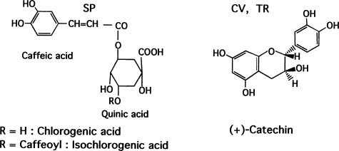
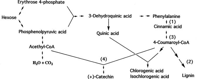
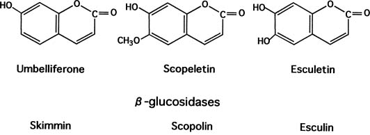
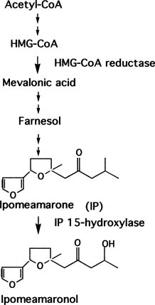
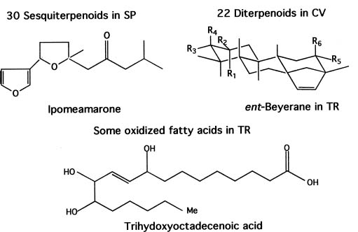
Abstract. This paper deals with a biochemical comparison of the postharvest metabolism and deterioration among three tropical tuberous crops: sweet potato, cassava, and taro, focusing on sweet potato. These often suffer from wounding and infection during harvest, transportation, and storage. In response to wounding or infection, the three crops underwent some similar metabolic alterations, such as in the production of stress compounds and changes in proteins, but they also showed some different reactions. In sweet potato and taro, wounding was often healed soon by the formation of a lignin layer succeeding production of polyphenols and changes in proteins. No such layer formed in cassava, which deteriorated at xylems soon after harvest in a phenomenon called vascular streaking, vascular discoloration, or physiological deterioration. When the three crops were attacked by microbes or insects, metabolic changes were induced more vigorously, including the induction of some enzymes, production of stress compounds such as coumarins and phytoalexins, and more enhancement of polyphenol production. However, in cassava, the stress compounds were also produced in the case of wounding.

Keywords: Colocasia esculenta; Coumarin; Ipomoea batatas; Manihot esculenta; Microbial deterioration; Physiological deterioration; Phytoalexin; Polyphenol; Storage protein; Tropical tuberous crops.

Abbreviations: SP, Sweet potato; CV, cassva; TR, taro; PD, physiological deterioration; MD, microbial deterioration; PAL, phenylalanine ammonia lyase.

Contents

Introduction 177

Materials and Classification 177

Response to Wounding 178

Response to Infection 178

Polyphenol Production 178

Production and Structures 178

Phenylalanine Ammonia Lyase and Peroxidase 179

Coumarin Production 179

Phytoalexin Production 179

Changes in Proteins 181

Chilling Injury 181

Conclusion 181

Literature Cited 182

Introduction

There are many kinds of tuberous crops in the tropics, some of which are utilized as dietary staples. However, much scientific research on these crops remains to be performed—including the field of postharvest biochemistry. A summary of the research which has been done, and of the questions yet to be elucidated on those crops, may be helpful in solving the world food shortage that is anticipated for the twenty-first century due to increases in the world population.

Materials and Classification

Postharvest metabolism and deterioration of sweet potato (SP, Ipomoea batatas [L.] Lam.), cassava (CV, Manihot esculenta Cranz), and taro (TR, Colocasia esculenta (L.) Schott, or C. antiquorum) after harvest are described from the biochemical point of view. The three produce tuberous organs under ground that contain large amounts of starch. All three originate in the tropical zone. However, the botanical classifications of class, family and genus, as well as chromosome and genome numbers, differ from each other (see Table 1). Hence, their natures are similar in some parts, but dissimilar in others. However, an understanding of the nature of one species of tuberous organ is useful in understanding the nature

Present address: 1-62-8, Karayama-cho, Chikusa, Nagoya, 464-0806, Japan. Tel: 81-52-781-6953; Fax: 81-52-781-6968; E-mail: iuritani@mb.infoweb.ne.jp


Botanical Bulletin of Academia Sinica, Vol. 40, 1999

Table 1. Botanical natures of the three tropical tuberous crops.

Common name Sweet potato Cassava Taro

Botanical name Ipomoea batatas Manihot esculenta Colocasia esculenta

Class Dicotiledon Dicotiledon Monocotiledon

Family Convolvulaceae Euphorbiaceae Araceae (Aroid)

Chromosome No. 15 18 22,26,28,38,42

Genom No. 6 2 2

Organ Root Root Corm (Base of stem)

of other species. Thus, some experimental results on the above three are compared with each other, focusing on sweet potato.

Response to Wounding

The tuberous organs are inclined to be mechanically wounded during harvest, transportation, and storage. When the organs of SP and TR were wounded, either naturally or artificially, injury was normally cured by the formaton of a lignin layer in the cells very close to the surfaces. On the other hand, CV organ is easily wounded during harvest because of the localized situation existing underground. The harvesting of CV roots after maturation caused vascular streaking or vascular discoloration (to brown or greenish brown) some days after harvest (Booth, 1976; Montaldo, 1973). The deterioration was called physiological deterioration (PD) (Rickard and Coursey, 1981; Rickard, 1985). The formation of lignin was weakly positive in the discolored part showing PD, according to a qualitative test using phloroglucinol saturated HCl (Rickard and Coursey, 1981; Rickard, 1985; Uritani and Reyes, 1982, 1984). When the roots were cut into the blocks artificially, PD appeared very soon and at a uniform pattern in the outer part of the secondary xylem parenchyma, called B-part on the cut surfaces, as shown in Figure 1 (Kojima and Uritani, 1983). Cut-wounded CV had almost no ability to form lignin layer when left at ambient conditions after harvest in the case of matured roots (Uritani et al., 1983; Uritani and Reyes, 1984).

Figure 2. Polyphenols in some tropical tuberous organs.

Response to Infection

The organs were sometimes infected by some pathogenic fungi from the wounded surfaces. When SP and TR tissues were infected by the SP-strain and TR-strain of Ceratocystis fimbriata (Ell. and Halst.) Elliot., black rot pathogen, respectively, the mycelia penetrated into the inner cells, and penetration ceased, mainly because of the production of phytoalexins (Kojima and Uritani, 1976). Then, lignin was formed. SP and TR were attacked only by their own strains (Kojima and Uritani, 1974). No specific strain of C. fimbriata pathogenic to CV has not been found. However, CV tissue was easily infected by some fungi such as Botriodiplodia theobromae Pat., normally after the occurrence of PD, and the damage was called microbial deterioration (MD) (Rickard and Coursey, 1981; Rickard, 1985).

Polyphenol Production

Production and Structures

In response to wounding or infection, polyphenols were produced in the tissue adjacent to the wounded surface or infected region. As indicated in Figure 2, these were chlorogenic acid, isochlorogenic acid, and the relatives in SP (Uritani and Muramatsu, 1953; Uritani and Miyano, 1955), and were (+)-catechin and the derivatives in CV and TR (Tanaka et al., 1983).

The amounts produced were much higher in SP than in CV, as indicated in Table 2. The table shows the amounts when polyphenols reached the maximum in wounded and infected tissues in SP and CV. The former was at µmol units per g. fresh wt., but the latter was at nmol units per g. fresh wt. (Akazawa and Uritani, 1961; Tanaka et al., 1983). The amounts of catechin produced

Figure 1. Physiological deterioration of cassava root block, in diameter of 5 to 6 cm. Left: The scheme of the block showing physiological deterioration; Right: the photograph. Cassava blocks were left in the laboratory condition (at 26 to 30°C and 78 to 92% of relative humidity) for 3 days, and physiological deterioration occurred in the B-part corresponding to the outer part of the secondary xylem parenchyma. A-part and C-part are the outer and inner parts of B-part, respectively.


Uritani — Postharvest biochemistry of tropical tuberous crops

Table 2. The amounts of polyphenols in wounded and infected tissues.

Sweet potato Cassava (µmol as chlorogenic acid) (nmol as (+)-catechin)

Wounded 14_16 48_59

Infected 34 460

and CV tissues and compared with the above results. The biosynthetic pathways of chlorogenic acid, catechins, and lignin are shown in Figure 3 for reference (see Edwards and Kessmann, 1992).

Coumarin Production

Bluish fluorescent coumarins were neither present in fresh SP, nor produced in response to wounding only. However, in response to infection by some fungi as C. fimbriata, these were produced at an early stage in SP tissue. We identified the main components as umbelliferone and scopoletin, and the minor ones as esculetin and the two b-D-glucosides such as scopolin and skimminn, as indicated in Figure 4 (Uritani and Hoshiya, 1953; Minamikawa et al., 1962, 1964). In TR, such fluorescent components did not seem to be formed in the tissue by wounding or infection. In CV, the components were absent in fresh tissue, but were produced in response to wounding, preceding the occurrence of PD. In tissue suffering from MD, fluorescent components were produced in the tissue adjacent to the infected region. We identified them as scopoletin, scopolin, and esculin, as shown in Figure 4 (Tanaka et al., 1983). Independently, Wheatley (1982) isolated the bluish fluorescent component in CV roots preceding PD appearance and identified it as scopoletin. It should be emphasized that the coumarins were produced in wounded CV tissue, just as in infected SP tissue, although less was produced. However, the amounts produced in both tissues were at nmol units per g fresh wt., as seen in Table 3 (Minamikawa et al., 1963; Tanaka et al., 1983).

Phytoalexin Production

Phytoalexins were not present in fresh SP, CV or TR tuberous tissues. Further, they were not produced normally by wounding of SP and TR. Phytoalexins were produced by infection with C. fimbriata and accumulated in infected regions. The components in SP were produced by a continuous injury such as a fungal infection or an insect larvae invasion by pests such as sweet potato weevil (Cylas formicarius Fabricius), West Indian sweet potato weevil (Euscepes postfasciatus Fairmaire), and sweet

in TR in response to wounding or infection have not yet been quantified, but the production in TR appeared to be higher than in CV, according to the qualitative test.

Polyphenols in these wounded tissues include the equal chemical skeleton, phenylpropanoid, which is involved in lignin biosynthesis. It is important to investigate the biosynthetic regulation of phenylpropanoid and lignin, especially in CV, considering almost no lignin-layer formation occurs in wounded CV tissue.

Phenylalanine Ammonia Lyase and Peroxidase

Preceding polyphenol production, several enzymes such as phenylalanine ammonia lyase (PAL) and trans-cinnamic acid 4-hydroxylase in the phenylpropanoid biosynthetic pathway were induced in wounded SP tissue (Tanaka et al., 1974; Tanaka and Uritani, 1977). PAL was induced very soon, and the activity peaked within 24 h, then gradually decreased. In wounded CV tissue, the activity was induced in a similar pattern though it peaked at a later stage, and the maximal activity was roughly the same as in SP (Tanaka et al., 1983).

Peroxidase participates in lignin formation. Hence, the activity was assayed in both wounded SP and CV tissues (Matsushita and Uritani, 1975; Tanaka et al., 1983). The activities were induced gradually in both SP and CV for longer than 3 days, and the level was a little higher in SP. The activities of PAL and peroxidase in both tissues, which were involved in the biosynthesis of their polyphenols and lignin, increased in a similar time course. However, much greater amounts of polyphenols and lignin were produced in SP tissue than in CV tissue. In addition to PAL and peroxidase, the activities of some enzymes that might be positioned at the rate-limiting steps, 4-coumarate CoA ligase for example, should also be assayed in wounded SP

Figure 3. The biosynthetic pathways of chlorogenic acid and isochlorogenic acid, catechins and lignin in plants. (1): phenylalanine ammonia lyase; (2): Peroxidase; (3): 4-coumarate CoA ligase; (4): chalcone synthase.

Figure 4. Coumarins in infected sweet potato and wounded or infected cassava.


Botanical Bulletin of Academia Sinica, Vol. 40, 1999

Table 3. The amounts of coumarins in infected sweet potato and wounded cassava.

Incubation days Umbelliferone Skimmin Scopoletin Scopolin Esculetin Esculin

nmol/g. fresh wt.

Diseased sweet potato

1 83 Small 6.6 Small Small _

2 135 Small 20 Small Small _

3 182 Small 60 Small Small _

Wounded cassava

1 _ _ 17 26 _ 22

2 _ _ 12 38 _ 42

3 _ _ Less Less _ Less

Small: Detected by chromatography qualitatively; Less: Decreased compared to the 2 day amounts; _: Not detected by the fluorescent assays used.

potato vine borer (Megastes grandalis). Toxic chemical treatments also produced phytoalexins (Uritani et al., 1960). In contrast, they were produced, in the case of CV, in the discolored region caused by either PD- or MD- induction.

These were toxic to the pathogenic fungi corresponding to SP, CV and TR.

In SP, about 30 sesquiterpenoids were identified by our group (Schneider et al., 1984) and Wilson's group (Wilson and Burka, 1979). Ipomeamarone is the main component, which was originally isolated by Hiura (1943) and recognized as the first example of a phytoalexin. The chemical structure was determined by Kubota et al. (see Figure 5). In CV, about 22 diterpenoids were identified (Sakai et al., 1986, 1994; Sakai and Nakagawa, 1988). These belong to 4 families, and the main family is the ent-beyerene family, including 10 components (see Figure 5). In TR, several oxidized fatty acids were produced, and one was identified as 9,12,13,-trihydroxy-(E)-10-octadecenoic acid as shown in Figure 5 (Masui et al., 1989). According to experiments on infected SP and TR tissues, the components were produced subsequently to the induction of the enzymes in the pathway. By way of illustration in SP, some enzymes in both steps before and after farnesol biosynthesis were induced. As examples of the respective enzymes, 3-hydroxy-3-methylglutaryl-CoA reductase

Figure 5. Chemical structures of main phytoalexins in some tropical tuberous organs.

Figure 6. The outline of the biosynthetic pathway of phytoalexins in sweet potato.


Uritani — Postharvest biochemistry of tropical tuberous crops

(Oshima and Uritani, 1968) and ipomeamarone 15-hydroxylase (Fujita et al., 1982) were isolated and characterized. The pathway involving the above enzymes is shown in Figure 6.

As indicated in Table 4, in the infected region of SP tissue, the production reached 2~3 %, 3 to 5 days after infection (Akazawa and Uritani, 1961). On the other hand, in wounded or infected CV tissue, the production in the discolored region was far less than in infected SP tissue (Sakai et al., 1994).

Changes in Proteins

In wounded and infected tissues of the above tuberous organs, many kinds of metabolism were activated, as mentioned above. This suggested that the zymograms of proteins from those tissues were changed in response to either wounding or infection. We indicated that the assumption was correct for the soluble protein fraction of SP, and the antigenic proteins called component A1 and A2 were present in the roots and changed to other proteins (Uritani and Stahmann, 1961). The proteins were proven to be storage proteins of 25 kDa by our colleagues (Li and Oba, 1985; Maeshima et al., 1985). Now, the proteins are called sporamin, and A and B are the main components. It is interesting that Hou and Lin (1993) and Wang and Yeh (1996) proved that trypsin inhibitors in SP are actually sporamins.

Nakamura's group determined the nucleotide sequence of sporamin (Hattori et al., 1985), and sporamin A, composed of 199 amino acids, was proven to be synthesized not only by transcription and translation, but also by post-translation processing through prepro-sporamin, then prosporamin, under the action of organelles such as endoplastic reticulum, Golgi body, and vacuole. In the process, glycosidation and dis-glycolation occurred (Nakamura et al., 1993).

In response to wounding and infection in SP root, the storage proteins were degraded to proteins of lower molecular weight (9,500 to 20,000), according to immunoelectrophoresis (Uritani and Stahmann, 1961; Li and Oba, 1985). These may be changed to other enzymatic proteins directly, or after the hydrolysis to small size peptides or amino acids. We have to elucidate more precisely how sporamins are converted to many kinds of enzymes.

In CV root, the three or four main soluble proteins were present (Shewry et al., 1992; Uritani et al., 1992). The CV proteins were degraded and converted to other proteins while the root was suffering from wounding or infection, according to the data on zymograms for the soluble proteins in healthy, wounded and infected CV tissues (Uritani et al., 1992). In TR tuber, the two to four main soluble proteins were present (Hirai et al., 1989, 1990), and the zymogram was changed in response to wounding and infection (Uritani et al., 1992). Hence, it was elucidated that the soluble proteins in these three tuberous organs changed according to an equal pattern in response to wounding and infection (Uritani et al., 1992).

Table 4. The amounts of phytoalexins in infected sweet potato and wounded cassava.

Incubation days Diseased sweet potato Wounded cassava

(mg/g fresh wt.) (µg/g fresh wt.)

1 5 14

2 16 163

3 32 283

Chilling Injury

The three crops originate in tropical regions and grow well in tropical climates. Hence, when the roots are stored at cold temperature, they suffer from chilling injury. The temperature that causes chilling injury differs among the three crops and their varieties. If storage temperature is high, the metabolism of the tuberous organs is activated, and more carbohydrates are consumed. Then, sprouting occurs in SP and TR during long periods of storage. The adequate storage temperature ranges for SP and CV are 9 to 11°C and 15 to 19°C, respectively. When CV roots were stored in the same range as SP roots, the CV roots sustained cold injuries and suffered from infection by non-pathogenic fungi. TR tubers, which have been acclimatized under the climate in Japan, can be stored successfully at even lower temperatures than would be healthy for SP roots. The difference in chilling injury between SP and CV coincided with the experimental data on the transition temperatures of the Arrhenius plots of succinoxidase activities of mitochondria isolated from both SP and CV roots (Maeshima et al., 1980).

Conclusion

Reactions of the three tuberous organs to wounding and infection were common in some cases, but unique in others. When mechanically wounded or infected, polyphenols were produced all in the organs, but the chemical components differed. Chlorogenic acids, isochlorogenic acid, and their relatives were produced in SP tissue. On the other hand, catechins were produced in CV and TR tissues. However, the phenylpropanoid skeleton is included in the polyphenols of the three organs. The bluish fluorescent coumarins were produced in infected SP tissue and in wounded and infected CV tissues, but no coumarin was found in that of infected TR. Phytoalexins were produced all in the organs, but the chemical structures differed from each other. However, all of the components should have been produced from cetyl-CoA, showing a lipophilic nature, while those in SP and CV belonged to terpenoids, and the one in TR was a kind of fatty acid. It is interesting that phytoalexins were produced only in the brownish regions of infected SP and TR tissues, but produced in the discolored region in CV suffering from not only MD, but also PD. The appearance of discoloration called PD was specific to CV tissue. However, lignin though hardly formed in CV on the wounded surface, formed quickly and in high density in the cases of SP and TR. If a lignin layer formed as quickly on the wounded


Botanical Bulletin of Academia Sinica, Vol. 40, 1999

surface of CV roots, this might inhibit PD and MD occurrence in CV, and enable the roots to be stored for a longer period. Further, this approach may lead to the elucidation of the mechanism on the occurrence of the vascular discoloration called PD. It must be important to analyze the rate limiting enzyme step(s) for lignin formation in CV roots when wounded.

Acknowledgements. In the preparation of this review, the author cited many research results performed by his colleagues who are or were engaged in biochemical works at Nagoya University, and he expresses his thanks to them. This article was based on a paper delivered at the International Symposium on Plant Senescence and Postharvest Biotechnology, Institute of Botany, Academia Sinica, December 1 to 2, 1997, under the invitation of Dr. Shang Fa Yang, Vice President of Academia Sinica, and Dr. Jei-Fu Shaw, Director of Institute of Botany, Academia Sinica. The author wishes to express his sincere thanks to them for inviting him to attend the Symposium and to submit a review to the Botanical Bulletin.

Literature Cited

Akazawa, T. and I. Uritani. 1961. Influence of environmental temperature on metabolic alterations related to disease resistance in sweet potato roots infected by black rot. Phytopathology 51: 668-674.

Booth, R.H. 1976. Storage of fresh cassava (Manihot esculenta). I. Postharvest deterioration and its control. Exp. Agric. 12: 103-111.

Edwards, R. and H. Kessmann. 1992. Isoflavonoid phytoalexins and their biosynthetic enzymes. In S.J. Gull, M.J. McPherson and D.J. Bowles (eds.), Molecular Plant Pathology, A Practical Approach, vol. II. Oxford University Press, Oxford, New York, Tokyo, pp. 45-62.

Fujita, M., K. Oba, and I. Uritani. 1982. Properties of a mixed function oxygenase catalyzing ipomeamarone 15-hydroxylation in microsomes from cut-injured and Ceratocystis fimbriata-infected sweet potato root tissues. Plant Physiol. 70: 573-578.

Hattori, T., T. Nakagawa, M. Maeshima, K. Nakamura, and T. Asahi. 1985. Molecular cloning and nucleotide sequence of cDNA for sporamin, the major protein of sweet potato tuberous roots. Plant Mol. Biol. 5: 313-320.

Hirai, M., K. Nakamura, and T. Sato. 1990. Gene cloning of storage proteins in taro. Abstracts on the Autumn Meeting of Japan. Breed Soc.

Hirai, M., T. Sato, and K. Takayanagi. 1989. Classification of Japanese cultivars of taro (Colocasia esculenta (L.) Schott) based on electrophoretic pattern of the tuber proteins and morphological characters. Jpn. J. Breed. 39: 307-317. (in Japanese)

Hiura, M. 1943. Studies on storage and rot of sweet potato (2). Rep. Gifu Agric. Coll. 50: 1-5. (in Japanese)

Hou, W.C. and Y.H. Lin. 1993. Dehydroascorbate reductase and monodehydroascorbate reductase activities of trypsin inhibitors, the major sweet potato (Ipomoea batatas [L.] Lam.) root storage protein. Plant Sci. 128: 151-158.

Kojima, M. and I. Uritani. 1974. The possible involvement of a spore agglutinating factor(s) in various plants in establishing host specificity by various strains of black rot fungus,

Ceratocystis fimbriata. Plant Cell Physiol. 15: 733-737.

Kojima, M. and I. Uritani. 1976. Possible involvement of furanoterpenoid phytoalexins in establishing host-parasite specificity between sweet potato and various strains of Ceratocystis fimbriata. Physiol. Plant Pathol. 8: 97-111.

Kojima, M. and I. Uritani. 1983. Changes of cyanide content and linamarase activity in wounded cassava roots. Plant Physiol. 72: 186-189.

Kubota, T. and T. Matsuura. 1953. Chemical studies on the black rot disease of sweet potato. J. Chem. Soc. Japan 74: 44-47, 101-109, 197-199, 248-251, 668-670. (in Japanese)

Li, H.S. and K. Oba. 1985. Major soluble proteins of sweet potato roots and changes in proteins after cutting, infection, or storage. Agric. Biol. Chem. 49: 737-744.

Maeshima, M., I. Uritani, and T. Asahi. 1980. Effect of temperature on the activities of cytochrome c oxidase and respiration in cassava root mitochondria. Agric. Biol. Chem. 44: 293-294.

Maeshima, M., T. Sasaki, and T. Asahi. 1985. Characterization of major proteins in sweet potato tuberous roots. Phytochemistry 24: 1899-1902.

Masui, H., T. Kondo, and M. Kojima. 1989. An antifungal compound, 9,12,13,-trihydroxy-(E)-10-octadecenoic acid, from Colocasia antiquorum inoculated with Ceratocystis fimbriata. Phytochemistry 28: 2613-2615.

Matsushita, K. and I. Uritani. 1975. Effects of cycloheximide, actinomycin D and ethylene on the increase and subsequent decrease in acid invertase activity in wounded sweet potato. Plant Cell Physiol. 16: 203-210.

Minamikawa, T., T. Akazawa, and I. Uritani. 1962. Isolation of esculetin from sweet potato roots with black rot. Nature 195: 726.

Minamikawa, T., T. Akazawa, and I. Uritani. 1963. Analytical study of umbelliferone and scopoletin synthesis in sweet potato roots infected by Ceratocystis fimbriata. Plant Physiol. 38: 493-497.

Minamikawa, T., T. Akazawa, and I. Uritani. 1964. Two glucosides of coumarin derivatives in sweet potato roots infected by Ceratocystis fimbriata. Agr. Biol. Chem. 28: 230-233.

Montaldo, A. 1973. Vascular streaking of cassava root tubers. Trop. Sci. 15: 39-46.

Nakamura, K., K. Matsuoka, F. Mukumoto, and N. Watanabe. 1993. Processing and transport to the vacuol of a precursor to sweet potato sporamin in transformed tobacco cell line BY-2. J. Exp. Bot. 44: 331-338.

Oshima, K. and I. Uritani. 1968. Enzymatic synthesis of a b-hydroxy-b-methylglutaric acid derivative by a cell-free system from sweet potato with black rot. J. Biochem. 63: 617-625.

Rickard, J.E. and D.G. Coursey. 1981. Cassava storage. Part I: Storage of fresh cassava. Trop. Sci. 23: 1-32.

Rickard, J.E. 1985. Physiological deterioration of cassava roots. J. Sci. Food Agric. 36: 167-176.

Sakai, T. and Y. Nakagawa. 1988. Diterpenic stress metabolites from cassava roots. Phytochemistry 27: 3769-3779.

Sakai, T., Y. Nakagawa, I. Uritani, and E.S. Data. 1986. Occurrence of various kinds of metabolites in physiologically and microbially damaged cassava (Manihot esculenta Crantz) Roots. Agric. Biol. Chem. 50: 2905-2907.


Uritani — Postharvest biochemistry of tropical tuberous crops

Sakai, T., Y. Nakagawa, I. Uritani, and E.S. Data. 1994. Occurrence and characteristics of stress metabolites in cassava roots. In I. Uritani, V.V. Garcia and E.M.T. Mendoza (eds.), Postharvest Biochemistry of Plant Food-Materials in the Tropics. Japan Scientific Societies Press, Tokyo, pp. 95-110.

Schneider, J.A., J. Lee, Y. Naya, K. Nakanishi, K. Oba, and I. Uritani. 1984. The fate of the phytoalexin ipomeamarone: furanoterpenes and butenolides from Ceratocystis fimbriata-infected sweet potatoes. Phytochemistry 23: 759-764.

Shewry, P.R., A. Clowes, A.S. Tatham, and J. Beeching. 1992. Opportunities for manipulating the amount and composition of proteins in cassava tuberous roots. In W.N. Roca and A.M. Thro (eds.), Proceedings of the First International Scientific Meeting of Cassava Biotechnology Network, Cartagena, Colombia, pp. 251-253.

Tanaka, Y. and I. Uritani. 1977. Synthesis and turnover of phenylalanine ammonia-lyase in root tissue of sweet potato injured by cutting. Eur. J. Biochem. 73: 255-260.

Tanaka, Y., M. Kojima, and I. Uritani. 1974. Properties, development and cellular- localization of cinnamic acid 4-hydroxylase in cut-injured sweet potato. Plant Cell Physiol. 15: 843-854.

Tanaka, Y., E.S. Data, S. Hirose, T. Taniguchi, and I. Uritani. 1983. Biochemical changes in secondary metabolites in wounded and deteriorated cassava roots. Agric. Biol. Chem. 47: 693-700.

Uritani, I. and E.D. Reyes (eds.). 1982. Bio-Resources Investigation on Production, Storage, Processing and Vegetation of Root Crops in The Tropics: 1981-Interim Report. Nagoya University Corporation, Nagoya, 108 pp.

Uritani, I. and E.D. Reyes (eds.). 1984. Tropical Root Crops: Postharvest Physiology and Processing. Japan Scientific Societies Press, Tokyo, 328 pp.

Uritani, I. and I. Hoshiya. 1953. Phytopathological chemistry of the black-rotted sweet potato. Part 6. Isolation of coumarin substances from sweet potato and their physiology. J. Agric. Chem. Soc. Japan 27: 161-164. (in Japanese)

Uritani, I. and K. Muramatsu. 1953. Phytopathological chemistry of black-rotted sweet potato . Part 4. Isolation and identification of polyphenols from the injured sweet potato (I). J. Agric. Biol. Chem. Japan 27: 29-33. (in Japanese)

Uritani, I. and M. Miyano. 1955. Derivatives of caffeic acid in sweet potato attacked by black rot. Nature 175: 812.

Uritani, I. and M.A. Stahmann. 1961. Changes in nitrogen metabolism in sweet potato with black rot. Plant Physiol. 36: 770-782

Uritani, I., E.S. Data, R.J. Villegas, P. Flores, and S. Hirose. 1983. Relationship between secondary metabolism changes in cassava root tissue and physiological deterioration. Agric. Biol. Chem. 47: 1591-1598

Uritani, I., M. Uritani, and H. Yamada. 1960. Similar metabolic alterations induced in sweet potato by poisonous chemicals and by Ceratocystis fimbriata. Phytopathology 50: 30-34.

Uritani, I., W. Takeuchi, Y. Kojima, M. Sasaki, S. Naito, K. Nagata, and V.V. Garcia. 1992. Some properties of proteins in taro corms and cassava roots. Jpn. J. Food Sci. Tech. 39: 945-950.

Wang, H.Y. and K.W. Yeh. 1996. Differences in trypsin inhibitory activities of sweet potato leaves and tuberous roots. Taiwania 41: 27-34.

Wheatley, C.C. 1982. Studies on Cassava Root Post-Harvest Physiological Deterioration. Ph.D. Thesis, University of London, London, 246 pp.

Wilson, B.J. and L.T. Burka. 1979. Toxicity of novel sesquiterpenoids from the stressed sweet potato (Ipomoea batatas). Food Cosmet. Toxicol. 17: 353-355.