Chang Taiwan dematiaceous hyphomycetes
Bot. Bull. Acad. Sin. (1999) 40: 247-250
Three dematiaceous hyphomycetes from Taiwan
Ho-Shii Chang
Institute of Botany, Academia Sinica, Taipei, Taiwan 115, Republic of China
(Received April 10, 1998; Accepted November 24, 1998)
Abstract. Three dematiaceous hyphomycetes, Virgatospora echinofibrosa, Helicoma depressispora, and Lylea catanulata are reported from Taiwan for the first time. The species are described and illustrated.
Keywords: Helicoma; Lylea; Taiwan; Virgatospora.
During our investigation of freshwater Ascomycetes on decaying woods and twigs, three interesting hyphomycetes were observed. They were described as follows.
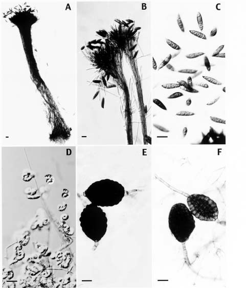
Virgatospora echinofibrosa Finley, 1967, Mycologia, 59: 538-541. (Figure 1A-C)
Conidiophores macronematous, synnematous, scattered, solitary, up to 1.3 mm high, 80 µm wide at the middle, and 130-145 µm wide at the base, individual conidiophores narrow, 1.5-2.7 µm, straight or flexuous, subhyaline to brown or blackish brown, smooth to finely echinulate, unbranched and tightly adpressed along most of their length but separating, splaying out and branching penicillately towards their apices. Conidiogenous cells monophialidic, determinate, cylindric to clavate. Conidia broadly fusiform with papillate, truncate ends, straight or curved, 3-septate, 35-48 × 9-15 µm. olivaceous grey, coarsely striate.
This stilbaceous hyphomycete has been indicated as an anamorph of Nectria spirostriata by Rossman (1983) and Matsushima (1993). A Myrothecium-state has also been reported as an anamorph of N. echinofibrosa by Rossman and Matsushima respectively. Our isolate growing on autoclaved sterilized corn leaf section placed on Sach's medium formed only Virgatospora-state synnemata and conidia. No teleomorph has been observed in our case.
Habitat. This fungus was isolated from the twig of an unknown tree collected from a stream at Wulai, Taipei county, Taiwan on September 25, 1996.
Helicoma depressispora Matsushima, Matsushima Mycological Memoirs No.7, p. 52, 1993. (Figure 1D-F)
Conidiophores micronematous to semi-macronematous. Conidia produced holoblastically on blunt-tipped denticles produced on lateral swellings of repent hyphae. Conidia pale yellow, circinate, coiled 1.25 to 1.75 times in a single plane, non-hygroscopic, 5-(6-7)-12 septate, slightly constricted at septa, 15-23 µm wide and 11-(14-15)-17 µm high;
conidial filament 4.5-7.0 µm wide. Chlamydospores intercalary, developing by cell division and enlargement, dictyosporous, constricted at septa, light brown at first, becoming black, 50-(75-85)-95 × 33-60 µm; outer wall of surface cells protuberant.
Two species of Helicoma, Helicoma chlamydosporum Shearer and H. depressispora form dictyochlamydospores. During our survey on microfungi on decayed twigs on streams we came across a fungal isolate that not only produced conidia of the genus Helicoma but also formed dictyochlamydospores. This fungus was identified as H. depressispora Matsushima. Matsushima (1993) first described this fungus, which he observed on decaying Palmae-petioles at Tambopata, Peru in 1990 and Rio Yuturi, Ecuador in 1992. In 1983 Schoknecht and Crane apparently also obtained this fungus, but they found only dictyochlamydospores which resembled those produced by H. depressispora, but with no helico-conidia.
Habitat. This fungus was observed on twigs collected at Wulai, Taipei county on July 9, 1995.
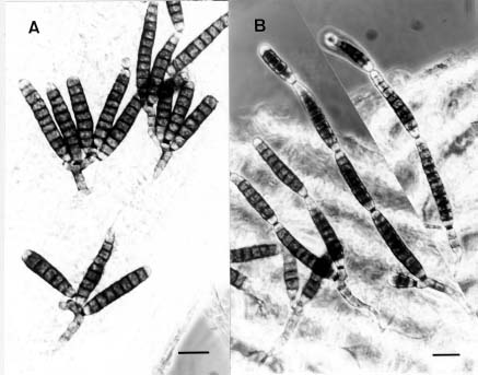
Lylea catenulata Morgan-Jones, Mycotaxon 3: 129-132, 1975. (Figure 2)
Conidiophores micronematous or semi-macronematous, inconspicuous, short, erect, cylindrical branches of the superficial mycelium, pale brown, smooth-walled, usually separated from the mycelium by a tansverse septum, 20-28 × 6-8 µm. Conidiophore growth ceases as the first conidium is formed. Conidiogenous cells monoblastic, integrate, frequently sympodial, cicatrized, scars prominent, dark, determinate or indeterminate. Conidia straight or slightly curved, cylindrical, obtuse at each end, catenate, dry, acrogenous, formed acropetally in chains, thick-walled with narrow cell lumina, guttulate, mid to dark brown smooth, (2-3) 6-8(13)-pseudosepta, 24-(50)-96 × 10-15 µm, formed in short acropetal chains. Successive conidia are formed apically from the terminal cell of the previously formed conidia and afterwards may be produced from the second and subsequent conidia at the terminal or intercalary loci.