Bot. Bull. Acad. Sin. (1999) 40: 259-265

Yang and Wang — Protease and amylase production of Streptomyces

Protease and amylase production of Streptomyces rimosus in submerged and solid state cultivations

Shang-Shyng Yang1,3 and Jan-Yi Wang2

1Department of Agricultural Chemistry, National Taiwan University, Taipei 106, Taiwan

(Received May 21, 1998; Accepted February 12, 1999)

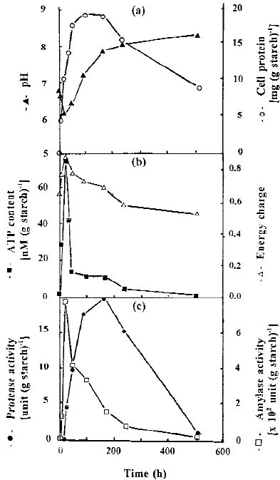
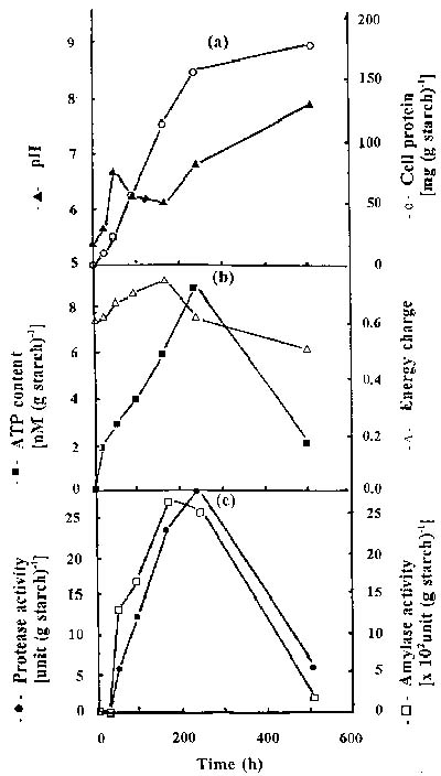
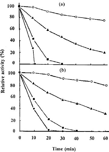
Abstract. The protease activity of Streptomyces rimosus TM-55 was first detected after 12 h of growth in submerged cultivation, and this activity peaked after 166 h of incubation. In solid state cultivation, protease was first secreted at 24 h, with the secretion peaking at 232 h. Amylase activity could be detected after 6 h in submerged cultivation, and it afterwards peaked after 48 h of incubation. In solid state cultivation, it began to be secreted at 24 h, and this secretion peaked after 180 h. Each gram of starch yielded 17.4 and 691.3 units of protease and amylase in submerged cultivation, respectively; whereas the values were 26.7 and 2,642.7 units in solid state cultivation. a-Amylase was the major amylase in both cultivation methods, and glucoamylase and debranching activity were minor components. Protease and amylase produced with both cultivation methods had a similar optimal pH, between 6.0 and 7.0, and optimal temperature, between 35 and 45°C. The enzyme activities produced in solid state cultivation were more stable with pH and temperature changes than those produced in submerged cultivation.

Keywords: Amylase; Protease; Streptomyces; Submerged and solid state cultivations.

Introduction

In Taiwan and other Asian countries, the Koji process has been used to produce various enzymes by growing molds on cereals or their brans. Although protease and amylase are mainly fungal and eubacterial products, the possibility of using streptomycetes for enzyme production has recently been investigated. Streptomyces species that produce proteases include S. clavuligerus, S. griseus, S. moderatus, S. rimosus, S. thermoviolaceus, and S. thermovulgaris (Pokorny et al., 1979; Renko et al., 1981, 1989; Chandrasekaran and Dhar, 1987; Bascaran et al., 1990; James et al., 1991; Muro et al., 1991; Yeoman and Edwards, 1994). Other hydrolases of Streptomyces species studied include aminopeptidase by S. fradiae, S. griseus, S. lividans, S. peptidofaciens, and S. rimosus (Vitale et al., 1986; Aphale and Strohl, 1993), chitinase by S. viridificans (Gupta et al., 1995), a-amylase by S. aureofaciens and S. rimosus (Vukeli et al., 1992; Cheng and Yang, 1995; Yang and Cheng, 1996), and b-glucosidase by Streptomyces sp. (Ozaki and Yamada, 1991).

Streptomyces species are heterotrophic feeders, and they can utilize both simple and complex molecules as nutrients. About three-fourths of the Streptomyces species may produce antibiotics. In addition to antibiotics, Streptomyces species liberate extracellular enzymes (Gupta et al., 1995). This work investigates the effect of sub

ú

c

merged and solid state cultivation techniques on growth, ATP content, and on the protease and amylase activities of Streptomyces rimosus.

Materials and Methods

Test Organism

Streptomyces rimosus TM-55 (CCRC 940061) was obtained from Dr. Thomas H. H. Ku, Cyanamid Taiwan Corporation.

Sweet Potato Residue and Peanut Meal Residue

Sweet potato residue and peanut meal residue were purchased from a local market in Taiwan and passed through a sieve (4 to 16 mesh) to remove dust and large aggregates. Sweet potato residue contained (w w-1) 14.0 to 16.1% moisture, 2.3 to 3.1% crude protein, 2.7 to 3.6% ash, 16.1 to 18.0% crude fibre, and 65.4 to 70.0% carbohydrate (mainly starch) (Yang, 1988). Peanut meal residue comprised (w w-1) 9.2 to 10.0% moisture, 49.4 to 51.4% crude protein, 5.4 to 6.1% ash, 6.7 to 7.5% crude fibre, 1.3 to 1.6% crude fat, and 25.2 to 26.2% nitrogen free extract.

Culture Media and Growth Conditions

Streptomyces rimosus was cultivated at 28°C on a slant containing (g l-1) soluble starch 10; yeast extract, 1; beef extract, 1; tryptone, 2; FeSO4•7H2O, 0.1; and agar, 20; at pH 7.2. Spores were harvested with a Tween 80 solution (5 ml, 0.05% v v-1), which was then adjusted with sterile water to give 107 to 108 spores per ml.

2Present address: National Laboratories of Foods and Drugs, Department of Health, Executive Yuan, Taipei 110, Taiwan.

3Correspondence author. Tel: 886-2-23621519; Fax: 886-2-23679827; E-mail: ssyang@ccms.ntu.edu.tw


Botanical Bulletin of Academia Sinica, Vol. 40, 1999

Submerged cultures contained (g l-1): soluble starch, 20; corn steep liquor (Sigma No. C-4648, contains solid 50%), 10; (NH4)2SO4, 6; CaCO3, 8; NaCl, 5; and soybean oil, 2; at pH 6.8 to 7.2. The broth was inoculated with 1.0 × 10⁶ spores per ml and shaken at 28°C and 250 rev min-1 for 1 to 9 days.

The solid substrate usually contained (g): sweet potato residue, 100; peanut meal residue, 20; MgSO4•7H2O, 2; and CaCO3, 1. The substrate was distributed into Erlenmeyer flasks, autoclaved, mixed thoroughly with the spores and the required amount of sterile water, and incubated statically at 28°C for 2 to 12 days with mixing once daily by rotating each flask. The depth of medium in each flask was about 2 cm. pH of the substrate was either measured directly by immersing the electrode into substrate or determined after mixing a portion with four times its volume of distilled water.

Extraction and Estimation of Adenine Nucleotides

After cultivation, cells were harvested by centrifugation at 6,000 g for 30 min. Cells from liquid medium or culture mass of solid medium were boiled with 0.02 M Tris-HCl buffer pH 7.6 for 10 min. The filtrate was used for adenosine nucleotide measurement. Firefly lantern extract (20 mg) containing luciferin and luciferase (Sigma) was dissolved in 5 ml of 0.02 M Tris-HCl buffer pH 7.6 and 1 ml of 0.01 M MgSO4•7H2O (dissolved in Tris-HCl buffer), and the mixture was stored at 4°C overnight. Before measurement, the enzyme solution was centrifuged at 3,000 g for 10 min. It was used within 2 h of preparation. Sample or adenosine nucleotide standard was mixed with the luciferin-luciferase mixture, and the luminescence was measured by ATP photometer (Turner TD-20e Luminometer, USA). Adenine nucleotide content was calculated from the standard curve of authentic compound. Energy charge was calculated as the mole fraction of ATP plus half the mole fraction of ADP, a measure of the amount of metabolically available energy stored in the adenylate pool (Karl, 1980; Sparling et al., 1986).

Extraction and Assay of Enzyme Activity

After cultivation, culture mass of solid medium was extracted with four volumes of 0.1% NaCl solution with shaking for 1 h at 25°C.

Protease activity of culture broth or culture mass extract was determined by a modified Anson's method (Yang and Huang, 1994). The reaction mixtures containing 1 ml of 1% casein in an appropriate buffer (pH 2.6: 0.1 M citric acid and 0.2 M NaH2PO4 McIlvaine buffer, pH 5.0 to 6.0: 0.1 M citric acid _ sodium citrate buffer, pH 6.7 to pH 7.5: 0.1 M Na2HPO4 and 0.1 M NaH2PO4 phosphate buffer, pH 7.6 to 8.5: 0.1 M Tris-HCl buffer, and pH 9.1 to 10.1: 0.1 M glycine-NaOH buffer) and 1 ml of enzyme solution were incubated at 37°C for 20 min, and the reaction was stopped with 3 ml of 10% trichloroacetic acid. The absorbance of the liberated tyrosine in the filtrate was measured at 280 nm. One unit of protease activity was defined as the amount of enzyme that produced an absorbance at 280 nm

equivalent to 1 µmole of tyrosine in one min under the assay conditions.

Amylase activity was measured by the dinitrosalicylic acid (DNS) method (Rick and Stegbauer, 1974). The reaction mixture contained 1 ml of 1% soluble starch in an appropriate buffer and 1 ml of enzyme solution. It was incubated at 25°C for 3 min. Two ml of DNS reagent were added, and the mixture was boiled for 5 min. After cooling, the reaction mixture was diluted with 16 ml of distilled water, and the absorbance was measured at 540 nm. One unit of amylase activity was defined as the amount of enzyme causing the release of 1 µmole of reducing sugars in one min under the assay conditions.

Alpha-amylase activity was assayed using a modification of the Phadebas amylase test (Pharmacia, Sweden) (Horn, 1990). Four ml of 0.02 M citrate buffer pH 5.0 and 0.2 ml of enzyme solution were preheated at 40°C for 5 min. One tablet of Phadebas was added to the reaction mixture, incubated at 40°C for 15 min, and stopped with 1 ml of 5 N NaOH. After centrifugation at 3,000 g for 5 min, the absorbance at 620 nm against the blank was measured. One unit of a-amylase activity was defined as the amount of enzyme hydrolyzing 1 µmole of glucose equivalents of starch in one min under the assay conditions.

Glucoamylase activity was determined by a modified Wilson and Ingledew method (1982). Five ml of soluble starch solution and 1 ml of enzyme solution were reacted at 50°C for 15 min, and the reaction was stopped by boiling for 5 min. After cooling, a 0.5 ml portion was reacted with 5 ml of peroxidase-glucose oxidase reagent (Sigma) at 40°C for 30 min, and the absorbance was measured at 450 nm. The absorbance of sample was converted to 1 mg of glucose in one min by using a standard curve.

Debranching activity was measured by a modified De Mot et al. method (1984). 0.5 ml of 1% pullulan and 0.2 ml of enzyme solution were reacted at 40°C for 45 min, 2.1 ml of DNS reagent was then added, and the reaction was stopped by boiling for 5 min. The reaction mixture was diluted with 11.55 ml of distilled water, and the absorbance was measured at 550 nm. The absorbance of sample was converted to 1 mg of glucose per min by using a standard curve.

Effect of pH on the Enzyme Activity

Effect of pH on enzyme activity was measured in pH 2.7, 5.0, 7.0 and 8.0 buffers at 37°C for 20 min with protease, and at 25°C for 3 min with amylase.

Effect of Temperature on the Enzyme Activity

Effect of temperature on enzyme activity was determined at 25, 35, 50, 60 and 78°C in pH 6.7 phosphate buffer. Test duration was 20 min for protease and 3 min for amylase, respectively.

Protein and Moisture Contents

Total nitrogen of cells from liquid medium and culture mass of solid medium was determined by a modified Kjeldahl method (Yang et al., 1991). Cell protein content


Yang and Wang — Protease and amylase production of Streptomyces

was calculated as 6.25 times the total nitrogen content. The moisture content of the culture mass was determined by drying the sample at 60°C under vacuum for 8 to 12 h to constant weight.

Results and Discussion

Enzymes Production in Submerged Cultivation

During the submerged cultivation of S. rimosus, protease activity was detected at 12 h and peaked at 166 h. The pH dropped from 6.7 to 6.1 after 24 h of cultivation and increased gradually to 8.2 at 480 h (Figure 1). Each gram of starch yielded 17.4 units of protease (i.e. each ml of culture broth supported the production of 0.4 units of protease). Amylase was secreted at 6 h, and had the highest activity at 48 h. Each gram of starch supported the production of 691.3 units of amylase (i.e. each ml of culture broth yielded 15.9 units of amylase). ATP content peaked at 24 h and then fell to a constant value between 240 and 504 h of cultivation. Energy charge increased from 0.68 to 0.90 at 24 h, and then decreased gradually to 0.57 between 240 and 504 h of incubation. ATP content is a potential index of microbial biomass (Karl and Holm-Hansen, 1978), and energy charge is a useful indicator of the energetic state of cells (Sivori, 1986). The energy charge of S. rimosus was very consistent with the energy charge of 0.87 in Acetobacter aceti (Bachi and Ettlinger, 1973) and Bacillus brevis (Fynn and Davison, 1976), the 0.76 to 0.79 in Clostridium kluyveri (Decker and Pfitzer, 1972), and the 0.8 to 0.9 in aerobically grown Saccharomyces cerevisiae (Ball and Atkinson, 1975). Cell growth had the log phase within 65 h, the stationary phase between 65 and 166 h, and the decline phase after 166 h of cultivation.

Enzyme Production in Solid State Cultivation

Protease activity was detected at 24 h and climaxed at 232 h (Figure 2). Each gram of starch yielded 26.7 units of protease (i.e. each gram of dry substrate supported the production of 15.8 units of protease). Amylase began to be secreted at 24 h with maximum secretion at 180 h. Each gram of starch yielded 2,642.7 units of amylase (i.e. each gram of dry substrate supported the production of 1,565 units of amylase). A very significant difference exists between submerged and solid state cultivation for protease and amylase production (p<0.01). The substrate concentration was low in submerged culture while the substrate accessibility was limited in solid state culture. Each liter of culture broth contained 22.4 to 23.6 g of starch in submerged culture while each liter of solid substrate contained 103.8 to 166.0 g of starch (the bulk density of solid substrate was between 0.50 and 0.80 g cm-3 in wet weight basis at 65% moisture content) in solid state culture. Therefore, the protease and amylase productivities per gram of starch in solid state cultivation were 53.4 and 282.3% higher than those in submerged culture, respectively. These results indicated that a high concentration of substrate in solid state cultivation

Figure 1. Time course of protease and amylase activities of S. rimosus in submerged cultivation at 28°C and 250 rev min-1. (a) pH and cell protein; (b) ATP content and energy charge; (c) Protease and amylase activities.

favored enzyme production. A similar result was also shown in the cellulase production of Trichoderma reesei. Cellulase productivity per gram of substrate in the solid state cultivation was 72% higher than that in the submerged culture (Chahal, 1985).

Amylase production in solid state cultivation took longer than that in submerged cultivation while the productivity of each gram of starch was about 2.82 fold higher than that in submerged cultivation. Amylase activity was connected with the substrate utilization by microbes while protease was associated with the growth of microbes. Therefore, amylase was secreted prior to protease.

Cell protein and moisture contents increased rapidly for the heavy growth and high metabolic activity of mycelia. The pH of the substrate first increased and then decreased for active growth. Matsushima et al. (1981) indicated that the pH drop in the acid protease production might be because of the accumulation of organic acid and


Botanical Bulletin of Academia Sinica, Vol. 40, 1999

the residue of sulfate ion in the utilization of ammonium sulfate. The pH lowering would be prevented when urea was used as the nitrogen source (Raimbault and Alazard, 1980; Yang, 1988). The pH of the substrate might be maintained with the supplement of alkali, an appropriate salt (Yang and Yuan, 1990), or other kinds of nitrogen sources (Nishio et al., 1981; Yang and Huang, 1994). Adenine nucleotide and ATP contents increased with the mycelial growth and then decreased rapidly in the slow growth period. Energy charge was high at 166 h incubation and then decreased gradually. Karl (1980) reported that energy charges are high in the log phase for heavy cell growth and low in the late stationary and decline phases for slow cell growth. Therefore, energy charge first increased and then decreased during a long cultivation period.

Protease activity in S. rimosus was produced at the middle of the exponential growth phase both in submerged and solid state cultivation. Enzymes produced by S. rimosus in solid state cultivation were more stable than those in submerged cultivation and could be temporarily stored without significant loss of activity. The difference might be because of cell stability. Cells were still active for 504 h of incubation in solid state cultivation whereas some cells lysis were observed for 166 h cultivation in submerged culture (Wang and Yang, 1995; Yang and Wang, 1996). Similar phenomena were also found in protease production of Aspergillus, Rhizopus, and Saccharomyces (Yang and Chiu, 1986; Yang and Huang, 1994), in the tetracycline production of Streptomyces viridifaciens (Yang and Ling, 1989) and the oxytetracycline productions of S. rimosus in submerged and solid state cultivations (Yang and Yuan, 1990; Yang and Swei, 1996).

Effect of pH and Temperature on Enzyme Activity

Protease and amylase activities were optimal at pH values of 6.0 to 6.7 in both cultivation methods. Protease activity was very sensitive to pH value, and it decreased sharply at pH 5.0 or pH 9.1. No enzymatic activity was found at pH values below 2.6 or higher than 10.1.

Figure 2. Time course of protease and amylase activities of S. rimosus in solid state cultivation at 28°C with mixing once daily. (a) pH and cell protein; (b) ATP content and energy charge; (c) Protease and amylase activities.

Table 1. Effect of pH on the enzyme activity of S. rimosus.

Submerged Solid state

pH Protease Amylase Protease Amylase

u (g starch)-1

2.6 0.0 0.0 0.0 0.0

5.0 6.5 258.0 6.2 1497.1

6.0 15.0 360.1 33.6 1568.7

6.7 17.5 397.2 35.2 1530.3

7.9 12.5 254.2 19.7 1377.1

9.1 9.0 154.2 13.2 1370.5

10.1 0.0 6.5 0.0 1277.2

Enzyme is diluted with nine volumes of buffer of different pH, and enzyme activity is measured at 37°C for 20 min for protease, and at 25°C for 3 min for amylase. pH 2.6 is maintained with McIlvaine buffer (0.1 M citric acid and 0.2 M NaH2PO4), pH 5.0 to 6.0 with 0.1 M citric acid _ sodium citrate buffer, pH 6.7 to 7.5 with 0.1 M Na2HPO4 and 0.1 M NaH2PO4 phosphate buffer, pH 7.6 to 8.5 with 0.1 M Tris-HCl buffer, and pH 9.1 to 10.1 with 0.1 M glycine-NaOH buffer.


Yang and Wang — Protease and amylase production of Streptomyces

However, amylase activity was still detectable at pH 10.1 in submerged cultivation, and it was moderate in solid state cultivation (Table 1). Streptomyces rimosus K-0K-02 had serine alkaline protease, leucine aminopeptidase, and trypsin-like protease that were stable from pH 4.5 to 9.0 (Renko et al., 1981, 1989; Vitale et al., 1986). Amylase

activity in solid state cultivation seems more stable with pH change than that in submerged culture.

Both enzymes had high activities between 35 and 50°C. Protease and amylase produced in submerged cultivation had lost some of their activities when the temperature was higher than 60°C (Table 2). Streptomyces rimosus K-0K-02 had a leucine aminopeptidase stable up to 70°C and had a trypsin-like protease that was stable up to 40°C (Vitale et al., 1986; Renko et al., 1989). Thermostability of amylase produced in solid state culture was higher than that of amylase produced in submerged culture (Figure 3). A similar phenomenon was also found with protease.

Effect of Cultivation Method on Amylases Production

Alpha-amylase was the major amylase in both cultivation methods while glucoamylase and debranching activity were much lower. Each gram of starch yielded 680 ± 60 units of a-amylase, 65 ± 5 units of glucoamylase, and 20 ± 2 units of debraching activity for 24 h of incubation in submerged cultivation. In solid state cultivation each gram of starch supported the production of 1,903 ± 268 units of a-amylase, 227 ± 40 units of glucoamylase, and 85 ± 6 units of debranching activity for 96 h of incubation. A similar tendency was also found in the cultivation of Schwanniomyces occidentalis in starch medium for amylase activity (Horn, 1990).

Alpha-amylase produced in both cultivation methods had the optimal pH at 5.0 to 6.0. a-Amylase activity in submerged cultivation was undetectable at pH higher than 9.1 or lower than 2.6 whereas the a-amylase produced in solid state cultivation had some activities at pH 9.1 or 2.6.

Enzyme production with solid state cultivation is more convenient than that with submerged culture for the utilization of starchy agricultural products. Hence, it is feasible to apply solid state cultivation as an economic alternative in producing value-added products and agricultural chemicals during the utilization and treatment of agricultural by-products.

Figure 3. Thermostability of amylase produced in different cultivation methods at pH 6.7 phosphate buffer. (a) Submerged cultivation; (b) Solid state cultivation. ¡, 40°C; 5, 50°C; n, 60°C; l, 70°C.

Table 2. Effect of temperature on the enzyme activity of S. rimosus.

Temperature Submerged Solid state

(°C) Protease Amylase Protease Amylase

u (g starch)-1

25 7.0 570.1 27.2 1890.0

30 19.3 870.2 40.1 1920.3

35 44.5 1090.4 84.6 2140.2

40 62.5 1320.2 88.8 2230.0

45 52.5 1235.5 84.8 2180.4

50 42.0 1060.3 75.4 2090.2

60 19.0 925.3 57.2 2050.3

76 10.3 390.6 17.3 2000.4

Enzyme is diluted with nine volumes of 0.1 M pH 6.7 phosphate buffer. Activity assay duration was 20 min for protease and 3 min for amylase.


Botanical Bulletin of Academia Sinica, Vol. 40, 1999

Acknowledgements. We thank the National Science Council of the Republic of China for its financial support (NSC 82-0409-B002-195 and NSC 83-0409-B002-064).

Literature Cited

Aphale, J.S. and W.R. Strohl. 1993. Purification and properties of an extracellular aminopeptidase from Streptomyces lividans 1326. J. Gen. Microbiol. 139: 417-424.

Bachi, B. and L. Ettlinger. 1973. Influence of glucose on adenine nucleotide levels and energy charge in Acetobacter acetic. Arch. Mikrobiol. 93: 155-164.

Ball, W.J. Jr. and D.E. Atkinson. 1975. Adenylate energy charge in Saccharomyces cerevisiae during starvation. J. Bacteriol. 121: 975-982.

Bascaran, V., V. Hardisson, and A. Brana. 1990. Regulation of extracellular protease production in Streptomyces clavuligerus. Appl. Microbiol. Biotechnol. 34: 208-213.

Chahal, D.S. 1985. Solid state fermentation with Trichoderma reesei for cellulase production. Appl. Environ. Microbiol. 56: 554-557.

Chandrasekaran, S. and S.C. Dhar. 1987. Multiple protease from Streptomyces moderatus. I. Isolation and purification of five extracellular proteases. Arch. Biochem. Biophys. 257: 395-401.

Cheng, C.W. and S.S. Yang. 1995. Amylase production of Streptomyces rimosus TM-55 and their 2-deoxyglucose mutants. Chin. J. Microbiol. Immunol. 28: 109-116.

De Mot, R., E. van Oudendijck, and H. Verachtert. 1984. Production of extracellular debranching activity by amylolytic yeasts. Biotechnol. Let. 6: 581-586.

Decker, K. and S. Pfitzer. 1972. Determination of steady-state concentrations of adenine nucleotides in growing C. kluyveri cells by biosynthetic labeling. Anal. Biochem. 50: 529-539.

Fynn, G.H. and J.A. Davison. 1976. Adenine nucleotide pool and energy charge during growth of a tyrothricin-producing strain of Bacillus brevis. J. Gen. Microbiol. 94: 68-74.

Gupta, R., R.K. Saxena, P. Chaturvedi, and J.S. Virdi. 1995. Chitinase production by Streptomyces viridificans: its potential on fungal cell wall lysis. J. Appl. Bacteriol. 78: 378-383.

Horn, C.H. 1990. Characterization of an amylolytic Schwanniomyces occidentalis mutant in relation to ethanol and protein production from grain sorghum. Ph. D. Thesis, Department of Microbiology and Biochemistry, University of the Orange Free State. Bloemfontein, South Africa.

James, P.D.A., M. Iqbal, C. Edwards, and P.G.G. Miller. 1991. Extracellular protease activity in antibiotic-producing Streptomyces thermoviolaceus. Curr. Microbiol. 22: 377-382.

Karl, D.M. 1980. Cellular nucleotide measurements and applications in microbial ecology. Microbiol. Rev. 44: 739-796.

Karl, D.M. and O. Holm-Hansen. 1978. Methodology and measurement of adenylate energy charge ratios in environmental samples. Marine Biol. 48: 185-197.

Matsushima, K., M. Hayakawa, M. Ito, and K. Stimada. 1981. Ecological study on the proteolytic enzymes of microorganisms. Part III. Features of the proteolytic enzymes system of hyper-acid-productive and non-acid-productive fungi. J. Gen. Appl. Microbiol. 27: 423-426.

Muro, T., T. Murakami, Y. Tominaga, T. Tokuyama, and S.

Okada. 1991. Purification and some properties of protease 1 having transfer action from Streptomyces griseus var. alcalophilus. Agric. Biol. Chem. 55: 307-314.

Nishio, N., H. Kurisu, and S. Nagai. 1981. Thermophilic cellulase production by Taralomyces sp. in solid state cultivation. J. Ferment. Technol. 59: 407-410.

Ozaki, H. and K. Yamada. 1991. Isolation of Streptomyces sp. producing glucose-tolerant b-glucosidases and properties of the enzymes. Agric. Biol. Chem. 55: 979-987.

Pokorny, M., Lj. Vitale, V. Turk, M. Renko, and J. Zuvani . 1979. Streptomyces rimosus extracellular proteases. 1. Characterization and evaluation of various crude preparations. Europ. J. Appl. Microbiol. Biotechnol. 8: 81-90.

Raimbault, M. and D. Alazard. 1980. Culture method to study fungi growth in solid fermentation. Europ. J. Appl. Microbiol. Biotechnol. 9: 199-209.

Renko, M., M. Pokorny, Lj. Vitale, and V. Turk. 1981. Streptomyces rimosus extracellular proteases. 2. Isolation and characterization of serine alkaline proteinase. Europ. J. Appl. Microbiol. Biotechnol. 11: 166-171.

Renko, M., Lj. Vitale, M. Kokalj, and M. Pokorny. 1989. Streptomyces rimosus extracellar proteases. 4, Trypsin-like proteinase. Appl. Microbiol. Biotechnol. 31: 38-44.

Rick, W. and H.P. Stegbauer. 1974. a-Amylase measurement of reducing groups. In H. V. Bergmeyer (ed.), Methods of Enzymatic Analysis, 2nd edn., vol. 2, Academic Press, New York.

Sivori, E. 1986. Adenylic nucleotides and energy charge during the embryonic development of Bufo arenarum. Comp. Biochem. Physiol. 85B: 573-576.

Sparling, G., P.T.W. Speir, and K.N. Whale. 1986. Changes in microbial biomass, ATP content, soil phosphomonoesterase and phosphodiesterase activity following air-drying of soils. Soil Biol. Biochem. 18: 363-370.

Vitale, Lj., M. Renko, B. Lenarcic, V. Turk, and M. Pokory. 1986. Streptomyces rimosus extracellar proteases. 3. Isolation and characterization of leucine aminopeptidase. Appl. Microbiol. Biotechnol. 23: 449-455.

Vukeli , B., A. Ritonja, M. Renko, M. Pokorny, and Lj. Vitale. 1992. Extracellular a-amylase from Streptomyces rimosus. Appl. Microbiol. Biotechnol. 37: 202-204.

Wang, J.Y. and S.S. Yang. 1995. Morphogenesis, biomass and oxytetracycline production of Streptomyces rimosus in submerged cultivation. Chin. J. Microbiol. Immunol. 28: 21-31.

Wilson, J.J. and M.W. Ingledew.1982. Isolation and characterization of Schwanniomyces alluvius amylolytic enzymes. Appl. Environ. Microbiol. 44: 301-307.

Yang, S.S. 1988. Protein enrichment of sweet potato residue with amylolytic yeasts by solid state fermentation. Biotechnol. Bioeng. 32: 886-890.

Yang, S.S., H.L. Chang, C.B. Wei, and H.C. Lin. 1991. Reduce waste production in the Kjeldahl methods. J. Biomass Energy Soc. China 10: 147-155.

Yang, S.S. and C.W. Cheng. 1996. Production, purification and characterization of a-amylase by Streptomyces rimosus. J. Chin. Agric. Chem. Soc. 35: 649-654.

Yang, S.S. and W.F. Chiu. 1986. Protease production with sweet potato residue by solid state fermentation. Chin. J. Microbiol. Immunol. 19: 276-288.

^

c

ó

c

ó


Yang and Wang — Protease and amylase production of Streptomyces

Yang, S.S. and C.I. Huang. 1994. Protease production by amylolytic fungi in solid state fermentation. J. Chin. Agric. Chem. Soc. 32: 589-601.

Yang, S.S. and M.Y. Ling. 1989. Tetracycline production with sweet potato residue by solid state fermentation. Biotechnol. Bioeng. 33: 1021-1028.

Yang, S.S. and J.Y. Wang. 1996. Morphogenesis, ATP content and oxytetracycline production by Streptomyces rimosus in solid state cultivation. J. Appl. Bacteriol. 80: 545-550.

Yang, S.S. and W.J. Swei. 1996. Oxytetracycline production by Streptomyces rimosus in solid state fermentation of corncob. World J. Microbiol. Biotechnol. 12: 43-46.

Yang, S.S. and S.S. Yuan. 1990. Oxytetracycline production with sweet potato residue by solid state fermentation. World J. Microbiol. Biotechnol. 6: 236-244.

Yeoman, K.H. and C. Edwards. 1994. Protease production by Streptomyces thermovulgaris grown on rapemeal-derived media. J. Appl. Bacteriol. 77: 264-270.

Streptomyces rimosus ¦b²GºA¤Î©TºA°ö¾i®É

¥Í²£³J¥Õú@¤Î¾ý¯»ú@
·¨²±¦æ ¤ý­sÅt

°ê¥ß¥xÆW¤j¾Ç¹A·~¤Æ¾Ç¨t

Streptomyces rimosus TM-55 ¦b²GºA°ö¾i®É¡A 12 ¤p®É«á§Y¥iÀË¥X³J¥Õú@¬¡©Ê¦Ó¦b°ö¾i 166 ¤p®É³Ì°ª­È¡F¦Ó¦b©TºA°ö¾i®É¡A«h»Ý 24 ¤p®É¤è¥i´ú¥X³J¥Õú@¬¡©Ê©M°ö¾i 232 ¤p®É«á¥i±o³Ì°ª¬¡©Ê¡C¦Ó¦b²GºA°ö ¾i®É¡A¾ý¯»ú@¥i¦b 6 ¤p®É«á´ú¥X¡A¦Ó¦b°ö¾i«á 48 ¤p®É¥i¹F³Ì¤j¬¡©Ê¡A¦ý¦b©TºA°ö¾i®É¡A«h»Ý 24 ¤p®É¤è¥i´ú±o¡A¦Ó¦b°ö¾i«á 180 ¤p®É¥i±o³Ì¤j¬¡©Ê¡C¨C§J¾ý¯»°ò½è¦b²GºA°ö¾i®É¤À§O¥i±o 17.4 ©M 691.3 ³æ¦ì³J¥Õú@©M¾ý¯»ú@¡F¦Ó¦b©TºA°ö¾i®É«h¤À§O¥i±o 26.7 ©M 2,642.7 ³æ¦ì»Ã¯À¬¡©Ê¡C a-¾ý¯»ú@¬°¥D­n¤§¾ý¯»ú@¡A ¦Ó¸²µå¿}¾ý¯»ú@©M¥h¤À¤ä¬¡©Ê«h¬°¦¸­n¾ý¯»ú@¡C¨âºØ°ö¾i¤è¦¡©Ò±o¤§¾ý¯»ú@©M³J¥Õú@¤§³Ì¾A¤ÏÀ³ pH ¤¶©ó 6 ©M 7 ¡A³Ì¾A¤ÏÀ³·Å«×¤¶©ó 35 ©M 45°C¡C¥H©TºA°ö¾i©Ò±o¤§»Ã¯À¨ä¹ï pH ¤Î·Å«×¤§Ã­©w©Ê¤j©ó²GºA°ö¾i ©Ò±o¡C

ÃöÁäµü¡G ¾ý¯»ú@¡F³J¥Õú@¡F©TºA°ö¾i¡F²GºA°ö¾i¡FStreptomyces¡C