Bot. Bull. Acad. Sin. (1999) 40: 267-273

Chen and Kuc' Ca2+-dependent excretion of salicylic acid

Ca2+-dependent excretion of salicylic acid in tobacco cell

suspension culture

Hsien-Jung Chen1 and Joseph Kuc' 2,3

1Postdoctorate, Institute of Botany, Academia Sinica, Nankang, Taipei 11529, Taiwan

2Professor Emeritus, Department of Plant Pathology, University of Kentucky, Lexington, Kentucky 40546, USA

(Received June 19, 1998; Accepted February 12, 1999)

Abstract. The radioactive 14C-Salicylic acid (SA) was used to monitor the metabolism of exogenous SA in tobacco cell suspension culture. Tobacco cells took up ca. 39.46% of the applied SA 5 min after the addition of a final 200 µM SA mixture containing [14C]SA to the cell suspension culture. Most of the SA absorbed by the cells was gradually lost to the culture medium, and 2.62% and 91.55% of the radioactivity were found in the cells and the culture filtrate, respectively, 5 h after treatment. Excretion of SA to the culture medium required external Ca2+ and was inhibited by EGTA pretreatment. The inhibition was reversible through the supplement of Ca2+ to the cell suspension culture. Based on the data, we conclude that SA excretion from the 200 µM-treated cells requires external Ca2+ and likely involves a putative Ca2+-dependent pathway in tobacco suspension culture.

Keywords: Ca2+; EGTA; SA loss; Salicylic acid; Tobacco cell suspension culture.

Introduction

SA, 2-hydroxybenzoic acid, is endogenous in many plants including tobacco, cucumber, rice, wheat, cotton, tomato, and Arabidopsis (Raskin et al., 1990). SA has been considered a natural growth regulator with numerous functions in plants (Raskin, 1992). It promotes bud formation and growth in tobacco callus culture (Lee and Skoog, 1965); induces flowering in Lemna gibba G3 and Lemna paucicostata 151 (Cleland and Ajami, 1974; Tanaka et al., 1979; Watanable and Takimoto, 1979); inhibits ethylene biosynthesis in pear cell suspension culture (Leslie and Romani, 1986); inhibits jasmonic acid biosynthesis and prevents wound inducible proteinase inhibitor gene expression in tomato (Pena-Cortes et al., 1993); regulates heat production in the inflorescence of an Arum lily (Raskin et al., 1987; Raskin et al., 1989); reverses ABA-induced stomatal closure (Rai et al., 1986); induces local resistance to TMV in Nicotiana tabacum cv. xanthi-nc, Samsun NN and White Burley (White, 1979); and functions as a factor for systemic acquired resistance (SAR) in tobacco (Malamy et al., 1990; Gaffney et al., 1993; Vernooij et al., 1994a; Vernooij et al., 1994b), cucumber (Metraux et al., 1990), and Arabidopsis (Delaney et al., 1994).

The concentration of endogenous SA varied significantly from tissue to tissue, and also from species to species. In thermogenic plants such as Dioon hildebrandtii, the amount of SA could be as high as 100 µg g-1 fresh weight in male cones. However, it could be

as low as less than 0.01 µg g-1 fresh weight in leaves of Nicotiana tabacum and Zea mays (Raskin et al., 1990). In plant-microbe interactions, the endogenous SA concentration could increase 10 to 20 fold in TMV-infected leaves (Malamy et al., 1992). SA can be found in nature in both free and conjugate forms during plant development (Cooper-Driver et al., 1972) and plant-microbe interactions (Malamy et al., 1992).

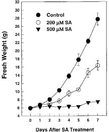
SA has been applied to tobacco cell suspension culture to study its effects on gene expression and cell physiology. Kapulnik et al. (1992) reported that 20 µM SA could induce cyanide-resistant respiration of tobacco cells in suspension culture. An SA-inducible gene encoding a 48 kDa MAP kinase protein has been isolated from tobacco. SA induction of the MAP kinase mRNA is dose-dependent with concentrations more than 50 µM, and the maximal induction could be found at the concentration of 500 µM in tobacco cell suspension (Zhang and Klessig, 1997). Kawano et al. (1998) reported that 500 µM SA could induce extracellular superoxide generation followed by an increase in cytosolic calcium ion in tobacco cell suspension culture. The acidic b-1,3-glucanase mRNA was induced by 200 µM and 20 µM SA in tobacco cell suspension culture (Chen, 1995). Therefore, the SA dose within 500 µM is appropriate and can be used to study its effects on particular gene induction and cell physiology in tobacco cell suspension culture.

In order to achieve the SA functions described above, SA has to enter the cells preceding the trigger of a particular gene expression. Ben-Tal and Cleland (1982) reported that Lemna gibba G3 plants took up nearly 90% of the applied 10 µM SA containing [14C]SA within 30 min. Rapid uptake of SA was also reported in tobacco suspen

3Corresponding author. Current address: 700 Front St., #1202, San Diego, CA 92101, U.S.A.


Botanical Bulletin of Academia Sinica, Vol. 40, 1999

sion culture with 20 µM SA containing [14C]SA. About 50% of the applied SA was absorbed by the cells within 10 min. Then the cellular [14C]SA radioactivity began to decline gradually within 2 h after treatment due to unknown mechanisms (Kapulnik et al., 1992). We report here that a similar quick uptake of exogenous SA by tobacco cells was also observed in suspension culture treated with 200 µM SA. More than 80% of the SA taken up by the cells was lost to the culture filtrate via a Ca2+-dependent SA excretion pathway in tobacco cell suspension culture.

Materials and Methods

Plant Materials and Chemicals

Tobacco cell suspension (Nicotiana tabacum cv. KY14) was subcultured every 7 days in a 1:2 (v:v) ratio with 3% (w/v) sucrose MS medium (pH 5.6) supplemented with 10 µg ml-1 thiamine-HCl, 1 µg ml-1 nicotinic acid, 1 µg ml-1 pyridoxine-HCl, 100 µg ml-1 myo-inositol, and 1 ppm 2,4-D. Cell suspension in a 1:1 (v/v) dilution ratio for 3 days was used for the experiments. Chemicals were purchased from the following companies: [7-14C] SA from New England Nuclear; MS salt mixture from GIBCO BRL; SA, EGTA and 2,4-D from Sigma. The SA and [14C]SA applied to the culture medium are nonionic, free acids.

Measurement of Tobacco Cell Growth

Twenty ml of tobacco cell suspension was mixed with 30 ml of fresh culture medium containing a final concentration of 0, 200 µM or 500 µM SA. Cells were harvested daily by vacuum filtration through Whatman No. 4 filter papers and briefly washed with phosphate buffer saline (NaCl 8 g l-1, KCl 0.2 g l-1, Na2HPO4 1.15 g l-1 and KH2PO4 0.2 g l-1, pH 7.5) for fresh weight measurement.

Measurement of [14C]SA Radioactivity

To determine the changes of SA amounts in the cells and the culture filtrate, 2 ml of cell suspension was mixed with 3 ml of fresh medium in a 50 ml Falcon disposable centrifuge tube. A final 200 µM SA mixture containing unlabeled SA and 0.76 µM [14C]SA (0.2 µCi) was added to the cell suspension. The cells and the culture filtrate were then collected separately at intervals after treatment. One ml of the culture filtrate was mixed directly with 15 ml of liquid scintillation fluid. The cells were collected for fresh weight measurement, mixed with 400 µl of 60% HClO4 and 100 µl of 30% H2O2, then incubated in an 80°C water bath for 2 h. Fifteen ml of liquid scintillation fluid was added to the vial. The radioactivity was determined with a PACKARD 2200CA Liquid Scintillation Analyzer.

Effect of Medium pH on SA Uptake

Tobacco cell suspension was mixed in a 1:1 (v:v) ratio with fresh culture media of various pH ranging from 3.5 to 8.5. A final 200 µM SA mixture containing [14C]

SA as described earlier was added to the cell suspension, and the cellular radioactivity was measured 5 min after treatment.

Chemical Treatments

Tobacco cell suspensions were pretreated with EGTA (a final concentration of 0.5, 1 or 5 mM) for 15 min prior to the addition of a final 200 µM SA mixture containing [14C]SA as described earlier. The cells were harvested for radioactivity determination at intervals after treatment.

For Ca2+ treatment, the solution containing a final 200 µM SA mixture as described earlier and Ca2+ (at final concentrations of 1, 2, 5, 10, 20 or 50 mM) was applied to the cell suspension, which had been pretreated with 5 mM EGTA for 15 min. The cells were collected for radioactivity measurement 5 h after treatment.

Tobacco cell suspensions were pretreated with 200 µM non-radioactive SA or SA analogs including acetylsalicylic acid (aspirin), 3,4-dihydroxybenzoic acid, 2,6-dihydroxybenzoic acid or 2,3-dihydroxybenzoic acid for 15 min prior to the addition of a final 200 µM SA mixture containing [14C]SA as described earlier. Cycloheximide (at final concentrations of 1, 2, 5, 10 or 30 µg ml-1) was fed together with the SA mixture to tobacco cell suspensions. Cellular radioactivity was determined 5 h after treatment.

Results

Excretion of SA from the 200 µM-Treated Cells to the Culture Medium

Tobacco cells in 200 µM SA experienced reduced fresh weights, to about 40% that of the untreated control, and little to no growth was found in 500 µM SA 7 days after subculture (Figure 1). The SA dose, 200 µM, which showed somewhat toxic to cell growth, was used for the following experiments.

Changes of SA amounts in the cells and the culture filtrate were monitored with radioactive [14C]SA. The radioactivity increased in the cells and decreased in the culture filtrate 5 min after 200 µM SA treatment, then decreased

Table 1. Changes of total radioactivity in the cells and the culture filtrate at intervals after the addition of a final 200 µM SA mixture containing [14C]SA to the tobacco cell suspensions. The data are expressed as means ± SE from three independent experiments. The [14C]SA radioactivity (417656 cpm) is converted into SA (138 µg).

Time after addition SA [µg (%)]

of 200 µM SA Culture filtrate Cells

0 h 137.99 ± 0.02 (99.72) 0.39 ± 0.28 (0.28)

5 min 59.82 ± 1.60 (43.23) 54.60 ± 4.39 (39.46)

1 h 86.55 ± 1.84 (62.55) 27.79 ± 5.67 (20.08)

2 h 89.20 ± 2.49 (64.46) 22.20 ± 6.99 (16.04)

3 h 114.56 ± 2.57 (82.79) 8.51 ± 0.20 (6.15)

4 h 117.75 ± 1.00 (85.09) 4.88 ± 0.93 (3.53)

5 h 126.68 ± 6.17 (91.55) 3.63 ± 1.30 (2.62)


Chen and Kuc' Ca2+-dependent excretion of salicylic acid

Figure 1. Tobacco cell growth curves in the presence of 0, 200 µM or 500 µM salicylic acid (SA) in the suspension cultures. The fresh weight of tobacco cells was measured daily after subculture. The data are expressed as means ± SE from three independent experiments, and the vertical bar of each datal point represents the size of SE.

Figure 2. Effect of medium pH on the uptake of SA by tobacco cells. The cellular [14C]SA radioactivity was measured 5 min after the addition of a final 200 µM SA mixture containing [14C]SA to the cell suspensions, which had medium pHs ranging from 3.5 to 8.5. The data are from three independent experiments.

in the cells and increased in the culture filtrate as a function of time within 5 h (Table 1). Tobacco cells took up about 39.46% of the applied [14C]SA 5 min after addition and most of the SA absorbed by the cells was gradually lost to the culture filtrate. Five hours after treatment, about 91.55% and 2.62% of the radioactivity was found in the culture filtrate and the cells, respectively (Table 1). The data suggest that excretion of SA to the culture medium is the major mechanism responsible for the decrease of cellular [14C]SA radioactivity. Decarboxylation of the [7-14C]carboxyl group of salicylic acid is another possibility contributing to the decrease of intracellular radioactivity. The radioactivity in the cells had been monitored up to 24 h, and no significant changes were found after 5 h (Chen, 1995). The recovery of radioactivity from different time points ranged from 80% to 95%. The 14CO2 derived from decarboxylated [7-14C] SA, which was not detected by the assay method used, or [7-14C] SA loss during experimental operation were the possible causes for the variation in recovery at different time points.

PH-Dependent Uptake of Exogenous SA

The cellular [14C]SA radioactivity from the suspension culture with various pH was measured 5 min after 200 µM SA addition. The uptake of SA and its accumulation in the cells was pH-dependent and inversely correlated with the increase of medium pH. The greatest radioactivity was found in the cells from medium pH 3.5 and was 7 to 8

times that of medium pH 8.5 (Figure 2). Since the pH of SA mixture was acidic, we monitored its effect on the medium pH change after 200 µM SA treatment. The medium pH dropped from 5.6 to about 4.7 immediately after SA addition, then increased to about the same level as untreated control (about pH 5.3) within 30 min (Chen, 1995).

Ca2+-Dependent Excretion of SA

Pretreatment with EGTA caused a higher retention of cellular [14C]SA radioactivity than the untreated control in a dose-dependent manner. The radioactivity in 5 mM EGTA-pretreated cells was about twelve times that of the untreated control 5 h after 200 µM SA addition (Figure 3). Supplement of Ca2+ (at final concentrations of 1, 2, 5, 10, 20 or 50 mM) to the cell suspension reversed the effect by 5 mM EGTA, and no significant difference of the cellular radioactivity was found between the 20 mM Ca2+ / 5 mM EGTA pretreatment and the untreated SA alone control (Figure 4). The data suggest a Ca2+-dependent excretion mechanism for SA in tobacco cell suspension culture.

Inhibition of SA Excretion by SA, Aspirin, and Cycloheximide

Pretreatment with 200 µM non-radioactive SA or aspirin increased the cellular [14C] SA radioactivity ca. fourfold the amount of the untreated control 5 h after the


Botanical Bulletin of Academia Sinica, Vol. 40, 1999

addition of a 200 µM SA mixture containing [14C]SA. Tobacco cells pretreated with SA analogs such as 2,3-dihydroxybenzoic acid, 3,4-dihydroxybenzoic acid, or 2,6-dihydroxybenzoic acid showed a level of cellular radioactivity similar to the untreated control (Figure 5A). The data indicate that excretion of SA from the treated cells is likely via an export pathway with specificity in tobacco cell suspension culture.

Cycloheximide caused a higher retention of the cellular [14C]SA radioactivity, 4 to 5 times that of the untreated control 5 h after 200 µM SA addition, and it was effective as low as 1 µg ml-1 (Figure 5B). The data suggest that excretion of SA from the treated cells requires de novo synthesized proteins.

Discussion

Tobacco cells rapidly accumulated tremendous amounts of the applied SA in a pH-dependent manner within 5 min after 200 µM SA addition (Table 1 and Figure 2). A similar pH-dependent uptake of SA and its accumulation was also previously reported in oat roots (Harper and Balke, 1981), renal proximal S2 and S3 tubules (Chatton and Roch-Ramel, 1991), Kidney epithelial cell line LLC-PK1 (Chatton and Roch-Ramel, 1992a), and MDCK cell monolayers (Chatton and Roch-Ramel, 1992b). Gutknecht (1990) demonstrated that the pH-dependent uptake is positively correlated to the amount of lipid-soluble, nonionic SA. The form of SA applied to the tobacco cell suspension culture was also nonionic, and a similar mechanism was suggested and possibly utilized in the study.

More than 80% of the absorbed SA was lost from the treated cells to the culture medium via a Ca2+-dependent excretion mechanism in 200 µM SA (Table 1, Figures 3 and 4). In lactating rat mammary tissue, SA stimulated external Ca2+ influx and calcium-dependent K+ efflux (Shennan, 1992). In tobacco cell suspension culture, treatment of 500 µM SA also rapidly increased cytosolic Ca2+ concentration within 10 seconds after application (Kawano et al., 1998). Therefore, it is possible that a calcium influx system is switched on shortly after the uptake of SA. This in turn activates a putative Ca2+-dependent SA excretion pathway, which functions to promote SA loss and lower toxic SA level in the cells (Figure 1). The toxic effects of SA on cell growth and the protective effects of calcium supplement had been reported previously in studies of tobacco cell suspension culture (Kapulnik et al., 1992) and daily salicylate-injected rats (Jastreboff and Brennan, 1994).

Excretion of SA was much reduced by the pretreatment with 200 µM non-radioactive SA and aspirin, but was only slightly affected by SA analogs such as 2,3-dihydroxybenzoic acid, 3,4-dihydroxybenzoic acid and 2,6-dihydroxy benzoic acid (Figure 5A). Takanaga et al. (1994) reported that the permeability coefficient and permeation rate of [14C]SA in human colon adenocarcinoma cell line Caco-2 were significantly reduced or competitively inhibited by non-radioactive SA, acetic acid, and benzoic

Figure 3. Effect of EGTA on the cellular [14C]SA radioactivity in 200 µM SA. Changes in cellular [14C]SA radioactivity were measured at intervals after the addition of a final 200 µM SA mixture containing [14C]SA to cell suspensions pretreated with EGTA (at final concentrations of 0.5, 1 or 5 mM) for 15 min. The cellular radioactivity 5 min after the addition of a final 200 µM SA mixture containing [14C]SA without EGTA was specified as 100%. The data are means from two independent experiments.

Figure 4. Reversal of the EGTA-induced repression of SA excretion by external Ca2+ in 200 µM SA. The cellular [14C]SA radioactivity was measured 5 h after the addition of a final 200 µM SA mixture containing [14C]SA alone (SA alone) or supplemented with Ca2+ (at final concentrations of 1, 2, 5, 10, 20 or 50 mM) to the cell suspensions pretreated with 5 mM EGTA. The data are chosen from a representative one of two independent experiments


Chen and Kuc' Ca2+-dependent excretion of salicylic acid

acid, all of which had been demonstrated to be transported in a carrier-mediated transporter mechanism. Our data agree with the report and suggest the possibility that a putative, carrier-mediated transporter with specificity may be responsible for the excretion of SA in tobacco cell suspension culture. SA excretion from the 200 µM-treated cells required de novo synthesized proteins (Figure 5B), possibly due to (1) an inducible carrier-mediated transporter for the excretion of SA, (2) the signals/signal transduction pathways that linked to activate the Ca2+-dependent SA excretion mechanism or (3) both.

We conclude here that excretion of exogenous SA from the 200 µM-treated cells to the culture medium requires external Ca2+ and involves a putative Ca2+-dependent export pathway, which likely functions to lower toxic SA level in the cells. Identification and isolation of the components may help unravel the mechanisms and function of SA excretion.

Literature Cited

Ben-Tal, Y. and C. Cleland. 1982. Uptake and metabolism of [14C]salicylic acid in Lemna gibba G3. Plant Physiol. 70: 291-296.

Chatton, J.-Y. and F. Roch-Ramel. 1991. Passive permeability of salicylic acid in renal proximal S2 and S3 tubules. J. Pharmacol. Exp. Ther. 256: 1112-1118.

Chatton, J.-Y. and F. Roch-Ramel. 1992a. Transport of salicylic acid through monolayers of kidney epithelial cell line (LLC-PK1). J. Pharmacol. Exp. Ther. 261: 518-524.

Chatton, J.-Y. and F. Roch-Ramel. 1992b. Nonionic diffusion of salicylic acid through MDCK cell monolayers. J. Pharmacol. Exp. Ther. 261: 1071-1078.

Chen, H.-J. 1995. Salicylic Acid and the Regulation Of b-1,3-Glucanase Gene Expression in Tobacco Cell Suspension Culture. Ph.D. thesis, University of Kentucky, Lexington, KY.

Cleland, C.F. and A. Ajami. 1974. Identification of the flower-inducing factor isolated from aphid honeydew as being salicylic acid. Plant Physiol. 54: 904-906.

Cooper-Driver, G., J.J. Corner-Zamodits, and T. Swain. 1972. The metabolic fate of hydroxybenzoic acids in plants. Z Naturforsch 27b: 913-916.

Delaney, T.P., S. Uknes, B. Vernooij, L. Friedrich, K. Weymann, D. Negrotto, T. Gaffney, M. Gut-Rella, H. Kessmann, E. Ward, and J. Ryals. 1994. A central role of salicylic acid in plant disease resistance. Science 266: 1247-1250.

Gaffney, T., L. Friedrich, B. Vernooij, D. Negrotto, G. Nye, S. Uknes, E. Ward, H. Kessmann, and J. Ryals. 1993. Requirement of salicylic acid for the induction of systemic acquired resistance. Science 261: 754-756.

Gutknecht, J. 1990. Salicylates and proton transport through lipid bilayer membrane: a model for salicylate-induced uncoupling and swelling in mitochondria. J. Membrane Biol. 115: 253-260.

Harper, J.R. and N.E. Balke. 1981. Characterization of the inhibition of K+ absorption in oat roots by salicylic acid. Plant Physiol. 68: 1349-1353.

Jastreboff, P.J. and J.F. Brennan. 1994. Alleviation of mortality induced by salicylate and stress. Experientia 50: 469-

Figure 5. Effects of non-radioactive SA, SA analogs and cycloheximide (CHX) on the cellular [14C]SA radioactivity 5 h after the addition of a final 200 µM SA mixture containing [14C]SA. (A) Effects of SA and SA analogs. The non-radioactive SA and SA analogs (at a final concentration of 200 µM) were added to the cell suspensions prior to the treatment with a 200 µM SA containing [14C]SA. The data are from three independent experiments. (1) no pretreatment control; (2) 3,4-dihydroxybenzoic acid; (3) 2,6-dihydroxybenzoic acid; (4) 2,3-dihydroxybenzoic acid; (5) acetylsalicylic acid (aspirin); (6) SA pretreatment. (B) Effect of cycloheximide. Cycloheximide (at final concentrations of 1, 2, 5, 10 or 30 µg ml-1) was applied together with the SA mixture to the cell suspension culture. The data are from three independent experiments.


Botanical Bulletin of Academia Sinica, Vol. 40, 1999

Arum lilies. Science 237: 1601-1602.

Raskin, I., I.M. Turner, and W.R. Melander. 1989. Regulation of heat production in the inflorescence of an Arum lily by endogenous salicylic acid. Proc. Natl. Acad. Sci. USA 86: 2214-2218.

Raskin, I., H. Skubatz, W. Tang, and B.J.D. Meeuse. 1990. Salicylic acid levels in thermogenic and non-thermogenic plants. Ann. Bot. 66: 369-373.

Raskin, I. 1992. Role of salicylic acid in plants. Annu. Rev. Plant Physiol. Plant Mol. Biol. 43: 439-463.

Shennan, D.B. 1992. Salicylate-induced cation fluxes across biological membranes. Biochem. Pharmacol. 44: 645-650.

Takanaga, H., I. Tamai, and A. Tsuji. 1994. pH-dependent and carrier-mediated transport of salicylic acid across Caca-2 cells. J. Pharm. Pharmacol. 46: 567-570.

Tanaka, O., C.F. Cleland, and W.S. Hillman. 1979. Inhibition of flowering in the long-day plant Lemna gibba G3 by Hunter's medium and its reversal by salicylic acid. Plant Cell Physiol. 20: 839-846.

Vernooij, B., L. Friedrich, A. Morse, R. Reist, R. Kolditz-Jawhar, E. Ward, S. Uknes, H. Kessmann, and J. Ryals. 1994a. Salicylic acid is not the translocated signal responsible for inducing systemic acquired resistance but is required in signal transduction. Plant Cell 6: 959-965.

Vernooij, B., S. Uknes, E. Ward, and J. Ryals. 1994b. Salicylic acid as a signal molecule in plant-pathogen interactions. Curr. Opin. Cell Biol. 6: 275-279.

Watanable, K. and A. Takimoto. 1979. Flower-inducing effects of benzoic acid and some related compounds in Lemna paucicostata 151. Plant Cell Physiol. 20: 847-850.

White, R.F. 1979. Acetylsalicylic acid (aspirin) induces resistance to tobacco mosaic virus in tobacco. Virology 99: 410-412.

Zhang, S. and D.F. Klessig. 1997. Salicylic acid activates a 48-kD MAP kinase in tobacco. Plant Cell 9: 809-824.

474.

Kapulnik, Y., N. Yalpani, and I. Raskin. 1992. Salicylic acid induces cyanide-resistance respiration in tobacco cell suspension cultures. Plant Physiol. 100: 1921-1926.

Kawano, T., N. Sahashi, K. Takahashi, N. Uozumi, and S. Muto. 1998. Salicylic acid induced extracellular superoxide generation followed by an increase in cytosolic calcium ion in tobacco cell suspension culture: The earlidst erents in salicylic acid signal transduction. Plant Cell Physiol. 39: 721-730.

Lee, T.T. and F. Skoog. 1965. Effects of substituted phenols on bud formation and growth of tobacco tissue cultures. Plant Physiol. 18: 386-402.

Leslie, C.A. and R.J. Romani. 1986. Salicylic acid: a new inhibitor of ethylene biosynthesis. Plant Cell Rep. 5: 144-146.

Malamy, J., J.P. Carr, D.F. Klessig, and I. Raskin. 1990. Salicylic acid: a likely endogenous signals in the resistance response of tobacco to viral infection. Science 250: 1002-1004.

Malamy, J., J. Hennig, and D.F. Klessig. 1992. Temperature-dependent induction of salicylic acid and its conjugates during the resistance response to tobacco mosaic virus infection. Plant Cell 4: 359-366.

Metraux, J.P., H. Signer, J. Ryals, E. Ward, M. Wyss-Benz, J. Gaudin, K. Raschdore, E. Schmid, W. Blum, and B. Inverardi. 1990. Increase of salicylicacid at the onset of systemic acquired resistance in cucumber. Science 250: 1004-1006.

Pena-Cortes, H., T. Albrecht, S. Prat, E.W. Weiler, and L. Willmitzer. 1993. Aspirin prevents wound-induced gene expression in tomato leaves by blocking jasmonic acid biosynthesis. Planta 191: 123-128.

Rai, V.K., S.S. Sharma, and S. Sharma. 1986. Reversal of ABA-induced stomatal closure by phenolic compounds. J. Exp. Bot. 37: 129-134.

Raskin, I., A. Ehmann, W.R. Melander, and B.J.D. Meeuse. 1987. Salicylic acid: a natural inducer of heat production in


Chen and Kuc' Ca2+-dependent excretion of salicylic acid

µÒ¯óÄa¯B°ö¾i²Ó­M»Ý­n¶tÂ÷¤l¤§¤ô·¨»Ä±Æ¥X§@¥Î

³¯Åãºa1Joseph Kuc'2'2

1 ¤¤¥¡¬ã¨s°|´Óª«¬ã¨s©Ò

2 Professor Emeritus, Department of Plant Pathology
University of Kentucky, Lexington, Kentucky 40546, U.S.A.
¥»¬ã¨s§Q¥Î©ñ®g©ÊºÒ 14 ¼Ð©w¤§¤ô·¨»Ä¡A±´°Q¥~¥[¤ô·¨»Ä¦bµÒ¯óÄa¯B°ö¾i²Ó­M¤§¥NÁ¹Lµ{¡C 200 µM ¤ô·¨»Ä³B²z¤§µÒ¯óÄa¯B°ö¾i²Ó­M 5 ¤ÀÄÁ¤º¬ù§l¦¬ 39.46% ¥~¥[ªº¤ô·¨»Ä¡A³Q§l¦¬ªº¤ô·¨»Ä¤j³¡¤À·|³Q ²Ó­M¦A±Æ¥X¦Ü°ö¾i²G¤¤¡A 5 ¤p®É«á¬ù 2.62% ¤Î 91.55% ªº¤ô·¨»Ä¤À§O¦s¦b©ó²Ó­M¤º¤Î°ö¾i°òÂo²G¤¤¡A ²Ó­M±Æ¥X¤ô·¨»Ä¤§¹Lµ{¨ü EGTA ¤§§í¨î¡A¦Ó¥~¥[¶tÂ÷¤l¥i¤ÏÂà EGTA ¤§§í¨î§@¥Î¡C¥Ñ¥H¤Wµ²ªG«Øij ¥~¥[¤ô·¨»Ä¥Ñ 200 µM ³B²z¤§µÒ¯óÄa¯B°ö¾i²Ó­M±Æ¥X®É»Ý­n¥~¨Óªº Ca2+ Â÷¤l¡A¦Ó¥B¥i¯à»P»Ý­n¶tÂ÷ ¤l¤§¤ô·¨»Ä±Æ¥X³~®|¦³Ãö¡C

ÃöÁäµü¡G ¶tÂ÷¤l¡F EGTA¡F¤ô·¨»Ä¡FµÒ¯óÄa¯B°ö¾i²Ó­M¡C