Bot. Bull. Acad. Sin. (1999) 40: 283-287

Chen and Kao — Excess Cu and leaf senescence

Effect of excess copper on rice leaves: evidence for involvement of lipid peroxidation

Li-Men Chen and Ching-Huei Kao1

Department of Agronomy, National Taiwan University, Taipei, Taiwan, Republic of China

(Received August 11, 1998; Accepted April 30, 1999)

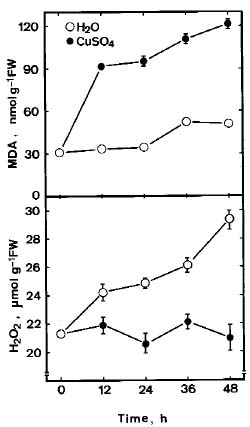
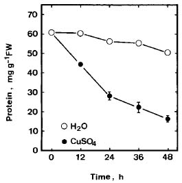
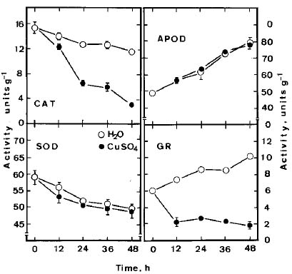
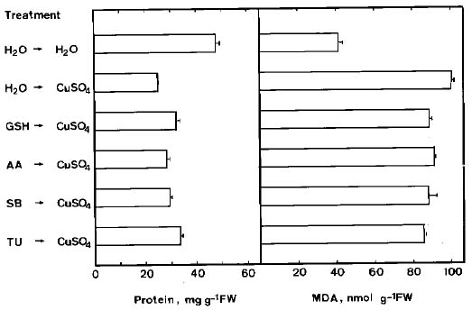
Abstract. Lipid peroxidation in relation to senescence of detached rice leaves caused by excess copper was investigated. Excess copper, which was found to promote senescence, increased the level of lipid peroxidation but not the level of H2O2. Catalase and glutathionine reductase activities were reduced by excess copper. Superoxide dismutase and ascobate peroxidase activities did not seem affected by excess copper. Free radical scavengers inhibited excess copper-promoted senescence and at the same time inhibited excess copper-induced lipid peroxidation, suggesting that lipid peroxidation induced by excess copper is mediated through free radicals.

Keywords: Copper; Lipid peroxidation; Leaf senescence; Oryza sativa.

Abbreviations: AA, ascorbic acid; APOD, ascobate peroxidase; CAT, catalase; GSH, reduced glutathione; GR, Glutathione reductase; MDA, malondialdehyde; SB, sodium benzoate; SOD, superoxide dismutase; TU, thiourea.

Introduction

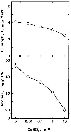
Although Cu is an essential micronutrient for plants, uptake of excess Cu can be harmful to most plants (Fernandes and Henriques, 1991). It has been reported that Cu mediated free radical formation in isolated chloroplasts (Scandmann and Boger, 1980), in intact roots (De Vos et al., 1993), in detached leaves (Luna et al., 1994), and in intact leaves (Weckx and Clijsters, 1996). Free radical-induced lipid peroxidation is considered to be an important mechanism of leaf senescence (Dhindsa et al., 1981; Kunnert and Ederer, 1985; Slater, 1972; Strother, 1988; Thompson et al., 1987). Excess Cu has been shown to induce leaf senescence (Chen and Kao, 1998; Jana and Choudhuri, 1982; Luna et al., 1994). It appears that Cu-induced leaf senescence is associated with lipid peroxidation. In this study, effects of Cu excess on the senescence, lipid peroxidation, and on some enzymes of activated oxygen metabolism in detached rice leaves were investigated.

Materials and Methods

Rice (Oryza sativa cv. Taichung Native1) was cultured as previously described (Kao, 1980 ). The apical 3-cm segments excised from the third leaves of 12-day-old seedlings were used. Briefly, rice seedlings were planted on a stainless net floating on half-strength Johnson's modified nutrient solution (pH 4.2) in a 500 mL beaker. The nutrient solution was replaced every three days. Rice plants were grown for 12 days in a greenhouse, where natural

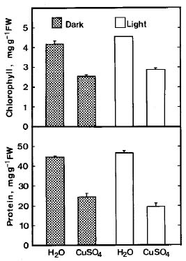
light was provided and the temperature was controlled at 30°C during the day and at 25°C at night. The apical 3 cm of the third leaf was used for the experiment. A group of 10 segments was floated in a Petri dish containing 10 mL of test solution. Incubation was carried out at 27°C in the light (40 µmol m-2 s-1) or in the dark.

Chlorophyll was determined according to Wintermans and De Mots (1965) after extraction in 96% (v/v) ethanol. For protein extraction, leaf segments were homogenized in 50 mM sodium phosphate buffer (pH 7.5). The extracts were centrifuged at 17,600 g for 20 min, and the supernatants were used for determination of protein by the method of Bradford (1976).

Malondialdehyde (MDA) was extracted with 5% (w/v) trichloroacetic acid and determined according to Heath and Packer (1968). MDA level is routinely used as an index of lipid peroxidation.

The H2O2 level was colorimetrically measured as described by Jana and Choudhuri (1981). H2O2 was extracted by homogenizing 50 mg leaf tissue with 3 mL of phosphate buffer (50 mM, pH 6.5). The homogenate was centrifuged at 6,000 g for 25 min. To determine H2O2 levels, 3 mL of extracted solution was mixed with 1 mL of 0.1% titanium sulfate in 20% (v/v) H2SO4. The mixture was then centrifuged at 6,000 g for 15 min. The intensity of the yellow colour of the supernatant at 410 nm was measured. H2O2 level was calculated using the extinction coefficient 0.28 µmol-1cm-1.

For extraction of enzymes, leaf tissues were homogenized with 0.1 M sodium phosphate buffer (pH 6.8) in a chilled pestle and mortar. The homogenate was centrifuged at 12,000 g for 20 min and the resulting supernatant was used for determination of enzyme activity. The whole

1Corresponding author. Fax: 886-2-2362-0879; E-mail: Kaoch@cc.ntu.edu.tw


Botanical Bulletin of Academia Sinica, Vol. 40, 1999

extration procedure was carried out at 4°C. CAT, SOD, APOD and GR were assayed as described previously (Chang and Kao, 1998). All data were expressed on the basis of gram fresh weight.

All experiments were repeated at least three times, and within each experiment treatments were replicated four times. Similar results and identical trends were obtained each time. The data reported here are from a single experiment.

Results

The senescence of detached rice leaves is characterized by a decrease in chlorophyll and/or protein levels (Kao, 1980). Chlorophyll and protein levels in detached rice leaves decreased with the increase of CuSO4 concentration in the light (Figure 1). It is obvious that the loss of chlorophyll is less sensitive than that of protein. The loss of chlorophyll and protein in detached leaves caused by CuSO4 in darkness was observed to be similar to that occurring in the light (Figure 2). However, Luna et al. (1994) reported that the rate of chlorophyll loss in oat leaves caused by CuSO4 in the light was more pronounced than in the dark. This discrepancy may be attributed to the different plant leaves used. We also observed that CuSO4 and CuCl2 were equally effective in enhancing the loss of chlorophyll and protein (data not shown) in detached rice leaves in the light, indicating that the loss of chlorophyll and protein is induced by Cu rather than by SO42- or Cl-.

Figure 3 shows the time courses of protein levels in detached rice leaves floating on water or CuSO4 (10 mM) in the light. It is clear that the promotion of protein loss (or senescence) by CuSO4 was evident 12 h after treatment. MDA level in CuSO4-treated detached rice leaves was observed to be higher than the water-treated controls throughout the entire duration of incubation (Figure 4). This shows that CuSO4 promoted senescence of detached rice leaves is linked to lipid peroxidation. Figure 4 also shows that H2O2 level increased significantly in detached rice leaves incubated in water. However, H2O2 level in CuSO4-treated detached rice leaves remained unchanged throughout the entire duration of incubation (Figure 4).

Lipid peroxidation is a free radical mediated process (Slater, 1984). The striking increase in lipid peroxidation in CuSO4-treated detached rice leaves may be a reflection of the decline of antioxidative enzymes. As shown in Figure 5, CuSO4-treated detached rice leaves had lower activities of CAT and GR than the controls. APOD and SOD in detached rice leaves do not seem to be affected by CuSO4 (Figure 5).

Figure 6 shows the effect of free radical scavengers such as ascorbic acid (AA), reduced glutathione (GSH), sodium benzoate (SB) and thiourea (TU) on CuSO4-promoted senescence and lipid peroxidation of detached rice leaves. It is clear that all tested free radical scavengers reduced senescence caused by CuSO4 and at the same time inhibited CuSO4-induced lipid peroxidation.

Figure 1. Effect of CuSO4 on chlorophyll and protein levels in detached rice leaves in the light. Detached rice leaves were incubated in solution containing 0-10 mM CuSO4. Chlorophyll and protein were determined 48 h after treatment. Vertical bars represent standard errors (n = 4). Only those standard errors larger than the symbol are shown.

Discussion

The present investigation shows that CuSO4 treatment resulted in an increased MDA level in detached rice leaves (Figure 4). This result supports the possibility that CuSO4-promoted senescence is mediated through lipid peroxidation, as suggested by Luna et al. (1994). This conclusion is supported further by the observation that free radical scavengers were able to inhibit senescence caused by CuSO4 and at the same time inhibit CuSO4-induced MDA level (Figure 6). The effects of CuSO4 on the loss of chlorophyll and protein could have resulted from the effects of free radicals produced by the treatment with Cu ions.

It has been demonstrated that excess Cu increased the activity of SOD in yeast and plant tissues (Chongpraditnum et al., 1992; Galiazzo et al., 1988; Gallego


Chen and Kao — Excess Cu and leaf senescence

Figure 2. Effect of CuSO4 in the light and in the dark on chlorophyll and protein levels in detached rice leaves. Detached rice leaves incubated in distilled water or 10 mM CuSO4 for 48 h. Vertical bars represent standard errors (n = 4).

Figure 4. Time courses of CuSO4 effect on MDA and H2O2 levels in detached rice leaves in the light. Detached rice leaves were incubated in distilled water or 10 mM CuSO4. Vertical bars represent standard errors (n = 4). Only those standard errors larger than the symbol are shown.

Figure 3. Time courses of the CuSO4 effect on protein level in detached rice leaves in the light. Detached rice leaves were incubated in distilled water or 10 mM CuSO4. Vertical bars represent standard errors (n = 4). Only those standard errors larger than the symbol are shown.

Figure 5. Time courses of CuSO4 effect on the activities of CAT, SOD, APOD and GR in detached rice leaves in the light. Detached rice leaves were incubated in distilled water or 10 mM CuSO4. Vertical bars represent standard errors (n = 4). Only those standard errors larger than the symbol are shown.


Botanical Bulletin of Academia Sinica, Vol. 40, 1999

Literature Cited

Bradford, M.M. 1976. A rapid and sensitive method for the quantitation of microgram quantities of protein utilizing the principle of protein-dye binding. Anal. Biochem. 72: 248-254.

Chang, C.J. and C.H. Kao. 1998. H2O2 metabolism during senescence of rice leaves: Changes in enzyme activities in light and darkness. Plant Growth Regul. 25: 11-15.

Chen, L.M. and C.H. Kao. 1998. Relationship between ammonium accumulation and senescence of detached rice leaves caused by excess copper. Plant Soil 200: 167-173.

Chongpraditnum, P., S. Mori, and M. Chino. 1992. Excess copper induced a cytosolic Cu, Zn-superoxide dismutase in soybean root. Plant Cell Physiol. 33: 239-244.

De Vos C.H.R., W.M. Ten Bookum, R. Vooijs, H. Schat, and L.J. De Kok. 1993. Effect of copper on fatty acid composition and peroxidation of lipids in the roots of copper tolerant and sensitive Silene cucubalus. Plant Physiol. Biochem. 31: 151-158.

Dhindsa, R.S., P. Plumb-Dhindsa, and T.A. Thorpe. 1981. Leaf senescence: Correlated with increased levels of membrane permeability and lipid peroxidation, and decreased levels of superoxide dismutase and catalase. J. Exp. Bot. 32: 93-101.

Fernandes, J.C. and F.S. Henrigues. 1991. Biochemical, physiological, and structural effect of excess copper in plants. Bot. Rev. 57: 246-273.

Foyer, C.H. and B. Halliwell. 1976. The presence of glutathione and glutathione reductase in chloroplast: a proposed role in ascorbic acid metabolism. Planta 133: 21-25.

Foyer, C.H., P. Descourvieres and K.J. Kunert. 1994. Protection against oxygen radicals: an important defence mechanism studied in transgenic plants. Plant Cell Eviron. 17: 507-523.

Galiazzo, F., A. Schiesser, and G. Rottilo. 1988. Oxygen-independent induction of enzyme activities related to oxygen metabolism in yeast by copper. Biochim. Biophys. Acta 965: 46-51.

Gallego, S.M., M.P. Benavides, and M.I. Tomaro. 1996. Effect of heavy metal ion excess on sunflower leaves: evidence for involvement of oxidative stress. Plant Sci. 121: 151-159.

Halliwell, B. and J.M.C. Gutteridge. 1989. Free Radicals in Biology and Medicine, 2nd edn. Clarendon, Oxford.

Heath, R.L. and L. Packer. 1968. Photoperoxidation in isolated chloroplasts. I. Kinetics and stochiometry of fatty acid peroxidation. Arch. Biochem. Biophy. 125: 189-198.

Jana, S. and M.A. Choudhuri. 1981. Glycolate metabolism of three submerged aquatic angiosperms during aging. Aquat. Bot. 12: 345-354.

Jana, S. and M.A. Choudhuri. 1982. Senescence in submerged aquatic angiosperms: Effect of heavy metals. New Phytol. 90: 477-484.

Kao, C.H. 1980. Senescence of rice leaves. IV. Influence of benzyladenine on chlorophyll degradation. Plant Cell Physiol. 21: 1255-1262.

Kunnert, K.J. and M. Edder. 1985. Leaf aging and lipid peroxidation: The role of antioxidents vitamine C and E. Physiol. Plant. 65: 85-88.

Figure 6. Effect of GSH, AA, SB and TU on protein and MDA levels in detached rice leaves treated with CuSO4. Detached rice leaves were pretreated with either distilled water, 5 mM GSH, 1 mM AA, 1 mM SB, or 5 mM TU for 12 h and then treated with either distilled water or 10 mM CuSO4 for 36 h in the light. Bars represent standard errors (n = 4).

et al., 1996; Luna et al., 1994). However, the results that we obtained with detached rice leaves in the light showed SOD activity did not seem to be affected by excess Cu2+ ions (Figure 5).

Glutathione (GSH) and ascorbate are the main antioxidants and are present in plant leaves (Foyer and Halliwell, 1976). Glutathione can react with singlet oxygen and hydroxyl radicals and protects the thiol groups of enzymes (Foyer et al., 1994). GR catalyzes the reduction of oxidized glutathionine (GSSG) in a NADPH-dependent reaction. GR, therefore, plays an essential role in the protection of chloroplasts against oxidative damage by maintaining a high GSH/GSSG ratio. In the present work, GR activity is decreased in detached rice leaves exposed to excess CuSO4 (Figure 5) suggesting a decrease in GSH/GSSG ratio. This would explain why CuSO4 treatment resulted in oxidative damage in detached rice leaves.

Activity of catalase, the enzyme responsible for eliminating H2O2, was lower in CuSO4-treated detached rice leaves than the water controls (Figure 5). However, H2O2 did not accumulate in CuSO4-treated detached rice leaves (Figure 4). Moran et al. (1994) also reported that H2O2 did not accumulate in water-stressed pea leaves. H2O2 can be used in an Fe- or Cu-catalyzed Haber-Weiss reaction. However, it is currently accepted that in vivo Cu is unlikely to cause Haber-Weiss reaction in the way that has been demonstrated for Fe (Halliwell and Gutteridge, 1989). H2O2 is unlikely being removed by APOD activity in detached rice leaves, because APOD activity in detached rice leaves is not effected by Cu (Figure 5). The production of H2O2 in leaves is mediated through glycolate oxidase. Whether CuSO4 treatment decreases glycolate oxidase activity in detached rice leaves remains to be seen.

Acknowledgements. This work was supported by a grant from the National Science Council Project, NSC 88-2313-B-002-066.


Chen and Kao — Excess Cu and leaf senescence

Luna, C.M., C.A. Gonzalez, and V.S. Trippi. 1994. Oxidative damage caused by an excess of copper in oat leaves. Plant Cell Physiol. 35: 11-15.

Moran, J.F., M. Becana, I. Iturbe-Ormaetxe, S. Frechilla, R.V. Klucas, and P. Apariciv-Tejo. 1994. Drought induces oxidative stress in pea plants. Planta 194: 346-352.

Sandmann, G. and P. Boger. 1980. Copper-mediated lipid peroxidation process in photosynthetic membranes. Plant Physiol. 66: 797-800.

Slater, T.F. 1972. What are free radicals? In J. R. Lagnado (ed.), Free Radical Mechanisms in Tissue Injury. Pion Ltd., London.

Slater, T.F. 1984. Free-radical mechanisms in tissue injury. Biochem. J. 222: 1-15.

Strother, S. 1988. The role of free radicals in leaf senescence. Gerontology 34: 151-156.

Thompson, J.E., R.L. Legge, and R.F. Barber. 1987. The role of free radicals in senescence and wounding. New Phytol. 105: 317-344.

Weckx, J.E.J. and H.M.M. Clijsters. 1996. Oxidative damage and defense mechanisms in primary leaves of Phaseolus vulgaris as a result of root assimilation of toxic amounts of copper. Physiol. Plant. 96: 506-512.

Wintermans, J.F.G.M. and A. De Mots. 1965. Spectrophotometric characteristics of chlorophyll a and b and their pheophytins in ethanol. Biochem. Biophys. Acta 109: 448-453.

¹L¶q»É¹ï¤ô½_¤ÁÂ÷¸­¤ù¤§®ÄÀ³¡G¯×½è¹L®ñ¤Æ§@¥Î°Ñ»P¤§µý¾Ú

³¯ÄR±Ó °ª´º½÷

°ê¥ß¥xÆW¤j¾Ç¹AÃÀ¾Ç¨t

¥»¬ã¨s±´°Q¹L¶q»É«P¶i¤ô½_¤ÁÂ÷¸­¤ù¦Ñ¤Æ»P¯×½è¹L®ñ¤Æ¶¡¤§Ãö«Y¡C¹L¶q»É³B²z¥i«P¶i¤ÁÂ÷¦Ñ¤Æ»P¼W ¥[¹L®ñ¤Æ§@¥Î¡A¦ý¤£·|¼W¥[¹L®ñ¤Æ²B§t¶q¡C¹L¶q»É³B²z§í¨î catalase »P glutathione reductase ¬¡©Ê¡A¦ý¤£ ¼vÅT superoxide dismutase ¤Î ascobate peroxidase ¬¡©Ê¡C¦Û¥Ñ°ò²M°£¾¯¥i§í¨î¹L¶q»É©Ò«P¶i¤§¸­¤ù¦Ñ¤Æ¡A ¦P®É§í¨î¹L¶q»É©Ò»¤¾É¤§¯×½è¹L®ñ¤Æ§@¥Î¡AÅã¥Ü¹L¶q»É©Ò³y¦¨ªº¯×½è¹L®ñ¤Æ§@¥Î«Y¥Ñ¦Û¥Ñ°ò©Ò¤Þ°_¡C

ÃöÁäµü¡G »É¡F¯×½è¹L®ñ¤Æ¡F¸­¤ù¦Ñ¤Æ¡F¤ô½_¡C