Bot. Bull. Acad. Sin. (1999) 40: 295-304

Tzeng et al. — Grapevine leafroll disease detection

Improvement of techniques for purification of leafroll

associated closterovirus from affected grapevines and the

preparation of antisera for disease indexing

Huey-Lin Chen Tzeng1, Moh-Jih Chen2, and Dean Der-Syh Tzeng2,3

1Department of Plant Pathology and Entomology, Taiwan Tobacco Research Institute, Tsao-Hu, Taichung 412, Taiwan

2Department of Plant Pathology, National Chung Hsing University, Taichung 402, Taiwan

(Received January 12, 1999; Accepted March 9, 1999)

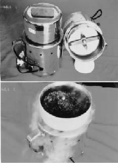
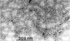
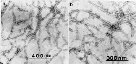
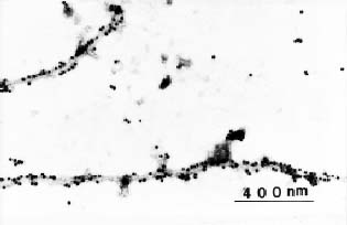
Abstract. An improved technique was developed for the purification of leafroll associated closterovirus (GLRaV) and successfully applied for the preparation of polyclonal and monoclonal antisera for the virus detection. The technique improvement mainly involves the use of a Chinese herb grinder for the homogenization of diseased sample. It requires very limited labor, is repeatable in regard to the virus yield, and can process considerable quantities within a short period. By this sample homogenization technique and a method modified from one in the available literature, GLRaV was successfully purified from diseased Kyoho, Black Queen, and Golden Mutscate grapevines cultivated in Taiwan. The electron microscopic examination of the obtained virus preparations revealed that the yield and purity were comparable or better than that reported by previous workers. With the obtained virus samples, polyclonal and monoclonal antisera were prepared. The results obtained from direct ELISA (enzyme linked immunosorbent assay) and indirect ELISA revealed that the prepared antisera detected mainly type III and type IV GLRaV. With the immunological method established, a disease survey was conducted between the years 1987 and 1994. A total of 204 grapevine cultivars or breeding lines were examined, and approximately 52% of them were found affected by GLRaV. Among the surveyed grapevines, about 30% were affected by Type III virus and another 31% by Type IV. A complexed infection of Types III and IV appeared to be common among Kyoho grapevines. The results also indicated the natural spread of GLRaV in tested field and suggested the need of a strict sanitation program for important breeding stock conservation.

Keywords: Antiserum preparation; Chinese herb grinder; Disease indexing; Grapevine leafroll disease; Virus purification.

Introduction

Leafroll is one of the most important diseases on cultivated grapevines and is a worldwide problem (Bovey et al., 1980). Research in recent years has indicated a consistent association of closterovirus particles with the leafroll affected grapevines. The etiological correlation of these closteroviruses and grapevine leafroll disease is now well accepted (Gugerli et al., 1984; Hu et al., 1989 & 1991; Zee et al., 1987; Zimmermann et al., 1988). Although it has been reported that mealybug (Pseudococcus spp.) (Engelbrecht and Kasdorf, 1990; Rosciglione and Gugerli, 1989; Tanne et al., 1989) and scale insect (Pulvinaria vitis L.) (Belli et al., 1994) may serve as agents for the disease transmission, it is generally believed that the widespread nature of this disease is primarily due to the use of diseased vines as propagating material. Strategies recommended for control of the disease in most grapevine growing countries mainly involves the use of virus free vines for the establishment or replacement of the vineyard.

The success of a clean seed program in the control of viral diseases depends greatly upon the technology and methodology available for the identification of the virus infection. The detection of GLRaV infection has traditionally been done solely by chip-bud grafting field indexing (Goheen, 1988). The main disadvantage of this methodology is that it requires a long process time and large amounts of field space and manpower. In addition, symptom development is often complicated by the presence of various biological and abiological stress factors (Egger et al., 1985; Ioannou and Gonsalves, 1991). Great improvement in the disease diagnosis was recently achieved by the successful development of virus specific antisera and the establishment of immunological detection techniques (Hu et al., 1989 and 1990a; Zee et al., 1987; Zimmermann et al., 1988 and 1990a,b). However, one of the major problems associated with the immunological detection of GLRaV is the limited availablity of antiserum; and what is even worse is the existence of at least seven different serotypes among the natural viral population on cultivated grapevines (Boscia et al., 1995; Choueiri et al., 1996; Hu et al., 1990a,b; Zimmermann et al., 1990a,b). In the Foundation Plant Material Service (FPMS) at the University of

3Corresponding author.


Botanical Bulletin of Academia Sinica, Vol. 40, 1999

California, Davis, in which indexing of grapevine leafroll virus infection has been one of main projects during the past decade, disease diagnosis still depends greatly on traditional field indexing although immunological detection has become a regularly practiced adjunct operation (Golino, 1992; Rowhani et al., 1992). Detection of the virus by RT-PCR (reverse transcription polymerase chain reaction) using specific primer pairs developed from the known nucleotide sequence of GLRaV has proved to be successful and highly sensitive (Liu, 1995). However, the application of such technology in disease indexing has been discouraged due to the need for a (at least partially) purified viral dsRNA as the template. Moreover, serotype specific primer pairs for known GLRaVs are still not available.

In Taiwan, a preliminary field survey conducted by Tzeng and Yeh in 1988 was the first to indicate the wide occurrence of leafroll infection and its substantial degenerative effects among the main grapevine cultivars growing in the field. The authors also indicated that the lack of precautions during the cultivar or breeding material introduction was an apparent reason for the prevelance of the disease. To safeguard the grape production industry, an extensive survey of disease distribution and the establishment of a virus free propagation material providing system are both essential. For fast disease indexing, a larger supply of antiserum suitable for the diagnosis of existing serotypes among the growing cultivars is urgently needed. The main objective of this study was to purify and characterize the viral agent from the regional grapevines and to prepare antiserum for disease survey. In the initial attempt, the protocol developed by Hu et al. (1989 and 1990a) was followed; the major difficulty of this purification scheme was the great need for manpower a thorough tissue homogenization requires. Even so, the virus yield was often not satisfactory. We describe herein an improved method for GLRaV purification using a specific model of Chinese herb grinder and its application in the preparation of antiserum for disease detection.

Materials and Methods

Biological Materials

Virus free LN-33 grapevines were introduced from FPMS of U. C. Davis and propagated in an isolated greenhouse. Golden Muscate, Kyoho, and Black Queen grapevine cultivars which were known to be affected by Type IV; Type III, IV; and Type III of GLRaVs respectively (Tzeng et al., 1994) were growing in the grapevine cultivar collection field plot in Taiwan Tobacco Research Institute (TTRI) at Tsao-Hu. The identity of the existing viral serotypes among these test grapevines were each confirmed by the conventional chip-bud grafting field indexing previously reported (Goheen, 1988) and by serological examinations using antisera of specific serotypes available (Hu et al., 1990a,b). The antisera used for the characterization of the affecting virus serotypes in the three main test grapevine cultivars included NY-1 (reactive specifically with GLRaV Type III), CA-4 (reactive specifically with GLRaV Type IV), and CA-

5 (reactive specifically with GLRaV Type II ) prepared by Professor Gonsalves' laboratory in Cornell University; and the Type I and GVA (grapevine virus A) antisera purchased from Bioreba Company. For virus purification, cuttings were harvested from the diseased grapevines during the dormant season from December to January; and the stem cortical tissues of these test materials were collected by a peelingknife and stored right away at -80°C until use.

Virus Extraction and Purification

A Chinese herb grinder (Figure 1, Model DM-6) obtained from Yo-Chi Machinery Inc. at Chang-Hwa was used for the pulverization of the test stem cortical tissues. In each operation, the grinding chamber of the machine was prefrozen with a suitable amount of liquid nitrogen. After loading the grapevine tissues, liquid nitrogen was added until a thick layer of frost formed on the chamber's outside wall (Figure 1). The pulverization was then performed with the chamber cover tightly closed. To avoid the danger of an explosive release of excessive nitrogen during the operation, it was necessary to make sure no liquid nitrogen remained in the chamber bottom before

Figure 1. The Chinese herb grinder (Yo-Chi, DM-6) used for the tissue pulverization in the performed tests. Right before the grinding operation, liquid nitrogen was applied to the sample loaded chamber until a thick frosty layer formed on the outer wall (lower picture).


Tzeng et al. — Grapevine leafroll disease detection

tightly closing the cover. To reduce the risk of heat generation, the operation was performed in an intermittent way; in each run, the machine was turned on no more than 3 seconds. A total of 20 to 30 seconds of machine operation was found sufficient to obtain finely pulverized test samples. After completion of the whole operation, the obtained sample powders were resuspended with 5X volume (w/v) of extraction buffer consisting of 0.5M Tris (pH 8.2), 15 mM 1,10-phenanthroline, 4% water insoluble polyvinyl polypyrrolidone (PVP), 0.5% bentonite, 0.2% 2-mercaptoethanol, 5% Triton X-100, 0.1% DIECA, and 0.01 M MgSO4•7H2O. For purification of the test virus, a method modified from the protocol described by Hu et al. (1990a) and Namba et al. (1991) was followed. Approximately 50 g fresh weight of the pulverized tissues were used in each operation. The sample suspension was stirred in a 4°C cold room for 30 min and squeezed through four layers of cheesecloth. The filtrates collected were allowed to settle in cold for about 20 min before being further clarified by centrifugation at 6000 g (Sorval SS-34 rotor) for 30 min. The supernatants were then laid on top of a 5 ml pad of TMS-20 buffer solution (a 0.1 M Tris-HCl buffer, pH 8.2, containing 20% sucrose and 0.01 M MgCl2) and centrifuged at 36,000 rpm (Hitachi RT-50 rotor) for 2 h to concentrate the contained virus particles. The resulting pellets were thoroughly dispersed in a 22 ml TMS-10 buffer (similar to TMS-20 buffer, but containing only 10% sucrose), dialyzed against the same buffer in the cold room for one day, and then settled in a slender tube (approximately 0.7 cm diameter) in a 4°C cold room for another 1-2 days. The clean upper phase (approximately 18 ml) was carefully removed by a pippet; and after supplementation with 2% Triton X-100, half the pellets (9 ml) were laid on top of a 3 ml Cs2SO4-sucrose cushion-step gradient (1 ml of 10, 22.5 and 30% Cs2SO4 each in TMS-10 buffer) and subjected to high speed centrifugation at 35,000 rpm (Hitachi SW-40 rotor) for 2.5 h at 6°C. The contents existing in the formed gradients were then collected by a 0.4 ml fraction series using an ISCO model 640 gradient fractionator. By electron microscopy or ELISA examination, the fractions which contained the test virus were identified, pooled, and dialyzed against TMS-10 buffer in a cold room overnight. After clarification by low speed centrifugation, further purification of the virus was achieved by a repeated Cs2SO4-sucrose density gradient centrifugation and fractionation.

Antiserum Preparation

For the antiserum preparation from rabbit, virus samples purified from diseased Kyoho (KH-1), and Black Queen (BQ-1) grapevines were used to immunize New Zealand white rabbit. The antigenic viral preparations (approximately 0.5 mg/ml) were emulsified thoroughly with either complete (for first injection) or incomplete (for the followed injections) Fruend's adjuvant for each injection. The immunization was performed by subcutaneous injection on the neck. After three consecutive weekly injections, a final booster injection was performed with the viral antigen without adjuvant emulsification on the ear

vein. The rabbit was then sacrificed for antiserum preparation one week after the booster injection, after the high titer of antiserum was assured.

As for the preparation of polyclonal and monoclonal antibodies from mouse, an immunological tolerance technique (Hsu et al., 1990) was applied. The viral antigenic sources were prepared as described above. As for the preparation of plant antigens (the tolerogens), stem cortical tissues collected from healthy LN-33 grapevines were processed the same way as those for GLRaV purification except that pellets obtained after differential centrifugation were resuspended with PBS to make a twofold concentration of plant antigens (i.e. a 10 g piece of stem cortical tissue would result in a 5 ml plant antigen preparation). For antiserum preparation, the pregnant Balb/C mice were checked daily for the baby birth. The new born baby mice were injected intraperitoneally with 25, 40, 100, and 200 µl of plant antigens (tolerogen) on days 1, 3, 5, and 7, respectively. Approximately 4 weeks after the immunological tolerance development pre-treatments, the viral antigens were used to immunize the test mice three consecutive times on the 5th, 8th, and 10th weeks. The viral antigens used for injection were emulsified with either complete (for first injection) or incomplete (for the subsequent injections) Freund's adjuvants. For antibody production, an additional booster injection was performed one week after the last injection. The mice were then sacrificed for spleen cell preparation about 3-4 days thereafter when a high titer of wanted antibody had been assured by ELISA. Continued hybridoma preparation was performed using the myeloma cell line P3/NS-1/1-Ag-4-1 obtained from the Molecular Virology Laboratory of the Department of Veterinery Medicine at National Chung Hsing University. The cell line was maintained in a complete medium containing RPMI-1640 (Boehringer Mannheim Biochemica, Germany), 1 mM L-glutamine, 1 mM pyruvate, 1 mM Na-pyruvate, 50 IU/ml penicillin, 150 mg/ml streptomycin, 0.25 mg/ml fungizone, and 15% fetal bovine serum. Fusion of the myeloma cells and the spleencytes was performed in the presence of 50% polyethylene glycol (MW. 1500, Boehringer Mannheim) according to Hsu et al. (1984). Production of antibody by the resulting hybridoma cells was determined by double antibody sandwich (DAS) indirect ELISA. Hybridoma cells secreting antibody reactive to crude extract of GLRaV affected Golden Muscate or Black Queen grapevines were cloned; these cells were further subcloned by the limiting dilution method for the isolation of single cell progenies. The monoclonal antibodies were produced either by ascites or by cultures of the hybridoma cell line in RPMIX-media or serum free media. For the polyclonal antibody production, the Balb/C mice immunized with viral antigen were primed with pristane and then injected intraperitoneally with 2 × 10⁶ NS-1 myeloma cells. For monoclonal antibody production, the plant antigen treated Balb/C mice (pretreated with healthy tolerogen) were primed with pristane and injected with 2 × 10⁶ hybridoma cells in a serum free RPMI medium. The ascitic fluid produced one week after injection of the myeloma or hybridoma cells were then harvested for the preparation of polyclonal and


Botanical Bulletin of Academia Sinica, Vol. 40, 1999

monoclonal antibodies, respectively. Purification of the immunoglobulin (IgG) was achieved by ammonium sulfate precipitation and affinity chromatography using a protein A-sepharose CL-4B column (Pharmacia Chemical Co.). The isotyping of the monoclonal antibodies obtained was done by indirect ELISA using the Isotyping ID Kit purchased from Southern Biotechnology Associates Inc.

Detection of GLRaV by ELISA Using the Prepared Antisera

For detection of GLRaV virus in diseased grapevine tissue using rabbit or mouse antisera produced by direct DAS ELISA as described by Clark and Adams (1977), the antisera were preabsorbed by healthy plant extracts before immunoglobulin purification according to the method described by Zee et al. (1987). The immunoglobulin of rabbit antisera was purified by ammonium sulfate precipitation and a subsequent DEAE cellulose (DEAE-Sephacel, Pharmacia Fine Chemicals, Piscataway, NJ) column chromatography. The microplates were coated first with these g-globulins at 2 µg/ml and incubated for 4 h at 37°C. Approximately 1 g test grapevine tissues were homogenized with 5 ml 0.5 M Tris buffer (pH 8.0). And after 2% non-fat milk was added, the homogenates were applied to the microplates as a second coating layer. The homogenates obtained from healthy grapevine tissues were used as a negative control. The purified immunoglobulins were conjugated to alkaline phosphatase, preabsorbed with healthy plant extracts, and subsequently applied to the microplates. After being incubated at 37°C for 2 h, the plates were thoroughly rinsed with washing buffer and reacted with p-nitrophenyl phosphate (1 mg/ml). The results were read by an ELISA reader at A405.

For an indirect ELISA, the plates were coated with partially purified virus collected from first Cs2SO4 density gradient centrifugation. After a washing with PBS-T and blocking with 2% non-fat milk (diluted with PBS), diluted antisera were added. The plates were then incubated for 2 h at 37°C; horseradish peroxidase labeled goat anti-mouse IgG and hydrogen peroxide-ABT were finally added for the color reaction.

For DAS indirect ELISA, the method reported by Vela et al. (1986) was used. Briefly, the Costa high bounding ELISA plates were coated with rabbit KH-1 or BQ-1 polyclonal IgG at 4°C overnight. After washing with PBS-T and blocking with 2% non-fat milk in PBS, crude extracts of stem cortical tissue of test grapevine were added. The plates were incubated at 4°C overnight. After a subsequent washing with PBS, the sample wells were further reacted with the prepared mouse polyclonal or monoclonal antibodies for 3 h at 37°C and washed again. The horseradish peroxidase labeled goat anti-mouse IgG (Sigma Chemical Co., diluted 1:1000) and hydrogen peroxide-ABT were then added in succession to continue the reaction. The results were read by ELISA reader at A405. Tissue extracts of healthy grapevine processed in the same way were used as control.

Immunoelectron Microscopy

Grids coated with formvar film and loaded with carbon were used for the experiment. Partially purified virus samples obtained after the differential centrifugation were coated on the grids, decorated with diluted antiserum or monoclonal antibody containing culture fluid, and then stained with 1% uranyl acetate. For immunogold labeling, grids with virus and antibodies were coated with antimouse IgG gold conjugate and then stained with 1% uranyl acetate. The negatively stained samples were then examined by a Jeol JEM-200CX model electron microscope.

Disease Indexing of Field Grapevines

Both direct and indirect DAS ELISA described were applied for the indexing of field grapevines for GLRaV infection. The tested grapevines included all cultivars and breeding lines collected in the preservation field at TTRI. Petioles and dormant canes were collected from the test grapevine cultivars during the late growing season both before and after pruning. The petioles were cut at both ends, and about 10-20 middle segments were collected from each test cultivar. The stem cortical tissues peeled off from 5-10 dormant canes of each test cultivar were also used as a comparison. For the performed ELISA tests, all the test tissues were cut into small pieces by scissors and ground into powder in the presence of liquid nitrogen by mortar and pestle. The powderized samples were suspended in 0.5 M Tris buffer and used for ELISA tests as described above.

Results

By the described sample grinding technique and the purification scheme, the target virus was successfully purified from GLRaV affected Kyoho, Black Queen, and Golden Muscate grapevines. The virus preparation resulting from the final Cs2SO4/ sucrose density gradient centrifugation indicated that the best yield and quality were consistently obtained from diseased Black Queen. The virus yield and purity obtained from Kyoho grapevine were slightly lower than those from Black Queen; and those from Golden Muscate appeared to be the lowest of the three cultivars. The virus yield of Golden Muscate was generally no more than 1/10 that of Black Queen as revealed by electron microscopy (EM). Since the virus content in diseased tissues was low, the partially purified virus preparations obtained from several sucrose cushion concentrations were pooled before subjecting them to the first cycle of Cs2SO4/ sucrose density gradient centrifugation. Examination by UV absorption together with EM of the collected samples indicated that the viruses were scattered in 3-4 fractions. The followed second run gradient separation further concentrates these virus samples; the resulting fraction peak correlates well with the existence of the test virus. Electron microscopy of the virus particles purified from diseased Black Queen grapevines by negative staining indicated that the preparation was largely free of observable particulate contaminants (Figure 2). The puri


Tzeng et al. — Grapevine leafroll disease detection

fied virus preparations were dialyzed against a 0.01 M MgCl2 amended 0.1 M Tris-HCl (pH 8.2), tittered and dispensed into equal size aliquots, and stored in a -80°C freezer before being used for antiserum preparation.

Production of Antisera and Immunological Detection

By the described immunization scheme, polyclonal antibodies against GLRaV on Kyoho (KH) and Black Queen (BQ) were successfully obtained from both rabbit (RPAb-KH and RPAb-BQ) and mice (MPAb-KH, and MPAb-BQ), respectively. For Golden Muscate (GM) grapevine, which was affected by Type-IV GLRaV, the polyclonal antiserum

was prepared only from mouse (MPAb-GM). The monoclonal antibodies were prepared from Kyoho cultivar. The immunoglobulins of these antisera were isolated, and by glutaraldehyde linkage, IgG-alkaline phosphatase conjugates of each respective antiserum were prepared. The titer of both rabbit polyclonal antibodies (RPAb-KH & BQ) exceeded 12,000 as determined by reaction to each own virus preparations. Using the direct DAS-ELISA already described, both antisera were found to be reactive with crude extracts obtained from either diseased Kyoho or Black Queen, but each failed to respond to those from either Golden Muscate or healthy control LN-33 (Table 1). Immunoelectron microscopy indicated that the RPAb-BQ antibody decorated the virus particles purified from diseased Black Queen (Figure 3A) very well just as did the NY-1 antiserum (Figure 3B) obtained from Cornell University.

With the application of an immunological tolerance technique, both polyclonal and monoclonal antisera against viruses of Kyoho, Black Queen, and Golden Muscate were successfully prepared. The monoclonal antibody producing hybridoma cell line KH-1 was selected for the reactivity trials performed. As shown in Table 2, the isotypes of KH-1 appeared to be a IgG2a, k-chain. The culture filtrate of KH-1 reacted well with GLRaV from Black Queen, but did not respond to that from Golden Muscate, nor to the crude extract of the healthy control LN-33. The detection by immuno-gold labeling using mouse polyclonal antibody prepared against Golden Muscate GLRaV (MPAb-GM) also supported this serotype specificity (Figure 4).

By indirect DAS ELISA with rabbit KH antiserum coating, the mouse polyclonal antibody prepared against GLRaV of Black Queen (MPAb-BQ) reacted well with par

Figure 2. Examination by electron microscopy the GLRaV purified from diseased Black Queen grapevine by the improved purification protocol described. The examined sample was obtained from the virus concentrate after the second Cs2SO4 density gradient centrifugation, and was negatively stained by 1% uranyl acetate.

Figure 3. Electron microscopy of the GLRaV partially purified from affected Black Queen grapevine stem cortical tissues. The applied samples were decorated either by rabbit polyclonal antiserum prepared in this investigation against the virus on Black Queen (a) or by NY-1 rabbit polyclonal antiserum obtained from Dr. Gonsalves (b), and then negatively stained with 1% uranyl acetate for the examination.


Botanical Bulletin of Academia Sinica, Vol. 40, 1999

Survey for GLRaV Infection Among Cultivars and Breeding Lines Collected in TTRI

A total of 204 cultivars or breeding lines in the grapevine cultivar collection field were tested. For the detection of Type III GLRaV infection, both direct ELISA using RPAb-BQ and RPAb-KH, and DAS indirect ELISA using RPAb-BQ, MPAb-BQ, and MAb-KH were performed. The number of reaction-positive grapevines was 61 which included: Barbera, Carignane, Pinot Noir, Royalty, Schwarzer Riesling, Missouri Riesling, Clairette Mazel, Red Moon, Seibel 10878, Cabernet Sauvignon, Black Queen, Champion, Carignane, Isabellaregia X Centennial, Buffalo, Fernao Pires, D X K 15, French Colombard, Himrod Seedless, Grand Noir, Olympia, Salamander, Inzolia, Lagrein, Super Hamburg, Van Buren, Kyoho, Muscadelle du Bordelais, Muscat Blanc, Royal Red, Muller Thurgau, Nebbiolo, Pedro Ximenes, Petite Bouschet, Petite Sirah, Pinor Blanc, Clairette, Pinot Noir, Shiraz, Black Hamburg, Cabernet Sauvignon, Chasselas, Red Veltliner, Flame Tokay, Foster, Golden Coin, Italia IP 65, Tinto Cao, Tunnat, Koshu, Servant, Muscate Malaga, White Riesling, Red Malaga, Rozaki, Kyoho, Red Malaga, Pione, Neo Muscate/5BB, Igawa 200/1202, and 8B. It is worth mentioning that, among these Type III GLRaV affected grapevines, 26 of them were introduced from U. C. Davis in 1982 through FPMS and were certified to be free from GLRaV infection at that time. These vines were: Carignane, Royalty, Seibel 10878, Van Buren, Foster, Carbernet Sauvignon, Carignane, Fernao Pires, French Colombard, Grand Noir, Inzolia, Muscadelle Du Bordelais, Muscate Blanc, Muller Thurgau, Nebbiolo, Pedro Ximenes, Petite Boushet, Petite Sirah, Pinor Blanc, Pinot Noir, Shiraz, Red Veltliner, Tinto Cao, Tunnat, Servant, and White Riesling.

For the detection of Type IV GLRaV, the direct ELISA using MPAb-GM or CA-4 antisera was performed. The following 64 grapevines in the same field showed positive reactivities: Golden Muscate, Barbera, Carignane, Grey

Figure 4. Electron microscopy of the GLRaV partially purified from affected Golden Muscate grapevine stem cortical tissues. The applied virus samples were first decorated by a mouse polyclonal antiserum prepared in this investigation against the virus on Golden Muscate; which were subsequently coated with antimouse IgG Gold conjugate and then negatively stained with 1% uranyl acetate for the proceeded examination.

tially purified GLRaVs from either diseased Kyoho or Black Queen (Table 3); but it failed to respond to those from Golden Muscate or crude extract of LN-33. By contrast, the MPAb-GM reacted positively with virus preparations from either Golden Muscate or Kyoho; however, it reacted poorly with that from Black Queen or the crude extract from LN-33. It was also noted during the experiment that while using these mouse antisera, the preabsorption treatment with a healthy plant component was apparently unnecessary. The range of application of monoclonal antibodies (MAb) obtained from the KH-1 hybridoma cell lines was similar to that of MPAb-BQ in regard to serotype specificity. MAb-KH1 reacted well with the GLRaVs from Kyoho and Black Queen grapevines (both known to be mainly affected by the Type III virus), but not with those from Golden Muscate (Type IV).


Tzeng et al. — Grapevine leafroll disease detection

Table 3. Detection of GLRaV in partially purified viral preparation obtained from diseased Kyoho (KH), Golden Muscate (GM), and Black Queen (BQ) grapevines by indirect ELISA using the polyclonal (MPAb) and monoclonal (MMAb) antibodies prepared from mice in this study. The ELISA plates were initially coated with partially purified virus obtained after the first Cs2SO4 density gradient centrifugation. The healthy LN-33 grapevine tissues were identically processed as a control. The serotypes of detected GLRaVs in tested samples were confirmed by each specific antiserum obtained from available sources as described in material and methods. Data presented are A405 values detected by an ELISA reader.

Plant materials Types of GLRaV MPAb-BQ MPAb-GM MMAb-KH1

Kyoho III & IV 1.21 0.70 1.02

Golden Muscate IV 0.10 0.86 0.03

Black Queen III 1.51 0.25 1.3

LN-33 (CK) _ 0.12 0.10 0.02

effect of the applied liquid nitrogen. The pulverization was performed with the chamber lid tightly closed; it is thus necessary to make sure no residual liquid nitrogen remains in the chamber bottom before tightening the chamber lid and turning on the machine. As a precaution against an extraordinary bursting release of leftover nitrogen, it is necessary to turn off the machine right after turning it on during the initial run. The running time for the subsequent intermittent grinding operations was generally 2-3 secs; the main consideration was the amount of burst out nitrogen. In the case of a suspicious bursting release of nitrogen, it is a good precaution to partially loosen the lid to release the build up internal pressure and retighten it again. No extraordinary strength is required, and up to a kilogram of test tissues can be processed within a couple of hours by a single person without extra help. The purity and identity of the resulting virus preparation was clearly indicated by the EM examination conducted (Figures 2 and 3). The success and repeatability of virus purification described above depicted the great value of the applied technique for GLRaV researches. During the course of this study, the same sample processing technique was also successfully applied to GLRaV dsRNA isolation (Tzeng et al., 1996) and as well to dsRNA isolation of citrus tristeza virus—another closterovirus prevailing in Taiwan (Tarng, 1994).

For the immunological detection of GLRaV, the available literature implicated the need for preabsorption, primarily to eliminate the substantial non-specific reaction. As for the rabbit polyclonal antibodies RPAb-KH and RPAb-BQ, their practical value for GLRaV indexing was clearly indicated by the reproducibility and high titer of the reactivity (Table 1). However, the need for preabsorption treatment reflected well the interference of plant contaminants in the virus preparation used for the antiserum production. This interference with immunological detection seemed to be a common problem associated with plant viruses, the purification of which is very difficult. GLRaV, unfortunately, is one of the typical examples (Hu et al., 1989). The mentioned preabsorption treatment with healthy plant components greatly improves the detection efficacy of the antiserum (Zee et al., 1987). An alternative resolution appears to be the employment of the immunological tolerance technique (Hsu et al., 1990). Immunological tolerance is defined as the development of immunological unresponsiveness of an animal to a spe

Riesling, B1106, C0406, E0632, Palomino, Salvador, Aleatico, Rulandor, Trollinger Klone 4-7, Aligote, T.G.-1 (GM X N), B0427/Dogridge, Missouri Riesling, Muscate Saint-Vallier, Clairette Mazel, Concord, Red Moon, Seibel 10878, Baco Blanc, Seibel 1000, Ives, Cabernet Sauvignon, Seibel 8357 (Colobel), Seibel 5279 (Aurora), Dutchess, Campbell Early, Ontario, Scarlet, Fock, Buffalo, Emerald Riesling, Feher-Szagos, Fernao Pires, Folle Blanche, Gros Guillanne, Igawa 17, Grand Noir, Green Hungarian, Green Veltliner, Grey Riesling, Mills, Unknown 2, Olympia, Salamander, Van Buren, Royal Red, Kyoho, Caino Gordo, Petite Sirah, Rubired, Shiraz, Seibel 10868, Italia IP 65, Trousseau, Anab-E-Shuhi, Koshu, Servant, Walsch Riesling, Pione, Neo Muscate/5BB, and the Unknown 4 cultivars. It was noted that 22 of these grapevines—Barbera, Carignane, Missouri Riesling, Clairette Mazel, Red Moon, Seibel 10878 (Chelois), Cabernet Sauvignon, Buffalo, Fernao Pires, Grand Noir, Olympia, Salamander, Van Buren, Royal Red, Petite Sirah, Shiraz, Servant, Koshu, Italia IP 65, Kyoho, Pione, and Neo Muscate/5BB—were suffering complexed infections of both Type III and Type IV viruses.

Discussion

The purity of virus preparation is a major determinative factor for both the specificity and reactivity of a prepared antiserum against the studied viral agent. Traditional GLRaV purification using motar and pestle for homogenization was very labor intensive, and the results were often quite discouraging. Using the DM-6 Chinese herb powderizing machine (Figure 1), we found that test grapevine stem cortical tissues immediately taken out from the -80°C freezer could be ground into fine powders within 30 secs. The efficiency of pulverization depends greatly on the maintenance of the superfrozen status of the test material throughout the grinding process. The danger is that the test samples will become defrosted by the heat generated during the process; the samples then tend to become soggy and brownish; and the yield of purified virus is quite low in this case. For the DM-6 model powderizing machine, it was noted that an application of 50 to 100 ml of liquid nitrogen was generally adequate for the desired purpose. After adding the liquid nitrogen, a slight swirling of the machine by hand was found to be helpful for the even distribution and better super-freezing


Botanical Bulletin of Academia Sinica, Vol. 40, 1999

cific antigenic substance which has been a target of the animal's immune system during its early growth stage. The phenomenon was first reported by Hanan and Oyama on rabbit in 1954 and later confirmed by Hirata et al. in 1960 on chicken. By preimmunization of new born Balb/C mice with healthy plant component, Hsu et al. (1990) demonstrated the development of immunological tolerance of test mice against certain tomato cultivars. Using these test mice, they were able to abolish the antigenic effect of the plant contaminants during a post-immunization treatment with a partially purified virus preparation. And by a modified use of conventional hybridoma technique, they successfully produced high titer antiserum specifically against tomato spotted wilt virus. In order to obtain a high reactivity antiserum without the mentioned interference for practical application to GLRaV disease indexing, the same immunological tolerance technique was applied in this investigation. The positive reactivities shown in Tables 2 and 3 and the lack of need for preabsorption treatment of the polyclonal or monoclonal mouse antibodies prepared, supported strongly the great value of this technique for immunological detection of the studied virus.

The reactivity of RPAb-BQ, RPAb-KH (Table 1), MPAb-BQ, and MMAb-KH1 (Table 3), resembled the NY-1 antiserum (Hu et al., 1990a) obtained from Cornell University indicating a GLRaV serotype III characteristics of these antisera produced. Likewise, the performance of MPAb-GM was similar to that of CA-4 (Hu et al., 1990b) indicating their specificity to serotype IV. The reactivity of MPAb-GM to diseased Kyoho grapevine indicated the presence of Type IV in addition to the Type III GLRaV. The same phenomenon was also observed in our previous work using NY-1 and CA-4 antisera as detection probes (Tzeng et al., 1994). In regards to the detection-methodologies applied, we noted that the highest sensitivity was consistently obtained by DAS indirect ELISA in which both RPAb and MPAb were applied (Table 3). The method was thus utilized for a subsequent experiment involving the indexing of GLRaV infection among field grapevines.

The conducted extensive field survey revealed that among the tested grapevines, approximately 30% were affected by the Type III virus, 31% were affected by Type IV, and about 9% were suffering complexed infection by both Types of virus. The detection of a high proportion of GLRaV infection among the collected cultivars or breeding stocks clearly reflected the seriousness of the studied disease and the urgent need for a sanitation programto get it under control. Further evidence of this urgent need is the observed disease spread among the grapevine collections in the test field plot. The prevalance of infection among these cultivation and breeding sources indicated their unsuitability for the attempted purpose and the need of prompt replacement by healthy breeding or propagation sources. During the course of this study, we found a few apparently virus free vines among the main grapevine cultivar Kyoho. Our efforts will be focused on the establishment and provision of virus free vine stocks.

Acknowdgements. The investigation was supported by a grant provided by the Council of Agriculture of Executive Yuan, Taiwan, R.O.C. The authors also greatly appreciate Dr. D. Gonsalves of Cornell University for the provision of NY-1, CA-4 and CA-5 GLRaV antisera.

Literature Cited

Belli, G., A. Fortusini, P. Casati, L. Belli, P.A. Bianco, and S. Prati. 1994. Transmission of a grapevine leafroll associated closterovirus by the scale insect Pulvinaria vitis L. Rivi. Patol. Veg. 4: 105-108.

Boscia, D., C. Greif, P. Gugerli, G.P. Martelli, B. Walter, and D. Gonsalves. 1995. Nomenclature of grapevine leafroll-associated putative closteroviruses. Vitis 34: 171-175.

Bovey, R., W. Gartel, W.B. Hewitt, G.P. Martelli, and A. Vuittenez. 1980. Virus and Virus-Like Diseases of Grapevines. Editions Payot Lausanne, 181 pp.

Choueiri, E., D. Boscia, M. Digiaro, M.A. Castellano, and G.P. Martelli. 1996. Some properties of a hitherto undescribed filamentous virus of the grapevine. Vitis 35: 91-93.

Clark, M.F. and A.N. Adams. 1977. Characteristics of the microplate method of enzyme-linked immunosorbent assay for the detection of plant viruses. J. Gen. Virol. 34: 475-483.

Egger, E., M. Borgo, and P. Antoniazzi. 1985. Behaviour of cv. Mission and other grapevine varieties used as leafroll indicator. Phytopathol. Medit. 24:141-143.

Engelbrecht, D.J. and G.G.F. Kasdorf. 1990. Transmission of grapevine leafroll disease and associated closteroviruses by the vine mealybug, Planococcus ficus. Phytophylacticia 22: 341-346.

Goheen, A.C. 1988. Leafroll, corky bark, and rupestris stem pitting. In R. C. Pearson and A. C. Goheen (eds.), Compendium of Grape Diseases, APS Press, St. Paul., pp. 52-53.

Golino, D.A. 1992. The Davis grapevine virus collection. Am. J. Enol. Vitic. 43: 200-205.

Gugerli, P., J. Brugger, and R. Bovey. 1984. L'enroulement de la vigne: Mise en evidence de particles virales et development d'une methode immuno-enzymatique pour le diagnostic rapide. Rev. Suisse Vitic. Arboric. Hortic. 16: 299-304.

Hanan, R. and J. Oyama. 1954. Inhibition of antibody formation in mature rabbits by contact with the antigens at the early age. J. Immunol. 73: 49-53.

Hirata, A.A., J.S. Garvey, and D.H. Campbell. 1960. Retention of antigen in tissue of serologically suppressed chickens. J. Immunol. 84: 576-581.

Hsu, H.T., J. Aebig, and W.F. Rochow. 1984. Differences among monoclonal antibodies to barley yellow dwarf viruses. Phytopathology 74: 600-605.

Hsu, H.T., Y.C. Wang, R.H. Lawson, M. Wang, and D. Gonsalves. 1990. Splenocytes of mice with induced immunological tolerance to plant antigens for construction of hybridomas secreting tomato spotted wilt virus-specific antibodies. Phytopathology 80: 158-162.

Hu, J.S., D. Boscia, and D. Gonsalves. 1990a. Use of monoclonal antibodies to characterize grapevine leafroll associated closteroviruses. Phytopathology 80: 920-925.


Tzeng et al. — Grapevine leafroll disease detection

Hu, J.S., D. Gonsalves, and D. Teliz. 1990b. Characterization of closterovirus-like particles associated with grapevine leafroll disease. J. Phytopathol. 128: 1-14.

Hu, J.S., D. Gonsalves, D. Boscia, M. Maixmer, and D.A. Golino. 1991. Comparison of rapid detection assays for grapevine leafroll disease associated closteroviruses. Vitis 30: 87-95.

Hu, J.S., D. Teliz, and D. Gonsalves. 1989. Detection of Closterovirus-Like Particles from Crude Plant Extracts with Immunosorbent Electron Microscopy (ISEM). Proc. 9th ICVG, Kiryat Anavim, Israel, Sept. 1987, pp. 207.

Ioannou, N. and D. Gonsalves. 1991. Grapevine Leafroll Disease in Cyprus: Incidence, Evaluation of Indicators and Serological Detection of a Closterovirus in Diseased Vines. Proc. 10th ICVG. Volos, Greece, Sept. 1990, pp. 251-258.

Liu, Y.C. 1995. Molecular Cloning of the Coat Protein Gene of Grapevine Leafroll-Associated Virus. M.S. Thesis, Department of Plant Pathology, National Chung Hsing University, 93 pp.

Namba, S., D. Boscia, O. Azzam, M. Maixner, J.S. Hu, D.A. Golinos, and D. Gonsalves. 1991. Purification and properties of closterovirus-like particles associated with grapevine corky bark disease. Phytopathology 81: 964-970.

Rosciglione, B. and P. Gugerli. 1989. Transmission of Grapevine Leafroll Disease and an Associated Closterovirus to Healthy Grapevine by the Mealybug Planococcus ficus Signoret. Proc. 9th ICVG, Kiryat Anavim, Israel, Sept. 1987, pp. 67-69.

Rowhani, A., D.A. Golino, and M. Cunningham. 1992. Comparison of bioassay in indexing and ELISA for the dection of grapevine leafroll virus in grapevine selections. Phytopathology 82: 1148. (Abstract)

Tanne, E., L. Naveh, and I. Sela. 1989. Serological and molecular evidence for the complexity of the leafroll disease of grapevine. Plant Pathol. 38: 183-189.

Tarng, Y.K. 1994. Molecular Cloning of Coat Protein Gene of

Citrus Tristeza Virus in Taiwan and its Application in Virus Detection. M.S. Thesis, Department of Plant Pathology, National Chung Hsing University, 90 pp.

Tzeng, H.-L.C. and H.M. Yeh. 1988. Survey of grapevine virus diseases in Taiwan. In Proceedings of a Symposium for Grapevine Research and Development in Taiwan. Taiwan Agriculture Research Institute, Taichung, pp. 124-130.

Tzeng, H.-L.C., M.J. Chen, and D.D.-S. Tzeng. 1994. The occurrence of grapevine leafroll disease among the main grapevine cultivars and breeding stocks in Taiwan. Pl. Pathol. Bull. 3: 156-167.

Tzeng, H.-L.C., M.J. Chen, and D.D.-S. Tzeng. 1996. An improved method for isolating closteroviral dsRNA from leafroll affected grapevines. Pl. Pathol. Bull. 5: 47-54.

Vela, C., M. Cambra, E.E. Cortes, E. Moreno, J.G. Miguet, C. Perez ed san Roman, and A. Sang. 1986. Production and characterization of monoclonal antibodies specific for citrus tristeza virus and their use for diagnosis. J. Gen. Virol. 67: 91-96.

Zee, F., D. Gonsalves, A.C. Goheen, K.S. Kim, R. Pool, and R.G. Lee. 1987. Cytology of leafroll-diseased grapevine and the purification and serology of associated closterovirus-like particles. Phytopathology 77: 1427-1434.

Zimmermann, D., B. Walter, and O. Le Gall. 1988. Purification de particules virales associes a ,'enroulement de la Vigne et mise au point d'un protocole ELISA permettant leur detection. Agronomie 8: 731-740.

Zimmermann, D., P. Bass, R. Legin, and B. Walter. 1990a. Characterization and serology detection of four closterovirus-like particles associated with leafroll disease on grapevine. J. Phytopathol. 130: 205-218.

Zimmermann, D., G. Sommermeyer, B. Walter, and M.H.V. van Regenmortel. 1990b. Production and characterization of monoclonal antibodies specific to closterovirus-like particles associated with grapevine leafroll disease. J. Phytopathol. 130: 277-288.


Botanical Bulletin of Academia Sinica, Vol. 40, 1999

¸²µå±²¸­¬r¯À¯f¯f¬r¯Â¤Æ§Þ³N¤§§ï¶i¤Î¨ä§Ü¦å²M¤§»s³Æ»PÀ³¥Î

³¯¼z¿Z1 ³¯¯ß¬ö2 ´¿¼w½ç2

1 µÒ°s¤½½æ§½µÒÃþ¸ÕÅç©Ò¯fÂή`¾Ç¨t

2 °ê¥ß¤¤¿³¤j¾Ç´Óª«¯f²z¾Ç¨t

¥»¬ã¨s¥D­n¥Øªº¦bµo®i¤@§ï¨}¤§§Þ³N¡A§Q¥Î¨ä¯Â¤Æ¸²µå±²¸­¯f¬r¡A¨Ã»s³Æ¨ä¹ïÀ³¦h¤¸¤Î³æ¤¸§Ü¦å ²M¡A¥H¨Ñ¯f®`ÀË´ú¤§¥Î¡C¦¹§ï¨}¤èªk¥]¬A§Q¥Î¤¤ÃÄ°®ª«¯»¸H¾÷¡A±N¿©¯f¸²µå¾ð¥Ö§÷®Æ©ó²GºA´á¤¤¿i¸H¡A¥Ñ ©ó¦¹¤@¤èªk¥u»Ý«Ü¦³­­ªº¤H¤O¡A¥B©óµuµu¤@¡B¨â¤ÀÄÁ¤º§Y¥i±N§÷®Æ¬ã¿i¦¨²Ó¯»¡AÀ³¥Î©ó¤j¶q¸²µå¾ð¥Ö§÷®Æ ¤§¬ã¿i®É¡A¥iÁקK¦]§÷®Æ¬ã¿i§xÃø¥B©Ò»Ý®É¶¡¸ûªø¡A¥H­P³y¦¨¯f¬r¤À¸Ñ¡B¦¬¶q´î§Cµ¥²{¶H¡A¹ï©ó¦Û¸²µå²Õ ´¤¤¯Â¤Æ¿@«×·¥§C¤§±²¸­¯f¬r¡A¯Â¤Æµ²ªG»P¦A²{©Ê§¡¥iÀò±o§ïµ½¡C§Q¥Î¦¹¤@¤èªk¡A¨Ã°Ñ·Ó¤Î­×¹¢«e¤H¤§¯f ¬r¯Â¤Æ¤èªk¡A¥»¬ã¨s¤w¦¨¥\¥Ñ¥xÆWªº¥¨®p¡B¶Â¦Z¤Îª÷­»µ¥¥D­n®â°ö«~ºØ¸²µå§÷®Æ¯Â¤Æ¥X±²¸­¯f¬r¡A¸g¹q¤l Åã·LÃèÀ˵øµo²{¡A©Ò¯Â¤Æ¯f¬r¤§¦¬¶q»P¯Â«×¡A½T»P«e¤H¬ã¨s©Ò±o¯f¬r»s³Æ¬Û·í©Î§ó¦n¡C©Ò¯Â¤Æ¤§¯f¬r¸gª` ®g¤j¥Õ¨ß¤Î Balb/C ¤p¥Õ¹«¡A¤w»s³Æ¦¨¥\³æ¤¸¤Î¦h¤¸§ÜÅé¡A§Q¥Îª½±µ¤Î¶¡±µ»Ã¯À³s±µ§ÜÅ餧¦å²MÀË´úªkÀË ©w¨Ãµo²{¡A¦M®`¥»¬Ù¸²µå¤§±²¸­¯f¬r¡A¥D­n¬°²Ä¤T»P²Ä¥|¦å²M«¬¡C§Q¥Î©Ò«Ø¥ßªº¦å²MÀ˶E§Þ³N¡A¥»¬ã¨s©ó 1987~1994 ¦~¶¡¡AÀË´úµÒ¸Õ©Ò©Ò«O¦sªº¸²µå«~ºØ¡Aµ²ªGµo²{ 204 ­Ó«O¦sªº¸²µå«~ºØ¤¤¡A¤j¬ù¦³ 50% ¿©±w±²¸­¯f¡A¨ä¤¤ 30% ·P¬V²Ä¤T«¬¡A¥t 31% ·P¬V²Ä¥|«¬¡A¥»¬Ù¥Ð¶¡¥D­n®â°ö«~ºØ¦p¥¨®pµ¥½Æ¦X·P¬V²Ä¤T¤Î ²Ä¥|«¬±²¸­¯f¬r¤§²{¶H¬Û·í´¶¹M¡C¦¹¥~½Õ¬dµ²ªG¨Ã«ü¥X¥»¯f¬r©ó¥Ð¶¡¦³©úÅ㽯©µ¤§²{¶H¡AÅã¥Ü¦b­«­nºØ·½ «O¦s¤W¡AÄY®æªº¯f­ìÀË´ú»P²M¼ä­]¤ì¤u§@·¥¬°¥²­n¡C


ÃöÁäµü¡G¦å²M»s³Æ¡F¤¤ÃÄ°®ª«¯»¸H¾÷¡F¯f®`À˶E¡F¸²µå±²¸­¯f¡F¯f¬r¯Â¤Æ¡C