Bot. Bull. Acad. Sin. (2000) 41: 73-80

Chen et al. Some chytrids of Taiwan

Some chytrids of Taiwan (III)

Shu-Fen Chen1,4, Mei-Lien Hsu2 and Chiu-Yuan Chien3

1 Department of Food Health, Chia-Nan Collage of Pharmacy and Science, Tainan 717, Taiwan

2 Taipei municipal Cheng-Kung High School, Taipei 100, Taiwan

3 Institute of Biological Sciences, National Taiwan Normal University, Taipei 117, Taiwan

(Received July 17, 1998; Accepted March 5, 1999)

Abstract. Ten species of monocentric and polycentric chytrids (Fungi, Chytridiomycota) from Taiwan were grown in pure culture and are described and illustrated. They are the Chytridialean species: Rhizophydium sphaerocarpum var. sphaerocarpum (Zopf) Fischer, R. melosirae Friedman, R. sphaerocarpum var. spirogyrae Barr, R. collapsum Karling, Cladochytrium replicatum Karling, C. hyalinum Berdan, Polychytrium aggregatum Ajello, and the Spizellomycetalean species: Gaertneriomyces spectabile (Uebelmesser) Chen and Chien, G. semiglobiferus (Uebelmesser) Barr, Spizellomyces palustris Barr. These species are all new to Taiwan, and G. spectabile is a new taxonomic combination.

Keywords: Chytridiales; Monocentric; Polycentric; Spizellomycetales; Taiwan.

Introduction

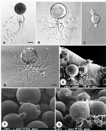
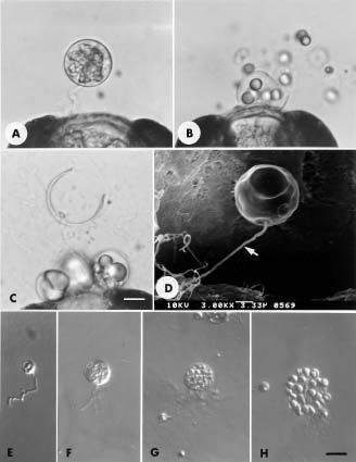
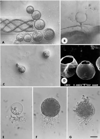
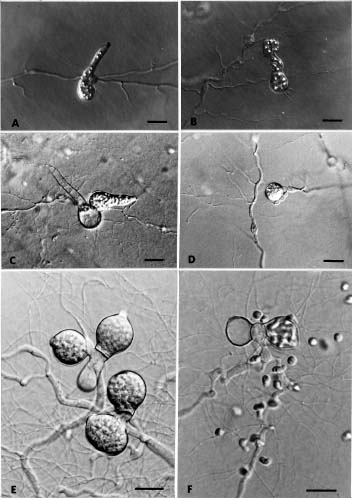
In a series of papers we have been reporting chytrid fungi of Taiwan (Chen and Chien, 1995; 1998). This paper adds information about 7 monocentric and 3 polycentric chytrids isolated from fresh water and soil. To document our reports we have isolated the fungi into pure culture and have photographed their developmental stages on natural substrates and on nutrient agar. The type of development of the thallus is an important character in the descriptions of Chytridialean families (Whiffen, 1944; Karling, 1977; Barr, 1980). The thallus of eucarpic chytrids is differentiated into a vegetative system and a reproductive organ. In monocentric species, the zoospore gives rise to a single sporangium or resting spore bearing a rhizoidal system. In polycentric species, a more extensive rhizoidal system (rhizomycelium) is estabilished, on which numerous sporangia or resting spores develop (Sparrow, 1960).

Ultrastructural studies of zoospores resulted in the establishment of the order Spizellomycetales, which was segregated from the Chytridiales by Barr in 1980. Four new genera were described in the new order, namely, Spizellomyces, Gaertneriomyces, Triparticalcar and Kochiomyces (Barr, 1980; 1984). Type species for these orders came primarily from species in the “Phlyctochytrium complex,” which consists of monocentric chytrids with multiple discharge papillae and rhizoids with swollen bases. Our observations of Phlyctochytrium spectabile Uebelmesser indicate that it also belongs in the order Spizellomycetales, in the genus Gaertneriomyces, and we

have made the new combination G. spectabile (Uebelmesser) Chen and Chien comb. nov.

Materials and Methods

Samples of water and soil were baited with pine pollen, grass leaves, and onion skin. Organisms were isolated and cultured on Emerson’s 1/4 YpSs agar containing antibiotics (Chen and Chien, 1998). Morphological characters and developmental stages were examined by using a light microscope and scanning electron microscope. Axenic cultures were kept on Emerson’s 1/4 YpSs medium in screw-cap tubes and transfered every three months. All pure cultures have been deposited at the Mycology Laboratory of the Institute of Biological Sciences, National Taiwan Normal University, Taipei, Taiwan, ROC.

Sparrow’s “Aquatic Phycomycetes” (1960), Karling’s “Chytridiomycetarum Iconorgraphia” (1977), and other’s were used as references for identification.

Species Descriptions

Gaertneriomyces spectabile (Uebelmesser) Chen and Chien comb. nov.

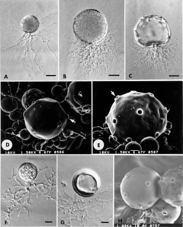
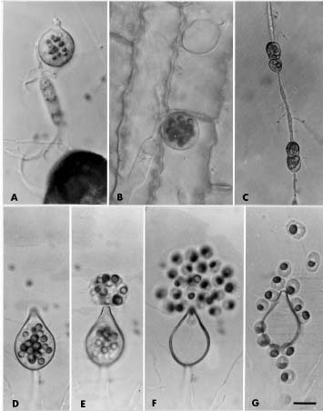
º Phlyctochytrium spectabile Uebelmesser, Arch. f. Mikrobiol. 25: 315, 1956. Figure 1A-E

On 1/4 YpSs agar: Sporangium spherical, 30-75 µm diam., with 2-10 or more prominent, hourglass-shaped papillae, with 5 × 5-12.5 µm plug within papillae; rhizoidal system consisting of a subsporangial globular apophysis, 5-10 µm diam., and moderately extensive, branched, delicate rhizoids; zoospores globose, 4-5 µm diam., or ovoid, with a small, inconspicious globule emerging singly from the discharge pore. Color of colony, white.

4Corresponding author. No. 181 Lane 482 Ta-T’ung Road, Sec. 2, Tainan 702, Taiwan. Tel : (06) 266-4911 ext. 340; Fax: (06) 266-6411.


Botanical Bulletin of Academia Sinica, Vol. 41, 2000

Specimen examined. YULIN HSIEN: Chiungpu, farm soil, 22 Jul 1992, NTNUS03c. Isolated on pine pollen from soil.

Notes. The hourglass-shaped papillae is the distinctive character. According to the ultrastructure of its zoospore (Chen, unpublished), this fungus should be placed in the Spizellomycetalean genus Gaertneriomyces.

Gaertneriomyces semiglobiferus (Uebelmesser) D. J. S. Barr, Can. J. Bot. 58: 2386, 1980.

º Phlyctochytrium semiglobiferum Uebelmesser, Arch. Mikrobiol. 25: 319, 1956. Figure 1F-H

On 1/4 YpSs agar: Sporangium spherical, 25-75 µm diam., with 2-7 prominent, 10-12.5 µm broad, hemispherical papillae; apophysis bulbous or inflated, rhizoidal system open and moderately extensive, with branches arising from the apophysis. Zoospores emerging singly, 4-6 µm diam., globular to amoeboid, with single globules. Resting spores spherical, 50-65 µm diam., with a smooth, thick wall, with 1 to 3 low papillae. Color of colony, white, center of colony, brown.

Specimen examined. YULIN HSIEN: Chiungpu, farm soil, 22 Jul 1992, NTNU S03a. Isolated on pine pollen from soil.

Notes. In some sporangia, apophysis arise from 2 or 3 positions, similar to Spizellomyces plurigibbosus. The ultrastructure of zoospore (Chen, 1996), however, is the Gaertneriomyces type (Barr, 1981). The thick-walled resting spore is the distinctive character (Sparrow, 1960).

Spizellomyces palustris Gaertner ex D. J. S. Barr, Can. J. Bot. 62: 1187, 1984.

º Phlyctochytrium palustre Gaertner, Arch. Mikrobiol. 21: 123, 1954 Figure 2A-G

On 1/4 YpSs agar: Sporangium spherical, 25-50 µm diam., or subspherical, with several discharge papillae. The main rhizoidal axis is bulbous or nearly tubular, occasionally with a taproot-like apophysis; rhizoidal system irregularly branched or somewhat dichotomously branched, richly and extensively branched; rhizoids taper evenly to blunt ends, occasionally uneven or constricted. Zoospores globose, about 5 µm diam., or irregularly shaped, (3.75-10)

Figure 1. A-E, Gaertneriomyces spectabile. A and B, developing sporangium; C, empty sporangium with four discharge pores; D and E, sporangium with hourglass-shaped (arrow) papillae or discharge pores. F-H, Gaertneriomyces semiglobiferus. F, sporangium with inflated apophysis; G, thick-walled resting spore with a discharge pore; H, sporangium with several discharge papillae. (Bar = 10 µm in A-C and F-G.)


Chen et al. Some chytrids of Taiwan

Figure 2. Spizellomyces palustris. A, sporangium with taproot-like apophysis; B, sporangium with extensive rhizoids; C, two small globular structures on the sporangium, looks like self parasitism; D, mature sporangium with several papillae; E, young sporangia, someone bearing small globular structures; F, discharging sporangium, amoeboid zoospores emerging; G, empty sporangium with several discharge pores. (Bar = 20 µm in A-D.)

× 15 µm; emerging individually from 5-10 discharge pores. Color of colony, white.

Specimen examined. YULIN HSIEN: farm soil, 22 Jul 1992, NTNUS03b. Isolated on pine pollen from soil.

Notes. Most sporangia with tubular or inflated main rhizoid, rhizoids evenly or constricted, similar to the S. palustris examined by Barr (1984). Physiological tests of our isolate, however, indicate that the maximum temperature for growth is nearly 45°C. Our isolate utilizes glucose, lactose, trehalose, cellobiose, and sucrose as well as N-acetyl-D-glucosamine (Chen, 1996). The ultrastructure of the zoospore is the Spizellomyces type (Chen, 1996). Occasionally 1 to 3 small globular structures occur on the outer surface of sporangia. Each globular structure extends a tubular structure into the sporangium.

Rhizophydium sphaerocarpum var. sphaerocarpum (Zopf) Fischer, Rabenhorst. Kryptogamen-Fl. 1(4): 95. 1892. Figure 3A-D

On pine pollen: Zoosporangium epibiotic or interbiotic, spherical, 7.5-47.5 µm diam. Rhizoids arising from a deli

cate main axis, on sporangium base. Zoospores ooze through a wide apical pore in a loose mass, empty sporangium urceolate with one apical pore. Resting spores spherical, 12.5-17.5 µm diam., with a large centric globule, smooth wall.

In 1/4 YpSs slush: Zoosporangium spherical, 22-27 µm diam., with a large apical or subapical exit pore, 9.9-12.4 µm diam., occasionally a very fine operculum-like structure was seen. Rhizoids from one fine axis, which is 19.7-54.3 µm long; extensive branching from distal end of rhizoidal axis.

On 1/4 YpSs agar: Young sporangium with several oil globules, one thread-like main axis extending from sporangium base, sparingly branched. Zoospores globular, 4-5 µm diam., emerging through the apical portion of wall, becoming amoeboid, then swimming away. Empty sporangium urceolate. Color of colony, buff to off-white.

Specimen examined. TAIPEI CITY: Yangmingshan Tatun Natural Park, water, 8 Apr 1991, Hsu01; TAIPEI HSIEN: Pinghsi, water from stream, 16 Jul 1992, NTNU101a; NANTOU HSIEN: Puli, waterfall, 20 Jul 1992,


Botanical Bulletin of Academia Sinica, Vol. 41, 2000

NTNU201b; CHIAYI HSIEN: Alishan, spring water, 4 Aug 1992, NTNU301a; ILAN HSIEN: Harpen, lake water, 27 Mar 1993, NTNU809a; TAIPEI HSIEN: Pinglin, stream water, 16 Oct 1993, NTNU2101a; TAOYUAN HSIEN: Tachi, pond water, 2 Jan 1994, NTNU2202a; TAICHUNG HSIEN: Tungshih woodland, lake water, 6 Sep 1994, NTNU2301a; TAICHUNG HSIEN: Hueysuen woodland, stream water, 21 Dec 1997, CSF3302a. Isolated on pine pollen from all water samples.

Notes. Ubiquitous. An urceolate empty sporangium and delicate slender rhizoids are the main characters. Physiological tests on our isolate indicate that the maximum temperature for growth is nearly 30°C. According to color of colony, this isolate is identified as R. sphaerocarpum var. sphaerocarpum. The above is similar to Barr’s (1970) report.

Rhizophydium melosirae Friedmann, Osterreich. Bot. Zeitschr. 99: 179, 1952. Figure 3E-H

On 1/4 YpSs agar: Sporangium spherical, 10-15 µm diam., wall thin, smooth, deliquescing completely upon discharge of the zoospores, rhizoids arising from a fine axis or globular swelling, 1-1.5 µm diam. Zoospores spherical, 2.5-3 µm

diam., with an anterior globule, liberated upon the deliquescence of the sporangium wall. Color of colony, white.

Specimen examined. YILAN HSIEN: Shunglienpyi, soil, 3 Oct 1992, NTNU602a. Isolated on pine pollen from soil.

Notes. Complete deliquescence of the sporangial wall upon discharge of the zoospores is the main character. This species is different from Rhizophydium patellarium. Only the upper two-thirds of the latter’s sporangial wall dissolves (Chen and Chien, 1998).

Rhizophydium sphaerocarpum var. spirogyrae D. J. S. Barr, Can. J. Bot. 48: 1607-1071, 1970. Figure 4A-D

On Spirogyra: Sporangium epibiotic, spherical. Rhizoid endobiotic, arising from a main axis and tapering, extending on Spirogyra cell. Zoospores discharge through wide apical portion, forming a temporarily vesiculate motionless group, operculum occasionally seen, become motile after discharge, soon swimming away. Zoospore spherical, 3.5-5 µm diam., flagellum about 25 µm long.

On 1/4 YpSs agar: Sporangia spherical, 7.5-47.5 µm diam., or ovate. Rhizoidal system with main axis, 3-7 µm diam., branch long, sparse. Zoospores discharge through wide

Figure 3. A-D, Rhizophydium sphaerocarpum var. sphaerocarpum. A, on pine pollen, an interbiotic young sporangium; B, mature sporangium; C, an empty sporangium; D, sporangium with a thread-like (arrow) main axis. E-H, Rhizophydium melosirae. E-G, developing sporangium; H, sporangial wall completely dissolves. (Bar = 10 µm in A-C and E-H.)


Chen et al. Some chytrids of Taiwan

Figure 4. A-D, Rhizophydium sphaerocarpum var. spirogyrae. A, on filaments Spirogyra, several young sporangia; B, on a filament of Spirogyra, empty sporangium with a large discharge pore; C, on 1/4 YpSs agar, three young sporangia; D, on 1/4 YpSs agar, two empty sporangia with a large discharge pore. E-G, Rhizophydium collapsum. E, young sporangium; F, mature sporangium; G, discharging sporangium. (Bar = 10 µm in A-C and E-G.)

apical portion, about 7.5-15 µm diam., sometimes with operculum beside exit pore. Zoospore spherical, 4-5 µm diam., with a lateral refractory globule. Resting spore not observed. Color of colony, buff to pale-yellow.

Specimen examined. TAIPEI CITY: Yangmingshan, stream water, 21 Jul 1993, NTNU1302. Isolated from Spirogyra in water.

Notes. This isolate is morphologically similar to Rhizophydium spherocarpum. Three varieties of R. sphaerocarpum are proposed (Barr, 1970; Huth and Gaertner, 1973). The physiological tests on our isolate indicate that the maximum temperature for growth is nearly 45°C. Compared with R. sphaerocarpum var. sphaerocarpum, they differ primarilly in the range of growth temperature. Moreover, our isolate is parasitic on Spirogyra, but also can be cultured on artificial medium. It is different from R. sphaerocarpum var. rhizoclonium (Huth and Gaertner, 1973) in its host, and from R.

sphaerocarpum var. sphaerocarpum in being unable to grow on pine pollen.

Rhizophydium collapsum Karling, Sydowia 17: 285, 1964. Figure 4E-G

In YPD broth: Sporangium spherical, 25-57.5 µm diam., wall delicate, with one to several papillae. Rhizoidal system arising from one to several points in close proximity on the sporangium base, delicate, short branch. Zoospores globose about 2.5 µm diam., release through several discharge pores, then wall collapses and dissolves. Color of colony, off-white.

Specimen examined. YILAN HSIEN: Harpen, pond water, 19 Feb 1993, NTNU806a. Isolated on pine pollen from water.

Notes. Zoospores discharge in Rhizophydium collapsum through several papillae, and then the sporan


Botanical Bulletin of Academia Sinica, Vol. 41, 2000

gial wall collapses and also partially dissolves. This species is different from R. patellarium by the number of discharge pores and differs from R. capillaceum in having the sporangial wall collapse and partially dissolve (Barr, 1971).

Cladochytrium replicatum Karling, Amer. J. Bot. 18: 538, 1931. Figure 5A-G

On grass leaf: Thallus intramaterial and extramaterial, eucarpic, polycentric. Rhizomycelium extensive, delicate, richly branched, with numerous fusiform, spindle-shaped, septate turbinate organs. Zoosporangium terminal or intercalary, apophysate or nonapophysate, spherical or ovoid, 10-32.5 µm diam., with one, occasionally two, short discharge tubes. Zoospore spherical, 5-7.7 µm diam., with a golden-brown globule; emerging in a gelatinous vesicle at the orifice, quiescent about 20 seconds previous to escaping from the vesicle and swimming away.

On 1/4 YpSs agar: Zoospore germinates into a polycentric thallus. Rhizomycelial growth on agar, mature zoosporangia at center of colony. Resting spore not observed. Center of colony, yellowish-orange, periphery white.

Specimen examined. TAIPEI CITY: Yangmingshan Tatun Natural Park, waterfalls, 13 Mar 1991, Hsu06a, 6 Oct 1991, Hsu06b; Yangmingshan Hsiangtien pool water, 5 Nov 1991, Hsu06c; TAIPEI HSIEN: Pingshi, stream water, 16 Jul 1992, NTNU101; YILAN HSIEN: Tahu, pond water, 3 Oct 1992, NTNU701; Harpen, pond water, 19 Feb 1993, NTNU806; Fushan Botanical Garden, stream water, 8 Dec 1993, NTNU2001; TAINAN HSIEN: Jen-te pond water, 17 Nov 1997, CSF3101c. Isolated on pine pollen, onion skin, and grass leaf from all water samples.

Notes. The rhizomycelium with septate tubinate organs and zoospore with a conspicious orange-brown globule are the main characters (Karling, 1935). It is ubiquitous in distribution.

Cladochytrium hyalinum Berdan, Amer. J. Bot. 28: 425, 1941. Figure 6A-D

On grass leaf: Thallus polycentric, delicate, hyaline. Zoosporangium terminal or intercalary, apophysate.

1/4 YpSs agar: Thallus polycentric, eucarpic. Rhizomycelium sparse, hyaline and extensive, with numer

Figure 5. Cladochytrium replicatum. A, on pine pollen, rhizomycelium with a spindle organ and a mature sporangium; B, on grass leaf, a mature sporangium and an empty sporangium; C, on 1/4 YpSs agar, rhizomycelium with two septate spindle organs; D-G, a series of the discharge process; E, vesicle forming on the discharge pore. (Bar = 10 µm in A-G.)


Chen et al. Some chytrids of Taiwan

Figure 6. A-D, Cladochytrium hyalinum. A, mature sporangium; B, zoospores discharging; C, empty sporangium and mature sporangium; D, a spherical resting spore on a multiple-celled spindle organ. E-F, Polychytrium aggregatum. E, rhizomycelium with two and three sporangia in aggregate; F, empty sporangium and discharging sporangium. (Bar = 20 µm in A-F.)

ous round, fusiform, or oval swellings. Zoosporangium terminal or intercalary, apophysate, subspherical, ovoid, bottle-gourd, or elongated, 30 µm diam.; exit tube single, with a gelatinous plug. Zoospores spherical, 8-10 µm diam., with a hyaline refractive globule; escaping in a globular mass, briefly quiescent previous to swimming away. Resting spore develops from a short branch arising directly from a multiple-celled spindle organ, usually spherical, about 20 µm diam., containing numerous hyaline refractive bodies. Color of colony, hyaline.

Specimen examined. TAIPEI CITY: Taipei Botanical Garden, pond water, 9 May 1994, NTNU402. Isolated on grass leaf from water.

Notes. Thallus is delicate and hyaline. Resting spore develops from a multiple-celled swollen structure that is the main character (Berdan, 1941).

Polychytrium aggregatum Ajello, Mycologia 34: 443, 1942. Figure 6E-F

In 1/4YpSs slush: Thallus polycentric, eucarpic. Rhizomycelium coarse, 4-5 µm diam., extensive, occasionally spiral circuitous. Sporangia in aggregates of two or more, terminal and intercalary, paired sporangia in different stages of development. At maturity, spherical, ovoid or fusiform, 25-27.5 µm diam., with an apical exit pore, about 5 µm diam.; proliferating. Zoospore delimited within the sporangium, partially emerging and forming a motionless, spherical mass at the mouth of the exit pore, then swimming away, others discharging individually, spherical, 5-7 µm diam. After a motile period zoospores become amoeboid and retract their flagella, settle down and germinate. Color of colony, white hyaline.

Specimen examined. TAIPEI CITY: Yangmingshan Chuanszu waterfalls, 6 Oct 1991, Hsu07; 2 Aug 1995,


Botanical Bulletin of Academia Sinica, Vol. 41, 2000

NTNU2502. Isolated on grass leaf, onion skin, and pine pollen from water samples.

Notes. This is a monotypic genus (Sparrow, 1960). The species is characterized by a coarse rhizomycelium, which lacks spindle organs or intercalary swellings, and has proliferating sporangia in aggregates of two or more. The above is similar to Ajello’s (1942) description, except that tuberculate zoosporangia were not observed on our isolate.

Discussion

Barr (1980) noted that members of the Spizellomyces live in soil. The species of this paper—Gaertneriomyces spectabile, G. semiglobiferus, and Spizellomyces palustris—and three species of another report (Chen and Chien, 1998)—S. punctatum, S. acuminatus, and S. pseudodichotomus—are all isolated from soils. This is strong evidence that members of this order are soil organisms rather than water organisms.

Literature Cited

Ajello, L. 1942. Polychytrium: A new cladochytriaceous genus. Mycologia 34: 442-451.

Barr, D.J.S. 1970. Two varieties of Rhizophydium sphaerocarpum (Chytridiales). Can. J. Bot. 48: 1067-1071.

Barr, D.J.S. 1971. Rhizophydium patellarium and R. capillaceum (Chytridiales). Can. J. Bot. 49: 1497-1502.

Barr, D.J.S. 1980. An outline for the reclassification of the

Chytridiales, and for a new order, the Spizellomycetales. Can. J. Bot. 58: 2380-2394.

Barr, D.J.S. 1981. Ultrastructure of Gaertneriomyces zoospore (Spizellomycetales, Chytridiomycetes). Can. J. Bot. 59: 83-90.

Barr, D.J.S. 1984. The classification of Spizellomyces, Gaertneriomyces, Triparticalcar, and Kochiomyces (Spizellomycetales, Chytridiomycetes). Can. J. Bot. 62: 1171-1201.

Berdan, H.B. 1941. A developmental study of three saprophytic chytrids I. Cladochytrium hyalinum sp. nov. Amer. J. Bot. 28: 422-438.

Chen, S.F. 1996. Studies on Morphology and Taxonomy of Chytridiales in Taiwan. Ph.D. Thesis, National Taiwan Normal University, Teipei, Taiwan, ROC.

Chen, S.F. and C.Y. Chien. 1995. Some chytrids of Taiwan (I). Bot. Bull. Acad. Sin. 36: 235-241.

Chen, S.F. and C.Y. Chien. 1998. Some chytrids of Taiwan (II). Bot. Bull. Acad. Sin. 39: 47-56.

Huth, K. and A. Gaertner. 1973. A new variety of Rhizophydium sphaerocarpum from the Weser estuary. Trans. Brit. Mycol. Soc. 61: 431-434.

Karling, J.S. 1935. A further study of Cladochytrium replicatum with special reference to its distribution, host range, and culture on artificial media. Amer. J. Bot. 22: 439-452.

Karling, J.S. 1977. Chytridiomycetarum Iconorgraphia. Luberecht J. Cramer. Monticello, New York, 414 pp.

Sparrow, F.K.Jr. 1960. Aquatic Phycomycetes. 2nd edn. The University of Michigan Press, 1187 pp.

Whiffen, A.J. 1944. A discussion of taxonomic criteria in the Chytridiales. Farlowia 1(4): 583-597.

»OÆW²£³ýµß (III)

³¯²Qªâ1 ³\¬ü½¬2 ²¬î·½3

1 ¹Å«nÃIJz¾Ç°|­¹«~½Ã¥Í¨t

2 »O¥_¥«¥ß¦¨¥\°ª¤¤
3 °ê¥ß¥xÆW®v½d¤j¾Ç¥Íª«¬ã¨s©Ò
¥»¤å´y­z¨Ã¹Ï»¡©ú¤CºØ³æ¤¤¤ß³ýµß¡GGaertneriomyces spectabile (Uebelmesser) Chen and Chien, G. semiglobiferus (Uebelmesser) Barr, Spizellomyces palustris Barr, Rhizophydium sphaerocarpum var. sphaerocarpum (Zopf) Fischer, R. melosirae Friedman, R. sphaerocarpum var. spirogyrae Barr, R. collapsum Karling ¥H¤Î¤TºØ¦h¤¤¤ß³ýµß¡GCladochytrium replicatum Karling, C. hyalinum Berdan, Polychytrium aggregatum Ajello. ¥þ³¡¬°»OÆW·s°O¿ýºØ¡A¨ä¤¤ Gaertneriomyces spectabile ¬°·s²Õ¦XºØ¡C

ÃöÁäµü¡G³ýµß¥Ø¡F³æ¤¤¤ß³ýµß¡F¦h¤¤¤ß³ýµß¡F¤p³ýµß¥Ø¡F»OÆW¡C