activities, H2O2 level, and cell wall peroxidase activity in roots

Bot. Bull. Acad. Sin. (2000) 41: 99-103

Chen et al. — Oxidative stress in copper-treated rice roots

Copper toxicity in rice seedlings: Changes in antioxidative enzyme activities, H2O2 level, and cell wall peroxidase activity in roots

Li-Men Chen, Chuan Chi Lin, and Ching Huei Kao1

Department of Agronomy, National Taiwan University, Taipei, Taiwan, Republic of China

(Received March 15, 1999; Accepted September 2, 1999)

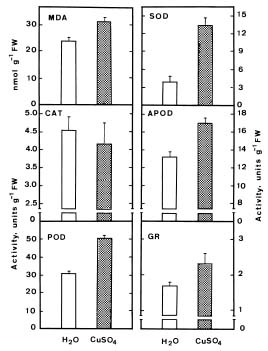
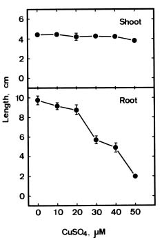
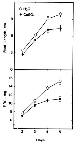
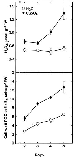
Abstract. The changes in lipid peroxidation, antioxidative enzyme activity, H2O2 level, and cell wall peroxidase activity in Cu-stressed roots of rice seedlings and their relation with root growth inhibition were investigated. CuSO4 was effective in inhibiting root growth but not shoot growth. Treatment with CuSO4 resulted in an increase in lipid peroxidation and modulated antioxidative enzyme activities in rice roots. CuSO4 increased the activities of superoxide dismutase, ascorbate peroxidase, glutathione reductase, and peroxidase, but had no effect on catalase. CuSO4 also increased H2O2 level and cell wall peroxidase in roots of rice seedlings. Exogenous application of H2O2 resulted in an inhibition of root growth. It appears that growth inhibition of root caused by Cu is associated with H2O2 dependent peroxidase-catalyzed formation of cross-linking among cell wall polymers.

Keywords: Copper; Lipid peroxidation; Oryza sativa; Oxidative stress; Root growth.

Abbreviations: APOD, ascorbate peroxidase; CAT, catalase; FW, fresh weight; GR, glutathione reductase; MDA, malondialdehyde; POD, peroxidase; SOD, superoxide dismutase.

Introduction

Copper (Cu) is an essential element for plant growth (Arnon and Stout, 1939), and it is important in various biochemical processes, but at toxic concentrations it interferes with numerous physiological processes (Fernandes and Henriques, 1991).

Cu is known to damage cell membranes by binding to the sulphydryl groups of membrane proteins and by inducing lipid peroxidation (De Vos et al., 1989; De Vos et al., 1992). Cu-mediated free radical formation has been demonstrated in isolated chloroplasts (Sandmann and Boger, 1980), in intact roots (De Vos et al., 1993), in leaf segments (Chen and Kao, 1999; Gallego et al., 1996; Luna et al., 1994) and in intact leaves (Weckx and Clijsters, 1996).

Cellular damage caused by free radicals might be reduced or prevented by a protective metabolism involving antioxidative enzymes such as SOD, APOD, GR, CAT and POD. SOD catalyzes the dismutation of two molecules of superoxide into oxygen and H2O2. APOD reduces H2O2 to water, with ascorbate as electron donor (Asada, 1992). GR plays a part in the control of endogenous H2O2 through an oxido-reduction cycle involving glutathione and ascorbate (Foyer and Halliwell, 1976; Smith et al., 1989). CAT and POD are implicated in removal of H2O2. It has been

reported that Cu increases the activities of antioxidative enzymes such as SOD (Chongpraditnum et al., 1992; Rama Devi and Prasad, 1998), POD (Karataglis et al., 1991), CAT (Rama Devi and Prasad, 1998), and APOD (Rama Devi and Prasad, 1998). It is well known that CAT and APOD play an important role in preventing oxidative stress by catalyzing the reduction of H2O2 (Weckx and Clijsters, 1996). Rama Devi and Prasad (1998) found that CAT and APOD activities were increased by Cu, suggesting that excess Cu may increase the production of H2O2. H2O2 is a necessary substrate for the cell wall stiffening process catalyzed by cell wall POD (Elstner and Heupel, 1976; Hohl et al., 1995; Schopfer, 1996), which is considered to be one of the mechanisms resulting in growth inhibition (Fry, 1986). The present investigation was designed to study the change in lipid peroxidation, antioxidative enzyme activities, H2O2 level and cell wall POD activity in Cu-stressed roots of rice seedling and their relation with root growth inhibition.

Materials and Methods

Rice (Oryza sativa L. cv. Taichung Native 1) seeds were sterilized with 2.5% sodium hypochlorite for 15 min and washed thoroughly with distilled water. These seeds were then germinated in a Petri dish (20 cm) containing distilled water at 37°C in the dark. After a 1-day incubation, uniformly germinated seeds were selected and transferred to Petri dishes (9.0 cm) containing two sheets of Whatman No. 1 filter paper moistened with 10 ml of distilled water

1Corresponding author. Fax: 886-2-23620879; E-mail: kaoch@ccms.ntu.edu.tw


Botanical Bulletin of Academia Sinica, Vol. 41, 2000

or test solution. Each Petri dish contained 20 germinated seeds. Each treatment was replicated 4 times. The germinated seeds were allowed to grow at 27°C in darkness, and 3 ml of distilled water or test solution was added to each Petri dish on day 3 of growth. All experiments described here were performed three times. Similar results of identical trends were obtained each time. The data reported here are from a single experiment.

The level of lipid peroxidation products in samples was expressed as 2-thiobarbituric acid reactive materials [aldehydes, mainly malondialdehyde (MDA) and endoperoxides (Buege and Aust, 1978)]. 2-Thiobarbituric acid-reactive materials in samples were assayed according to the modified method of Heath and Packer (1968).

The H2O2 level was colorimetrically measured as described by Jana and Choudhuri (1981). H2O2 was extracted by homogenizing 50 mg roots with 3 ml of phosphate buffer (50 mM, pH 6.8). The homogenate was centrifuged at 6,000 g for 25 min. To determine H2O2 levels, 3 ml of extracted solution was mixed with 1 ml of 0.1% titanium chloride in 20% (v/v) H2SO4 and the mixture was then centrifuged at 6,000 g for 15 min. The intensity of the yellow colour of the supernatant at 410 nm was measured. H2O2 level was calculated using the extinction coefficient 0.28 µmol-1 cm-1.

For extraction of antioxidative enzymes, roots were homogenized with 0.1 M sodium phosphate buffer (pH 6.8) in a chilled pestle and mortar. The homogenate was centrifuged at 12,000 g for 20 min and the resulting supernatant was used for determination of enzyme activity. The whole extraction procedure was carried out at 4°C. CAT, POD, SOD, APOD and GR were assayed as described previously (Chang and Kao, 1998).

Cell walls were prepared by homogenizing roots in ice cold phosphate buffer (50 mM, pH 5.8) using a pestle and mortar. The homogenate was centrifuged at 1,000 g, and washed at least four times with 50 mM phosphate buffer (Lee and Lin, 1995). The pellet was collected and used as cell wall fraction. POD ionically bound to the cell walls was extracted with 1 M NaCl. Cell walls prepared as described above were incubated in 1 M NaCl for 2 h with shaking at 30°C and centrifuged at 1,000 g. The supernatant was used to assay POD activity. POD activities were measured using a modification of the procedure described by Curtis (1971). The assay medium contained 50 mM phosphate buffer (pH 5.8), 7.2 mM guaiacol, 11.8 mM H2O2 and 0.1 ml enzyme extract in a final assay volume of 3.0 ml. The reaction was initiated by the addition of H2O2 and the change in absorbance at 470 nm was measured. Activity was calculated using the extinction coefficient (26.6 mM-1 cm-1 at 470 nm) for tetraguaiacol per min.

Results

Figure 1 shows the effect of CuSO4 on the growth of rice seedlings. Increasing concentrations of CuSO4 from 20 to 50 µM progressively decreased root length. However, no reduction of shoot length by CuSO4 was

observed. The differential effect of Cu on root and shoot growth could be accounted for by the fact that Cu is accumulated mainly in roots and to a minor extent in shoots (Fernandes and Henriques, 1991). Figure 2 shows time courses of the effect of CuSO4 (30 µM) on root length and root FW. As judged by root length and root FW, the reduction of root growth was evident 2 days after treatment.

Figure 3 demonstrates that CuSO4 treatment resulted in a significant increase in MDA level, an indicator of lipid peroxidation. Also shown in Figure 3 is the increase in POD, SOD, APOD and GR activities in roots of rice seedlings treated with CuSO4. However, CuSO4 had no effect on the activity of CAT in roots of rice seedlings. Similar results were obtained when enzyme activities were expressed on the basis of dry weight (data not shown).

Figure 4 shows time courses of endogenous H2O2 level and cell wall POD activity in roots of rice seedlings in the presence and absence of CuSO4 (30 µM). It is clear that CuSO4-treated roots had higher H2O2 level and cell wall POD activity than water controls. Exogenous application of H2O2 (10 mM) resulted in a reduction of root growth and an increase in endogenous H2O2 level in roots of rice seedlings.

Discussion

Although Cu can interfere with a number of physiological processes, the primary site of Cu toxicity is probably at the cell membrane level (De Vos et al., 1989). This is also evident from our study, in which Cu treatment resulted in an increase in lipid peroxidation in rice roots. The metal-induced lipid peroxidation is mostly attributed to increased production of free radicals (Halliwell and Gutteridge, 1984; Aust et al., 1985). Our results indicate that excess Cu increased oxidative stress, as is evident from increased lipid peroxidation (Figure 3). De Vos et al. (1989), Rama Devi and Prasad (1998), and Mazhoudi et al. (1997) reported a similar increase in lipid peroxidation when plants were treated with Cu.

The protective mechanisms adapted by plants to scavenge free radicals and peroxides include several antioxidative enzymes such as SOD, APOD, GR, CAT and POD. The antioxidative enzymes are important components in preventing the oxidative stress in plants as is based on the fact that the activity of one or more of these enzymes is generally increased in plants when exposed to stressful conditions (Allen, 1995). Overexpression of genes encoding these enzymes in several transgenic plant species conferring protection against free radicals has also been demonstrated (Allen, 1995). In the present study, Cu treatment resulted in an increase in the activities of SOD, APOD, GR and POD (Figure 3), which can be considered as an indirect evidence for enhanced production of free radicals under Cu stress. The increase of SOD, APOD and POD has been reported with Cu (Chongpraditnum et al., 1992; Karataglis et al., 1991; Mazhoudi et al., 1997; Rama Devi and Prasad, 1998). However, Mazhoudi et al. (1997) reported that CAT and


Chen et al. — Oxidative stress in copper-treated rice roots

Figure 1. Effect of CuSO4 on the growth of rice seedlings. Seedling growth was measured after 5 days of treatment. Vertical bars represent standard errors.

Figure 3. Effect of CuSO4 (30 µM) on the level of MDA, and the activities of CAT, POD, SOD, APOD and GR in roots of rice seedlings. MDA level and antioxidative enzyme activities were measured after 5 days of treatment. Vertical bars represent standard errors.

Figure 2. Time course of CuSO4 effect on root length and root FW of rice seedlings. Rice seedlings were treated with distilled water or 30 µM CuSO4. Vertical bars represent standard errors.

Figure 4. Changes in H2O2 level and cell wall POD activity in roots of rice seedlings in the presence and absence of CuSO4 (30 µM). Vertical bars represent standard errors.


Botanical Bulletin of Academia Sinica, Vol. 41, 2000

APOD activities were not affected by Cu. We also found no change in CAT activity (Figure 3). Such a variation in response of these enzymes to Cu stress could be due to the variability of plant species in producing free radicals (Mazhoudi et al., 1997). APOD and POD are known to play an important role in reducing oxidative stress by catalyzing the reduction of H2O2 (Weckx and Clijsters, 1996). Thus, the increase in the activities of APOD and POD by Cu (Figure 3) suggests increased production of H2O2. In the present study, CuSO4 treatment indeed increases the production of H2O2 in roots (Figure 4).

It has been postulated that the action of POD located in the cell wall would be to confer rigidity to the cell wall and prevent later expansion involved in growth (Fry, 1986). This cell wall stiffening process appears to involve oxidative coupling, dependent on H2O2 (Fry, 1986). CuSO4 treatment increases cell wall POD activity and H2O2 level in roots of rice seedlings (Figure 4). It has been shown that H2O2 caused a rapid cross-linking of cell wall polymers (Schopfer, 1996). If H2O2 plays an important role in the cell wall stiffening process, it is expected that H2O2 would inhibit root growth. This is indeed the case. Exogenous application of H2O2 inhibited root growth of rice seedlings (Figure 5). Thus, CuSO4-induced inhibition in root growth of rice seedlings is likely due to cell wall stiffening related to H2O2 dependent POD-catalyzed formation of cross-linking among cell wall polymers.

Acknowledgements. This work was supported by a grant from the National Science Council Project, NSC 88-2313-B-002-066.

Literature Cited

Allen, R.D. 1995. Dissection of oxidative stress tolerance using transgenic plants. Plant Physiol. 107: 1049-1054.

Arnon, D.I. and P.R. Stout. 1939. The essentiality of certain elements in minute quantity for plants with special reference to copper. Plant Physiol. 14: 371-375.

Asada, K. 1992. Ascorbate peroxidase- a hydrogen peroxide scavenging enzyme in plants. Physiol. Plant. 85: 235-241.

Aust, S.D., L.A. Marehouse, and C.E. Thomas. 1985. Role of metals in oxygen radical reactions. J. Free Radic. Biol. Med. 1: 3-25.

Buege, J.A. and S.D. Aust. 1978. Microsomal lipid peroxidation. Methods Enzymol. 52: 302-310.

Chang, C.J. and C.H. Kao. 1998. H2O2 metabolism during senescence of rice leaves: Changes in enzyme activities in light and darkness. Plant Growth Regul. 25: 11-15.

Chen, L.-M. and C.H. Kao. 1999. Effect of excess copper on rice leaves: evidence for involvement of lipid peroxidation. Bot. Bull. Acad. Sin. 40: 283-287.

Chongpraditnum, P., S. Mori, and M. Chino. 1992. Excess copper induces a cytosolic Cu, Zn-superoxide dismutase in soybean root. Plant Cell Physiol. 33: 239-244.

Curtis, C.R. 1971. Disc electrophoretic comparisons of protein and peroxidases from Phaseolus vulgaris leaves infected with Agrobacterium tumefaciens. Can. J. Bot. 49: 333-337.

De Vos, C.H.R., H. Schat, R. Vooijs, and W.A.O. Ernst. 1989. Copper induced damage to the permeability barrier in roots of Silene cucubalus. J. Plant Physiol. 135: 164-169.

De Vos, C.H.R., M.J. Vonk, and H. Schat. 1992. Glutathione depletion due to copper induced phytochelatin synthesis causes oxidative stress in Silene cucubalus. Plant Physiol. 98: 853-858.

De Vos, C.H.R., W.M. Ten Bookum, R. Vooijs, H. Schat, and L.J. De Kok. 1993. Effect of copper on fatty acid composition and peroxidation of lipids in roots of copper tolerant and sensitive Silene cucubalus. Plant Physiol. Biochem. 31: 151-158.

Elstner, E.F. and A. Heupel. 1976. Formation of hydrogen peroxide by isolated cell walls from horseradish (Armoracia lapathifolia Gilib). Planta 193: 283-289.

Fernandes, J.C. and F.S. Henriques. 1991. Biochemical, physiological, and structural effects of excess copper in plants. Bot. Rev. 57: 246-273.

Foyer, C.H. and B. Halliwell. 1976. The presence of glutathione and glutathione reductase in chloroplasts: a proposed role in ascorbic acid metabolism. Planta 133: 21-25.

Fry, S.C. 1986. Cross-linking of matrix polymers in the growing cells of angiosperm. Annu. Rev. Plant Physiol. 37: 165-186.

Gallego, S.M., M.P. Benavides, and M.L. Tomaro. 1996. Effect of heavy metal ion excess on sunflower leaves: evidence for involvement of oxidative stress. Plant Sci. 121: 151-159.

Halliwell, B. and J.M.C. Gutteridge. 1984. Oxygen toxicity, oxygen radicals, transition metals and disease. Biochem. J. 219: 1-14.

Figure 5. Changes in root growth and endogenous H2O2 level in roots of rice seedlings grown in the presence or absence of exogenous H2O2 (10 mM). Vertical bars represent standard errors.


Chen et al. — Oxidative stress in copper-treated rice roots

Heath, R.L. and L. Packer. 1968. Photoperoxidation in isolated chloroplasts. I. Kinetics and stoichiometry of fatty acid peroxidation. Arch. Biochem. Biophys. 25: 189-198.

Hohl, M., H. Greinev, and P. Schopfer. 1995. The cryptic growth response of maize coleoptile and its relationship to H2O2-dependent cell wall stiffening. Physiol. Plant. 94: 491-498.

Jana, S. and M.A. Choudhuri. 1981. Glycolate metabolism of three submerged aquatic angiosperms during aging. Aquat. Bot. 12: 345-354.

Karataglis, S., M. Moustakas, and L. Symeonidis. 1991. Effect of heavy metals on isoperoxidases of wheat. Biol. Plant. 33: 3-9.

Lee, T.-M. and Y.-H. Lin. 1995. Changes in soluble and cell wall-bound peroxidase activitieswith growth in anoxia-treated rice (Oryza sativa L.) coleoptiles and roots. Plant Sci. 106: 1-7.

Luna, C.M., C.A. Gonzalez, and V.S. Trippi. 1994. Oxidative damage caused by an excess of copper in oat leaves. Plant Cell Physiol. 35: 11-15.

Mazhoudi, S., A. Chaoui, M.H. Ghorbal, and E. El Ferjani. 1997. Response of antioxidative enzymes to excess copper in tomato (Lycopersicm esculentum, Mill). Plant Sci. 127: 129-137.

Rama Devi, S. and M.N.V. Prasad. 1998. Copper toxicity in Ceratophyllum demersum L. (Coontail), a free floating macrophyte: Response of antioxidant enzymes and antioxidants. Plant Sci. 138: 157-165.

Sandmann, G. and P. Boger. 1980. Copper-mediated lipid peroxidation processes in photosynthetic membranes. Plant Physiol. 66: 797-800.

Schopfer, P. 1996. Hydrogen peroxide-mediated cell-wall stiffening in vitro in maize coleoptile. Planta 199: 43-49.

Smith, I.K., T.L. Vierheller, and C. Thorne. 1989. Properties and functions of glutathione reductase in plants. Physiol. Plant. 77: 449-456.

Weckx, J.E.J. and H.M.M. Clijsters. 1996. Oxidative damage and defense mechanisms in primary leaves of Phaseolus vulgaris as a result of root assimilation of toxic amounts of copper. Physiol. Plant. 96: 506-512.

¤ô½_¥®­]»É¤§¬r®`¡G§Ü®ñ¤Æ»Ã¯À¬¡©Ê¡B¹L®ñ¤Æ²B§t¶q

»P²Ó­M¾À¹L®ñ¤Æ»Ã¯À¬¡©Ê¤§ÅܤÆ

³¯ÄR±Ó ªL¶Çµa °ª´º½÷

°ê¥ß¥xÆW¤j¾Ç¹AÃÀ¾Ç¨t

¥»¬ã¨s¥D­n±´°Q»É³B²z¤ô½_¥®­]¹ï®Ú¤º¯×½è¹L®ñ¤Æ§@¥Î¡B§Ü®ñ¤Æ»Ã¯À¬¡©Ê¡B¹L®ñ¤Æ²B§t¶q»P²Ó­M¾À ¤º¹L®ñ¤Æ»Ã¯À¬¡©ÊÅܤƤ§¼vÅT¡A¦P®É±´°Q³o¨ÇÅܤƻP»É§í¨î®Ú¥Íªø¶¡¤§Ãö«Y¡C²¸»Ä»É³B²z¥i¼W¥[¤ô½_¥®­] ®Ú¯×½è¹L®ñ¤Æ§@¥Î»P§ïÅܧܮñ¤Æ»Ã¯À¤§¬¡©Ê¡C²¸»Ä»É¥i¼W¥[¤ô½_¥®­]®Ú superoxide dismutase¡Aascorbate peroxidase¡Aglutathione reductase »P peroxidase ¬¡©Ê¡A¦ý¤£¼vÅT catalase ¬¡©Ê¡C²¸»Ä»É³B²z¦P®É¥i¼W¥[®Ú¤§¹L®ñ¤Æ²B§t¶q»P®Ú¤º²Ó­M¾À¹L®ñ¤Æ»Ã¯À¬¡©Ê¡C¥~¥[¹L®ñ¤Æ²B³B²z¥iÅãµÛªº§í¨î¤ô½_¥®­]®Ú¥Íªø¡C»É©Ò§í ¨î¤§¤ô½_¥®­]®Ú¥Íªø«Ü¥i¯à»P¸g¥Ñ¹L®ñ¤Æ²B¤Î¹L®ñ¤Æ»Ã¯À©Ò¶Ê¤Æ¤§²Ó­M¾ÀÅܵw¦³Ãö¡C

ÃöÁäµü¡G»É¡F¯×½è¹L®ñ¤Æ§@¥Î¡F¤ô½_¡F®ñ¤Æ°f¹Ò¡F®Ú¥Íªø¡C


Botanical Bulletin of Academia Sinica, Vol. 41, 2000