solanacearum in soil samples by polymerase chain reaction

Bot. Bull. Acad. Sin. (2000) 41: 121-128

Lee and Wang — Detection of R. solanacearum by PCR

The design of specific primers for the detection of Ralstonia solanacearum in soil samples by polymerase chain reaction

Yung-An Lee1 and Chi-Chung Wang

Department of Biology, Fu Jen Catholic University, Hsin Chuang 24205, Taipei, Taiwan, Republic of China

(Received March 24, 1999; Accepted October 1, 1999)

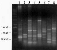
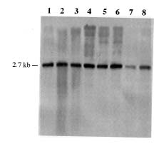
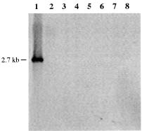
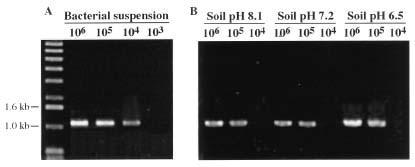
Abstract. A 0.7-kb DNA fragment, amplified by the randomly amplified polymorphic DNA (RAPD) method from Ralstonia solanacearum total DNA, was cloned and evaluated as a specific DNA probe. This 0.7-kb DNA fragment hybridized to a 2.7-kb EcoRI fragment in the EcoRI-digested total DNA of R. solanacearum. The 2.7-kb EcoRI fragment was also cloned and hybridized only to R. solanacearum but not to other Pseudomonas spp., pathovars of Xanthomonas campestris, and Erwinia spp. tested. The DNA sequence of this 2.7-kb fragment was obtained and used to design specific oligonucleotide primers for polymerase chain reaction (PCR) amplification. The primers amplified the same 1.1-kb PCR product from all R. solanacearum strains tested and failed to amplify DNA from any other plant pathogenic bacterial strains tested. DNA isolated from several saprophytic bacteria did not produce any PCR products with these primers. This specific PCR for R. solanacearum was also performed from colonies grown on BG medium with similar results. The sensitivity of the PCR assay using the specific primers was about 20 cells. The PCR assay was used to detect R. solanacearum in soil using these primer sets. No PCR product could be found when soil extract containing R. solanacearum was used directly in the assay. DNA extraction from soil was needed for the success of PCR assay. A simple method for DNA extraction from soil for PCR assay was developed and can hasten the detection of R. solanacearum in soil.

Keywords: Erwinia; Polymerase chain reaction; Pseudomonas; Ralstonia solanacearum; Xanthomonas.

Introduction

Ralstonia solanacearum (synonym Pseudomonas solanacearum E. F. Smith) (Yabuuchi et al., 1995) causes bacterial wilt, one of the most important and widespread bacterial diseases of crops in the world. This pathogen has been recorded on several hundred species representing 44 families of plants (Buddenhagen and Kelman, 1964; Hayward, 1991). In Taiwan, bacterial wilt has been reported on tomato, tobacco, potato, sweet pepper, eggplant, bird-of-paradise (Yang et al., 1980), perilla (Perilla crispa) (Hong et al., 1990), radish (Lin et al., 1994), anthurium plants (Su and Leu, 1995), and eustoma (Chao et al., 1995). Ralstonia solanacearum can be spread within and between countries by soil, by water, and by latently infected planting materials (Ciampi et al., 1980; Hayward, 1991). Accordingly, rapid and highly sensitive detection methods of R. solanacearum are required for quarantine to reduce field losses and limit the spread of bacterial wilt (Seal et al., 1992a, 1993; Seal, 1995).

The success of quarantine procedures relies on the use of rapid and sensitive detection techniques. Traditional methods for detecting R. solanacearum depend on a series of biochemical tests on purified colonies. This iden

tification procedure is too time consuming for quarantine and other diagnostic laboratory purposes. Nucleic acid probes and polymerase chain reaction (PCR), on the other hand, have been widely reported as tests that can be performed without culturing. Diagnostic tests based on PCR amplification of plant pathogens from environmental samples have shown that rapid and specific detection of very low numbers of the pathogen can be achieved (Seal et al., 1992a, 1993; Seal, 1995).

Development of a DNA probe specific for R. solanacearum is complicated by the genetic diversity of this species. Ralstonia solanacearum strains represent a heterogeneous group that has been subdivided informally into five races on the basis of host range (Buddenhagen et al., 1962; Buddenhagen, 1986), or five biovars based on the ability to oxidize certain disaccharides and hexose alcohols (Hayward, 1964; He et al., 1983). In addition, there is a phylogenetic dichotomy in R. solanacearum, which can be separated into two divisions based on the analysis of restriction fragment length polymorphism patterns (Cook et al., 1989), PCR amplification with tRNA consensus primers (Seal et al., 1992b), and rRNA sequences (Li et al., 1993; Seal et al., 1993; Seal et al., 1999; Taghavi et al., 1996). Division I contains all members of race 1 biovars 3, 4 and 5, and division II contains all members of race 1 biovar 1 and races 2 and 3. Strains of R. solanacearum in Taiwan are all race 1, and most are biovar 3 though some are biovar 4 (Hsu, 1991). These strains are all assigned to

1Corresponding author. Fax: 886-2-2902-1124; E-mail: bio1007@fujens.fju.edu.tw


Botanical Bulletin of Academia Sinica, Vol. 41, 2000

division I. Although DNA fragments specific for R. solanacearum and even race 3 strains have been cloned by subtractive hybridization, they did not specifically hybridize to all R. solanacearum strains tested (Cook and Sequeira, 1991; Seal et al., 1992a; Opina et al., 1997). For effective quarantine and detection measures against R. solanacearum, it is necessary to develop a specific and sensitive diagnostic test for R. solanacearum strains in Taiwan. We describe here the use of the randomly amplified polymorphic DNA (RAPD) technique to identify an R. solanacearum-specific DNA fragment. This DNA fragment was subcloned, the DNA sequences were determined, and specific oligonucleotide primers were designed for detection of R. solanacearum by PCR. Furthermore, a simple method for DNA extraction from soil for PCR assay was developed to detect the existence of R. solanacearum in soils.

Materials and Methods

Bacterial Strains and Culture Conditions

The strains of R. solanacearum, Pseudomonas syringae, Xanthomonas, and Erwinia used in this study, their pathovar designations, and sources are listed in Table 1. Ralstonia solanacearum was cultured in BG media (Boucher et al., 1985) at 30°C. Escherichia coli DH5a (BRL

Life Technologies, Inc., Gaithersburg, MD), X. campestris, Erwinia spp. and P. syringae were cultured in Luria-Bertani agar or broth medium (Sambrook et al., 1989) at 37°C for E. coli and at 28°C for the other species. Ampicillin (50 µg/ml) was added as necessary to maintain selection of the resistance marker in pBluescript SK(+) (Stratagene Co., La Jolla, CA).

General DNA Manipulations

Mini-scale preparations of E. coli plasmid DNA, total genomic DNA isolation of plant pathogenic bacteria, restriction endonuclease treatments, DNA ligation, transformation, and agarose gel electrophoresis were done as described by Sambrook et al. (1989). PCR-amplified DNA fragments used as probes were recovered from agarose by using the QIAEX II Gel Extraction Kit (QIAGEN Inc. Chatsworth, CA) and labeled with digoxigenin-11-dUTP (DIG) using a PCR DIG probe synthesis kit (Boehringer Mannheim Biochemicals, Indianapolis, IN). The PCR for preparation of DIG-labeled DNA probes was performed in an air thermal cycler (Rapidcycler, Idaho Technology, ID) programmed for denaturation at 94°C for 2 min and then for 35 cycles of 0 sec at 94°C, 0 sec at 50°C, and 35 sec at 72°C. Prehybridization, hybridization, and washing for Southern hybridization with DIG-labeled probes were performed at 68°C according to the

Table 1. Bacterial strains and hybridization results of the 2.7 kb EcoRI fragment of R. solanacearum to the bacteria.

Positive species and strainsa

R. solanacearum strainsb:

from tomato: PS21; PS-S4; PS-S32; PS-S80; PS68; PS72; PS95.

from tobacco: PS31; PS48.

from potato: PS60.

from sweet pepper: PS87; PS96.

from eggplant: PS99; PS100.

from anthurium: PS-AU003.

from New Zealand flax: PS-PDN002; PS-PDN003.

from cat-tail willow: PS-CTW009.

from eustoma: PS-ES001; PS-ES008.

from rose: PS-RO005; PS-RO006.

Negative species and strainsa

Pseudomonas species and strainsc

P. cepacia PC22; P. gladioli UCBPP550; P. syringae pv. apii PA102; P. syringae pv. tabaci PT124; P. syringae pv. eriobotryae UCBPP258; P. syringae pv. atrofaciens UCBPP260; P. syringae pv. coronafaciens UCBPP470; P. syringae pv. glycinea UCBPP527.

X. campestris pathovarc

X. campestris pv. betlicola XB104; X. campestris pv. campestris XCC1-1; X. campestris pv. dieffenbachiae XD113; X. campestris pv. oryzicola XO111; X. campestris pv. pelargonii XP15; X. campestris pv. phlei XP3-5; X. campestris pv. punicae XP177; X. campestris pv. spermacoces XS8-9; X. campestris pv. vesicatoria XV123.

Erwinia spp.c

E. amylovora EA169; E. carotovora subsp. atroseptica UCBPP149; E. carotovora subsp. carotovora Erc1; E. cypripedii EC155; E. quercina EQ101; E. rhapontici ER102; E. rubrifaciens ER103.

aThe positive strains have hybridization signal with 2.7-kb EcoRI DNA fragment in Southern blot hybridization. The negative strains have no signal. The hybridization method is described in Materials and Methods.

bThe strains were obtained from Department of Plant Pathology, National Chung Hsing University, Asian Vegetable Research and Development Center and Department of Pesticide Application, Taiwan Agricultural Chemicals and Toxic Substances Research Institute, Taiwan.

cThe strains were obtained from the International Collection of Phytopathogenic Bacteria, University of California, Berkeley, and American Type Culture Collection, Rockville, MD, USA.


Lee and Wang — Detection of R. solanacearum by PCR

manufacturer’s protocol. Double-stranded sequencing was performed by the dideoxy chain termination method (Sanger et al., 1977) adapted for the AutoRead Sequencing Kit (Pharmacia Biotech, Uppsala, Sweden) with standard universal forward and reverse M13 primers.

Primer and PCR Conditions

For RAPD analysis, PCR amplifications were carried out in 10-µl volumes which contained 10 ng of genomic DNA, 3 mM MgCl2, 0.5 µM primer, 0.4 U of Taq DNA polymerase (DyNAzyme II, Finnzymes Oy, Finland), 200 µM (each) deoxynucleoside triphosphate (dNTP) in 50 mM Tris HCl (pH 8.3), and 500 µg/ml BSA. Amplification was performed in an air thermal cycler programmed for two cycles of 70 sec at 94°C, 17 sec at 42°C, and 80 sec at 72°C; 35 cycles of 10 sec at 94°C, 17 sec at 42°C, and 80 sec at 72°C; and a final extension for 4 min at 72°C. Fifteen different arbitrary primers from 15-22 bases in length were used. Oligonucleotide primers were synthesized with a DNA synthesizer by the Agricultural Biotechnology Laboratories, National Chung Hsing University, Taiwan. Amplified DNAs were detected by electrophoresis in 0.8% agarose gels.

For specific amplification of R. solanacearum, two oligonucleotide primers, BP4-R and BP4-L, were designed from the nucleotide sequence of 2.7-kb EcoRI fragment cloned from R. solanacearum PS68. BP4-R (5'-GACGACATCATTTCCACCGGGCG-3') and BP4-L (5'-GGGTGAGATCGATTGTCTCCTTG-3') delineated a 1,102- bp fragment. DNA was amplified in 10-µl of reaction mixture prepared as described above. PCR condition was programmed for denaturation at 94°C for 2 min and then for 35 cycles of 0 sec at 94°C, 0 sec at 50°C, and 70 sec at 74°C. To determine the minimum number of R. solanacearum that could be detected by PCR, 10-fold serial dilutions of PS68 strain were made. Two µl aliquots from each tube of the dilution series were added directly to the PCR mixture. Aliquots from undiluted and serial dilutions of cultures were spread on BG plate to count the bacteria, and colony-forming units (cfu) was determined.

Soil and Soil Inoculation

Soil samples were collected from Yang-Ming mountain and Hsin-Chuang agriculture areas in northern Taiwan. Surface soil samples (depth, 0 to 15 cm) were collected, sieved (mesh size 2 mm), and stored under field-moist conditions at 4°C. The soil samples had pH range from 6.5 to 8.1. Subsamples (1 g) were seeded with 0.1-ml decimal dilutions of R. solanacearum culture which originally contained 107 cfu/ml. The inoculated soil samples were incubated for one hour at room temperature to equilibrate and allow for binding of the bacterial cells to the soil colloids.

Extraction of DNA from Soil for PCR Reaction

All soil samples were quantified as wet weight throughout the experiments. Total DNA was extracted from soil samples by a rapid method modified from the DNA isola

tion method developed by Chowdhury (1991). Inoculated soil samples (0.2 g) were suspended in 0.5 ml of distilled water by vortexing and allowed to stand for 5 min. The supernatant was transferred to a new microfuge tube and an equal volume of phenol:chloroform:isoamyl alcohol (25:24:l, v/v/v; pH 7.6) was added, after which they were mixed by vortexing at the maximum speed for 1 min and centrifuged at 16,000 g for 10 min. After the aqueous phase was transferred to a new tube, 0.1 volume of 3 M sodium acetate (pH 5.2) and 0.6 volume of isopropanol were added. The solution was mixed well and centrifuged at 16,000 g for 15 min. DNA pellets were washed with 70% ethanol twice, vacuum dried, and resuspended in 20 µl of TE (10 mM Tris, 1 mM EDTA, pH 8.0) buffer. Two µl of this DNA solution were taken for further PCR analysis.

Results

RAPD Analysis and Selection of a DNA Fragment Specific to R. solanacearum

To identify primers that generated RAPD patterns characteristic of R. solanacearum, 15 different primers were tested with DNA extracted from R. solanacearum PS68 and several pseudomonads. The amplification pattern obtained with primer R-424 on the DNA extracts of R. solanacearum PS68 was reproducible and revealed one major product of 0.7-kb. The sizes of the products amplified from pseudomonads ranged from 0.3 to 2.8 kb (Figure 1). All of the R. solanacearum strains tested by RAPD with R-424 primer exhibited the same pattern (data not shown). The 0.7-kb DNA fragment was eluted from agarose gel, la

Figure 1. Band patterns obtained after RAPD amplifications of R. solanacearum and other plant pathogenic bacteria strains with the oligonucleotide R-424. A 0.7-kb DNA fragment was amplified with DNA from R. solanacearum. Lanes: 1, R. solanacearum PS68; 2, Pseudomonas cepacia PC22; 3, P. syringae pv. apii PA102; 4, P. syringae pv. tabaci PT124; 5, P. syringae pv. eriobotryae UCBPP258; 6, P. syringae pv. atrofaciens UCBPP260; 7, P. syringae pv. glycinea UCBPP527; 8, P. gladioli UCBPP550. The size of the band is indicated on the left.


Botanical Bulletin of Academia Sinica, Vol. 41, 2000

beled with DIG by PCR with the R-424 primer, and used as a probe against EcoRI-digested total DNAs of R. solanacearum and several pseudomonads. The result showed that the 0.7-kb DNA fragment hybridized to a 2.7-kb EcoRI fragment in the genomic DNA of R. solanacearum PS68, but not with those of any pseudomonads tested (Figure 2).

To isolate and identify the 2.7-kb EcoRI fragment responsible for this specific hybridization, the region of an agarose gel containing 2.7-kb fragments of EcoRI-digested genomic DNA of R. solanacearum PS68 was excised. The DNAs were recovered from the gel, ligated to EcoRI-digested pBluescriptSK(+), and used to transform competent E. coli DH5a cells. Plasmid DNAs were isolated from 80 transformants and, after digestion with EcoRI and agarose gel electrophoresis, were hybridized with DIG-labeled 0.7-kb fragment. Three positive clones were selected. The 2.7-kb EcoRI fragment in one of the positive clones was eluted, labeled by the random primer method, and used to probe EcoRI-digested genomic DNAs from R. solanacearum PS68 and several pseudomonads. The probe hybridized only to R. solanacearum PS68 but not to the pseudomonads tested. The same probe was also used to hybridize genomic DNAs from other plant pathogenic bacteria, such as pathovars of Xanthomonas campestris, Erwinia spp., and several strains of soil saprophytic bacteria, and no hybridization signal was found (Table 1). Because longer DNA fragments contain more genetic information, the 2.7-kb fragment was used instead of the 0.7-kb fragment in further analysis.

Figure 3. Hybridization of the 2.7-kb EcoRI fragment to EcoRI-digested total genomic DNA of different strains of R. solanacearum. DNA (0.5 µg) was loaded into each lane, electrophoresed in 0.8% agarose, and hybridized with the digoxigenin-11-dUTP-labeled probe. Lanes: 1, R. solanacearum PS-31; 2, PS-60; 3, PS-87; 4, PS-AU003; 5, PS-PDN002; 6, PS-CTW009; 7, PS-ES001; 8, PS-RO005. The size of the band is indicated on the left.

To determine if the specificity of the 2.7-kb EcoRI fragment for R. solanacearum PS68 was indicative of a broader specificity for all strains isolated in Taiwan, the 2.7-kb fragment was used as a hybridization probe against total DNA extracted from all strains of R. solanacearum shown in Table 1. It was found that the 2.7-kb fragment hybridized with genomic DNAs of all R. solanacearum strains tested. Representative results are shown in Figure 3.

Specific Primers for Detection of R. solanacearum by PCR

The positions of restriction endonuclease sites and DNA sequence of the 2.7-kb EcoRI fragment were determined. Based on these DNA sequences, two oligonucleotides (BP4-R and BP4-L) were synthesized and used for subsequent PCR amplification tests. To determine the specificity of these primers, PCRs were carried out with DNA of all of the strains listed in Table 1. The PCR amplification using BP4-R and BP4-L primers generated an approximately 1.1 kb amplified DNA fragment for all R. solanacearum strains tested. No amplification was observed with any other Pseudomonas, Xanthomonas, or Erwinia strains tested.

Identical results were obtained when this specific PCR was performed with R. solanacearum PS68 colonies grown on BG medium. To determine the sensitivity of detection of the PCR assay with primers BP4-R and BP4-L, 2-µl samples of serial dilutions of R. solanacearum PS68 culture were used as template for the PCR. The tests were repeated six times, and the sensitivity of detection was determined as 104 cfu per ml of bacterial suspension on the

Figure 2. Hybridization of the 0.7-kb fragment amplified from R. solanacearum with R-424 to EcoRI-digested total genomic DNA of R. solanacearum and other plant pathogenic bacteria strains. DNA (0.5 µg) was loaded into each lane, electrophoresed in 0.8% agarose, and hybridized with the digoxigenin-11-dUTP-labeled probe. Lanes: 1, R. solanacearum PS68; 2, P. cepacia PC22; 3, P. syringae pv. apii PA102; 4, P. syringae pv. tabaci PT124; 5, P. syringae pv. eriobotryae UCBPP258; 6, P. syringae pv. atrofaciens UCBPP260; 7, P. syringae pv. glycinea UCBPP527; 8, P. gladioli UCBPP550. The size of the band is indicated on the left.


Lee and Wang — Detection of R. solanacearum by PCR

Figure 4. Detection of R. solanacearum by PCR-specific reaction with BP4-R and BP4-L primers on (A) suspensions of R. solanacearum strain PS68 (cfu/ml) and on (B) crude DNA extracted from three soil samples with pH from 6.5 to 8.1 inoculated with different amounts of PS68 (cfu/g). Molecular sizes are given in kb.

ethidium bromide-stained gel (Figure 4A). Since only 2 µl of bacterial suspension were used for PCR, this method was able to detect 20 cells.

PCR assay for the Detection of R. solanacearum in Soil

No PCR product could be found when soil extract containing R. solanacearum was used directly in the PCR assay. DNA extraction from soil was needed for the success of PCR assay. The quality and quantity of DNA, obtained by using our simple DNA extraction method from soil, can be used for PCR analysis. Following the direct DNA extraction from each inoculated soil, a 2-µl aliquot from 20 µl of crude DNA was used as a template for PCR reaction, resulting in an amplification of 1.1-kb fragment by BP4-R and BP4-L primers as shown in Figure 4B. The sensitivity of detection was determined as 105 cfu per gram of soil on the ethidium bromide-stained gel. However, since only 0.2 gram of soil sample and 0.1 volume of the DNA extraction solution was used for PCR, this method was actually able to detect 2 × 10³ cells. No amplification of target sequence was observed in uninoculated soils.

Discussion

DNA-based methods have provided powerful tools to identify and detect microorganisms with high sensitivity and specificity. The RAPD procedure was used to find a specific 0.7-kb DNA fragment unique to R. solanacearum. By using this 0.7-kb fragment as a probe, a 2.7-kb EcoRI fragment from R. solanacearum PS68 was identified and cloned. The usefulness of this 2.7-kb EcoRI fragment for diagnostic and detection tests may depend on the frequency with which R. solanacearum strains have homology to the fragment. Since strains of division II of R. solanacearum—including race 1 biovar 1 and races 2 and 3—have not yet been found in Taiwan, they were not tested in this study. Nevertheless, the tested strains of

R. solanacearum were isolated from 10 different host plants, and were therefore considered to be representative samples of this species in Taiwan. It was found that the 2.7-kb fragment hybridized with genomic DNAs of all R. solanacearum strains tested. An intriguing question with respect to this specific DNA concerns its function. Analysis of the 2.7-kb EcoRI DNA fragment revealed no significant relatedness to any known sequences present in the EMBL/GenBank Data Library. The DNA fragment was found to contain at least one potential open reading frame which specifies a protein of 472 amino acids (data not shown). This potential product also showed no significant similarity to any known proteins in the protein data banks. Furthermore, since it was possible that pathogen-specific DNA might encode information required for disease, the DNA region is being deleted to assess the effect of the deletion on pathogenicity or virulence of R. solanacearum.

The PCR primers designed in this study were shown to be specific to R. solanacearum and produced a more easily detected PCR product than the previous PCR primers used. By constructing specific primers BP4-R and BP4-L based on the sequence of 2.7-kb EcoRI DNA fragment, R. solanacearum could be detected between 10 and 50 cells in suspensions, without the need for prior enrichment or cultivation. Cook and Sequeira (1991) utilized a subtraction hybridization procedure to enrich R. solanacearum race 3-specific sequences and obtained a probe that reacted with all race 3 strains but with only 5 of 90 non-race 3 strains tested. This probe can not be used in Taiwan because strains of R. solanacearum isolated in Taiwan are all race 1. Two specific primer sets, PS96-H/PS96-I and 759/760, have been designed for detection of R. solanacearum (Opina et al., 1997; Seal et al., 1992a). These primer sets, however, amplified 148-bp and 281-bp PCR products from R. solanacearum DNA, respectively, which are mobilized together with oligonucleotide primers and bacterial RNAs and have to be electrophoresed on a high


Botanical Bulletin of Academia Sinica, Vol. 41, 2000

percentage of agarose gel (e.g. 1.5-2.0%). The PCR primers BP4-R and BP4-L designed in this study yielded a 1.1-kb product that can be easily visualized on a normal 0.8- 1.0% agarose gel.

Soil is a vehicle for the spread of R. solanacearum, and a high level of wilting occurred in soils previously planted with susceptible plant cultivars (Hayward, 1991). For effective quarantine measures and disease control, it is important to develop a specific, rapid, and sensitive detection method for R. solanacearum in soil. The application of PCR in soil analyses is limited by the labor-intensive extraction of total DNA from soil. In this paper we modified a DNA isolation method to isolate total DNA from soil. The method was originally developed by Chowdbury (1991) for a rapid plasmid isolation, but it can also be used to extract chromosomal DNA from cells. Isopropanol was used to precipitate DNA instead of ethanol, because it is more selective for DNA during precipitation (Sambrook et al., 1989) and can remove PCR inhibitors (Hänni et al., 1995). The DNA extracted from soil by this simple method can be directly used for PCR analysis, although the sensitivity on DNA extracted from soil samples (2 × 10³ cells) was lower than that on bacterial suspension (20 cells). The decreased sensitivity could be attributed to binding of the bacterial cells to the soil colloids and PCR-interfering compounds present in the soil (Tsai and Olson, 1992). The sensitivity of PCR for soil samples may be improved by using selective enrichment methods (Brook et al., 1997; Ito et al., 1998). Selective enrichment medium can be added into soil suspension. After incubation to increase bacterial cell number, total DNA can be easily extracted by this method for further PCR analysis.

In conclusion, a DNA probe and a PCR specific to R. solanacearum are described here. The use of this PCR and the simple DNA extraction method from soil offer a rapid method for the identification and detection of R. solanacearum and can be employed in analyzing and monitoring plant materials and soil where R. solanacearum is usually implicated.

Acknowledgements. The authors thank Dr. Shih-Tien Hsu, Dr. Kuo-Ching Tzeng (Department of Plant Pathology, National Chung Hsing University, Taichung, Taiwan), Dr. Jaw-Fen Wang (Asian Vegetable Research and Development Center, Tainan, Taiwan), Chiu-Chu Su (Department of Pesticide Application, Taiwan Agricultural Chemicals and Toxic Substances Research Institute, Taichung, Taiwan) for donating bacterial strains, and Dr. Ya-Chun Chang (Department of Plant Pathology, National Taiwan University, Taipei, Taiwan) for helpful discussions and critical reading of the manuscript. This research was supported by a grant from the National Science Council Project NSC 84-2321-B-030-006-B13.

Literature Cited

Boucher, C.A., P.A. Barberis, A.P. Trigalet, and D.A. Demery. 1985. Transposon mutagenesis of Pseudomonas solanacearum: isolation of Tn5-induced avirulent mutants. J. Gen. Microbiol. 131: 2449-2457.

Brook, M.D., B. Currie, and P.M. Desmarchelier. 1997. Isolation and identification of Burkholderia pseudomallei from soil using selective culture techniques and the polymerase chain reaction. J. Appl. Microbiol. 82: 589-596.

Buddenhagen, I.W. 1986. Bacterial wilt revisited. In G.J. Persley (ed.), Bacterial Wilt Disease in Asia and the South Pacific. Proceedings of an international Workshop, PCARRD, Los Banos, Philippines: Australia Centre for International Agricultural Research Proceedings 13, pp. 126-143.

Buddenhagen, I.W. and A. Kelman. 1964. Biological and physiological aspects of bacterial wilt caused by Pseudomonas solanacearum. Annu. Rev. Phytopathol. 2: 203-230.

Buddenhagen, I.W., L. Sequeira, and A. Kelman. 1962. Designation of races in Pseudomonas solanacearum. Phytopathology 52: 726. (Abstract)

Chao, Y.C., W.J. Liang, L.L. Huang, W.C. Ho, and C.H. Tsai. 1995. Bacterial wilt of Texas blue bell (Eustoma grandiflorum L.). Plant Pathol. Bull. (Taiwan) 4: 193-195.

Chowdhury, K. 1991. One step miniprep method for the isolation of plasmid DNA. Nucleic Acids Res. 19: 2792.

Ciampi, L., L. Sequeira, and E.R. French. 1980. Latent infection of potato tubers by Pseudomonas solanacearum. Am. Potato J. 57: 377-386.

Cook, D., E. Barlow, and L. Sequeira. 1989. Genetic diversity of Pseudomonas solanacearum: detection of restriction fragment length polymorphisms that specify virulence and the hypersensitive response. Mol. Plant-Microbe Interact. 2: 113-121.

Cook, D. and L. Sequeira. 1991. The use of subtractive hybridization to obtain a DNA probe specific for Pseudomonas solanacearum race 3. Mol. Gen. Genet. 227: 401-410.

Hänni, C., T. Brousseau, V. Laudet, and D. Stehelin. 1995. Isopropanol precipitation removes PCR inhibitors from ancient extracts. Nucleic Acids Res. 23: 881-882.

Hayward, A.C. 1964. Characteristics of Pseudomonas solanacearum. J. Appl. Bact. 27: 265-277.

Hayward, A.C. 1991. Biology and epidemiology of bacterial wilt caused by Pseudomonas solanacearum. Annu. Rev. Phytopathol. 29: 65-87.

He, L.Y., L. Sequeira, and A. Kelman. 1983. Characteristics of strains of Pseudomonas solanacearum from China. Plant Dis. 67: 1357-1361.

Hong, W.F., S.T. Hsu, and K.C. Tzeng. 1990. Bacterial wilt of perilla caused by Pseudomonas solanacearum. Plant Pathol. Bull. (Taiwan) 32: 327-328.

Hsu, S.T. 1991. Ecology and control of Pseudomonas solanacearum in Taiwan. Plant Prot. Bull. (Taiwan) 33: 72-79.

Ito, S., Y. Ushijima, T. Fujii, S. Tanaka, M. Kameya-Iwaki, S. Yoshiwara, and F. Kishi. 1998. Detection of viable cells of Ralstonia solanacearum in soil using a semiselective medium and a PCR technique. J. Phytopathol. 146: 379-384.

Li, X., M. Dorsch, T. Del Dot, L. I. Sly, E. Stackebrandt, and A.C. Hayward. 1993. Phylogenetic studies of the rRNA group II Pseudomonas based on 16S rRNA gene sequences. J. Appl. Bact. 74: 324-329.

Lin, C.H., S.T. Hsu, and K.C. Tzeng. 1994. Radish (Raphanus sativus L.), a new host of Pseudomonas solanacearum in Taiwan. Plant Pathol. Bull. (Taiwan) 3: 147-155.

Opina, N., F. Tavner, G. Holloway, J.-F. Wang, T.-H. Li, R.


Lee and Wang — Detection of R. solanacearum by PCR

Maghirang, M. Fegan, A.C. Hayward, V. Krishnapillai, W.F. Hong, B.W. Holloway, and J.N. Timmis. 1997. A novel method for development of species and strain-specific DNA probes and PCR primers for identifying Burkholderia solanacearum (formerly Pseudomonas solanacearum). As. Pac. J. Mol. Biol. Biotechnol. 5: 19-33.

Sambrook, J., E.F. Fritsch, and T. Maniatis. 1989. Molecular cloning. A Laboratory Manual, 2nd ed. Cold Spring Harbor Laboratory, Cold Spring Harbor, New York.

Sanger, F., S. Nicklen, and A.R. Coulson. 1977. DNA sequencing with chain-terminating inhibitors. Proc. Natl. Acad. Sci. USA 74: 5463-5467.

Seal, S.E. 1995. PCR-based detection and characterization of Pseudomonas solanacearum for use in less developed countries. EPPO Bull. 25: 227-231.

Seal, S.E., L.A. Jackson, and M.J. Daniels. 1992a. Isolation of a Pseudomonas solanacearum-specific DNA probe by subtraction hybridization and construction of species-specific oligonucleotide primers for sensitive detection by the polymerase chain reaction. Appl. Environ. Microbiol. 58: 3751-3758.

Seal, S.E., L.A. Jackson, and M.J. Daniels. 1992b. Use of tRNA consensus primers to indicate subgroups of Pseudomonas solanacearum by polymerase chain reaction amplification. Appl. Environ. Microbiol. 58: 3759-3761.

Seal, S.E., L.A. Jackson, J.P.W. Young, and M.J. Daniels. 1993. Differentiation of Pseudomonas solanacearum, Pseudomonas syzygii, Pseudomonas pickettii and the Blood Disease bacterium by partial 16S rRNA sequencing-construction of

oligonucleotide primers for sensitive detection by polymerase chain reaction. J. Gen. Microbiol. 139: 1587-1594.

Seal, S.E., M. Taghavi, N. Fegan, A. C. Hayward, and M. Fegan. 1999. Determination of Ralstonia (Pseudomonas) solanacearum rDNA subgroups by PCR tests. Plant Pathol. 48: 115-120.

Su, C.C. and L.S. Leu. 1995. Bacterial wilt of anthurium caused by Pseudomonassolanacearum. Plant Pathol. Bull. (Taiwan) 4: 34-38.

Taghavi, M., C. Hayward, L.I. Sly, and M. Fegan. 1996. Analysis of the phylogenetic relationships of strains of Burkholderia solanacearum, Pseudomonas syzygii and the blood disease bacterium based on 16S rRNA gene sequences. Int. J. Syst. Bacteriol. 46:10-15.

Tsai, Y.-L. and B.H. Olson. 1992. Detection of low numbers of bacterial cells in soils and sediments by polymerase chain reaction. Appl. Environ. Microbiol. 58: 754-757.

Yang, T.H., S.T. Hsu, and K.C. Tzeng. 1980. Studies on bird-of-paradise strains of Pseudomonas solanacearum-physiological characteristics, pathogenicity and lectin induction. J. Agric. For. (Taiwan) 29: 119-133.

Yabuuchi, E., Y. Kosako, I. Yano, H. Hotta, and Y. Nishiuchi. 1995. Transfer of two Burkholderia and an Alcaligenes species to Ralstonia gen. Nov.: Proposal of Ralstonia pickettii (Ralston, Palleroni and Doudoroff 1973) comb. Nov., Ralstonia solanacearum (Smith 1896) comb. Nov. and Ralstonia eutropha (Davis 1969) comb. Nov. Microbiol. Immunol. 39: 897-904.


Botanical Bulletin of Academia Sinica, Vol. 41, 2000

¥H»E¦X»Ã¯À³sÂê¤ÏÀ³ÀË´ú¤gÄ[¤º«C¬\¯fµßªº±M¤@©Ê¤Þ¤lªº³]­p

§õ¥Ã¦w ¤ý±Ò¥ò

¨p¥ß»²¤¯¤j¾Ç¥Íª«¾Ç¨t

¹ï«C¬\¯fµßªº°ò¦]²Õ®Ö»Ä (genomic DNA) ¥H¥ô·N©ñ¤j¦h«¬©Ê®Ö»Ä (RAPD) §Þ³N¡A¶i¦æ»E¦X»Ã¯À³sÂê¤ÏÀ³ (PCR) ªº¤ÀªR¡Aµo²{¥iÂX¼W¥X¤@¬q 0.7-kb ªº PCR ²£ª«¡C¥H¦¹ PCR ²£ª«°µ¬°±´°w¡A»P¸g EcoRI »Ã¯À³B²zªº«C¬\¯fµßªº°ò¦]²Õ®Ö»Ä¡A¶i¦æ«n¤è¤óÂø¦X¤ÏÀ³¡A¥i¦b 2.7-kb ³B¦³Âø¦X°T®§¡C±N¦¹ 2.7-kb EcoRI ®Ö»Ä¤ù¬q¿ï´Þ¥X«á¡A¦A¥H¦¹¤ù¬q°µ¬°±´°w¶i¦æÂø¦X¤ÏÀ³¡Aµo²{¥u¦³«C¬\¯fµßªº°ò¦]²Õ®Ö»Ä¥i²£¥ÍÂø¦X°T ®§¡A¦Ó¨ä¥L´Óª«¯f­ì²Óµß¦p Pseudomonas spp.¡BXanthomonas campestris ¯f­ì¤pºØ¡BErwinia spp. µ¥§¡µL°T®§²£¥Í¡C±N¦¹ 2.7-kb EcoRI ®Ö»Ä¤ù¬q¸g®Ö»Ä©w§Ç¡A¨Ã¨Ì¸Ó§Ç¦C³]­p¥X¤@²Õ PCR ¤Þ¤l¡A¦¹²Õ¤Þ¤l¹ï«C¬\¯fµß¨ã¦³±M¤@©Ê¡A¥iÂX¼W¥X 1.1-kb ªº PCR ²£ª«¡A¥B PCR ¤ÏÀ³ªº±Ó·P«×¥i¥H¹F¨ì 20 ²Ó­M¡C¦¹ PCR ¤ÏÀ³¥ç¥Î©óÀË´ú¤gÄ[¤ºªº«C¬\¯fµß¡A­Yª½±µ¥H§t¦³«C¬\¯fµßªº¤gÄ[µÑ¨ú²G¶i¦æ PCR ¤ÏÀ³¡Aµo²{µL¥ô¦ó PCR ²£ª«¡C­Y±q§t¦³«C¬\¯fµßªº¤gÄ[©â¨ú®Ö»Ä«á¦A¶i¦æ PCR ¤ÏÀ³¡A«h¥iÂX¼W¥X 1.1-kb ªº PCR ²£ª«¡C¬°¥[³t¹ï¤gÄ[¤º«C¬\¯fµßªºÀË´ú¤u§@¡A¤w¬ãµo¥X¤@­Ó±q¤gÄ[©â¨ú®Ö»Ä¥H¶i¦æ PCR ¤ÏÀ³ªºÂ²©ö¤èªk¡C

ÃöÁäµü¡G³n»G¯fµß¡F»E¦X»Ã¯À³sÂê¤ÏÀ³¡F°²³æ­Mµß¡F«C¬\¯fµß¡F¶À­ì±ìµß¡C