Bot. Bull. Acad. Sin. (2000) 41: 129-137

Yang and Yang — Xanthomonas campestris pv. citri recA gene

Construction and characterization of a recA mutant of Xanthomonas campestris pv. citri

Yen-Chun Yang and Mei-Kwei Yang1

Department of Biology, Fu Jen University, 510 Chun-Chen Road, Taipei 242, Taiwan, Republic of China

(Received August 23, 1999; Accepted October 25, 1999)

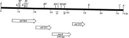
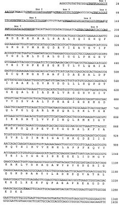
Abstract. The recA gene of Xanthomonas campestris pathovar citri (X. c. pv. citri) was cloned and sequenced. Nucleotide sequence analysis revealed an open reading frame of 1,032 bp that encodes a 344-amino acid protein and which shows a high degree of homology to recA genes from other bacteria. The cloned recA gene of X. c. pv. citri restored to a recA deletion mutant of Escherichia coli the ability to resist killing induced by methylmethane sulfonate or ultraviolet radiation, indicating that the cloned gene was functional in E. coli. A recA mutant of X. c. pv. citri was constructed by gene replacement and was shown both not to possess detectable DNA recombination activity and to be markedly more sensitive to DNA-damaging agents than is wild-type X. c. pv. citri. Transformation of recA mutants of E. coli and X. c. pv. citri with a plasmid containing X. c. pv. citri recA conferred the ability to produce a 37-kDa protein that reacted with antiserum to E. coli RecA protein. Two additional open reading frames were also identified in the recA region of the X. c. pv. citri genome: One located immediately upstream from recA that encodes a 213-amino acid protein with a high degree of similarity to the LexA protein of E. coli, and another located immediately downstream of recA that encodes a 153-amino acid protein with sequence similarity to the RecX protein of Pseudomonas aeruginosa.

Keywords: Complementation; DNA repair; Homologous recombination; RecA.

Abbreviations: X. c. pv. citri, Xanthomonas campestris pathovar citri; LB, Luria-Bertani; PCR, polymerase chain reaction; UV, ultraviolet; CFU, colony-forming unit; MMS, methylmethane sulfonate; ORF, open reading frame; Kmr, kanamycin-resistance gene; RF, replicative form.

Introduction

Xanthomonas campestris pv. citri, a Gram-negative, non-spore-forming bacterium, and one of approximately 140 pathovars of X. campestris (Verniere et al., 1993), causes citrus canker disease and thereby imposes an enormous economic burden worldwide. To date, only one gene, referred to as pthA, has been associated with the virulence of this organism (Lawson et al., 1989; Swarup et al., 1991, 1992); additional virulence genes remain to be identified. However, molecular studies of X. c. pv. citri have been hampered by the lack of recombination-deficient mutants. Thus, it has been difficult to maintain recombinant plasmids in X. c. pv. citri without the occurrence of deletions or rearrangements in the plasmid, which are likely the result of homologous recombination.

The RecA protein plays a major role in homologous recombination (Camerini-Otero and Hsieh, 1995; Clark, 1973; Cox, 1991; Kowalczykowski et al., 1994; Ogawa and Ogawa, 1986; Roca and Cox, 1990). More than 60 recA genes have been isolated and characterized from a wide variety of organisms, including X. c. pv. oryzae and X. c. pv. campestris (Karlin and Brocchieri, 1996; Lee et al., 1996;

Lloyd and Sharp, 1993; Miller and Kokjohn, 1990; Rabibhadana et al., 1993). We have now cloned, sequenced, and characterized the recA gene of X. c. pv. citri as well as constructed a recA mutant of this organism.

Materials and Methods

Bacterial Strains and Plasmids

The bacterial strains and plasmids used in this study are listed in Table 1. Both X. campestris and E. coli strains were grown in Luria-Bertani (LB) medium at 28°C and 37°C, respectively. Chloramphenicol, kanamycin, and gentamycin were added to LB medium at final concentrations of 30, 50, and 15 µg/ml, respectively.

DNA Manipulation and Hybridization

Plasmid DNA was isolated from bacterial cultures by the method of Birnboim and Doly (1979), and was further purified by ethidium bromide-CsCl density gradient centrifugation. Chromosomal DNA was isolated from X. c. pv. citri as described by Pitcher et al. (1989). Restriction enzyme digestions and ligation were carried out according to standard methods described by Sambrook et al. (1989). The competent X. c. pv. ciri cells were prepared and subjected to electroporation by the method of Yang

1Corresponding author. Tel: +886-2-29031111 ext. 2462; Fax: +886-2-29021124; E-mail: bio1004@mails.fju.edu.tw


Botanical Bulletin of Academia Sinica, Vol. 41, 2000

Table 1. Bacterial strains and plasmids.

Strain or plasmid Relevant genotype or characteristics Source or reference

Strain

Escherichia coli

JC10287 AB1157 derivative; D(srl-recA) 304 Czonka and Clark, 1979

AB1157 recA+ strain Bachmann, 1971

X. campestris pv. citri

XW47 Virulent citrus canker type strain Wu et al., 1985

XCK75 recA mutant derived from XW47 This study

Plasmid

pBCSK+ Phagemid derived from pUC19; used as cloning vector Stratagene

pUC4-KIXX E. coli gene cartridge vector Pharmacia

PUCGM Derivative of pUC19, Gmr Schweizer, 1993

pHC8 Derivative of pRK415, oriV trfA Tcr Chen, 1994

pUC4G Plasmid pUC4-KIXX with Kmr replaced by Gmr This study

pXC560 pBCSK+ with an inserted 560-bp PCR fragment containing This study

a portion of X. c. pv. citri recA

pAP74 pBCSK+ with an inserted 7.4-kb ApaI fragment containing This study

X. c. pv. citri recA

pAP74K pAP74 with the recA gene interrupted by Kmr This study

pEG40 pHC8 with an inserted 4.0-kb EagI fragment containing This study

X. c. pv. citri recA

pCF4G pUC4G containing the 1.5-kb NlaIV fragment from This study

RF DNA of bacteriophage cf

Gmr and Tcr, gentamycin- and tetracyclin-resistance genes, respectively.

et al. (1991). Southern blot analysis was also performed as described previously (Southern, 1975; Yang and Yang, 1997). DNA probes were labeled with digoxigenin by random priming (Feinberg and Vogelstein, 1983) with the use of a DIG DNA labeling kit (Boehringer Mannheim). Colony hybridization was performed as described (Grunstein and Hogness, 1975).

PCR Amplification

Comparison of the nucleotide sequences of recA from various bacterial species revealed several highly conserved regions. Two oligonucleotides based on these conserved sequences were synthesized and used as primers for polymerase chain reaction (PCR) amplification of a portion of the recA gene of X. c. pv. citri. The forward primer (primer 1, 5'-CGGAATTCTCGGGCAAGACCACC-3') and reverse primer (primer 2, 5'-TAGCAAGCTTGTTCTT GACCACCTT-3') correspond to amino acids 67 to 73 (Asp-Ser-Ser-Gly-Lys-Thr-Thr) and 246 to 251 (Lys-Val-Val-Lys-Asn-Lys), respectively, of the RecA protein of E. coli; EcoRI and HindIII restriction sites were incorporated at the 5' ends of primers 1 and 2, respectively, to simplify cloning. PCR was performed in a total volume of 100 µl containing 100 ng of X. c. pv. citri genomic DNA, PCR buffer, 2.5 U of Taq DNA polymerase, 2.5 mM MgCl2, 0.2 mM of each deoxynucleoside triphosphate, and 20 pmol of each primer for 35 cycles of 94°C for 1 min, 60°C for 1.5 min, and 72°C for 2 min.

DNA Sequencing and Nucleotide Sequence Analysis

DNA sequences were determined by the dideoxy chain termination method (Sanger et al., 1977) with Pfu DNA polymerase (Stratagene, La Jolla, CA). Computer analysis of nucleotide and amino acid sequences was performed with the PC/GENE package version 6.85 (IntelliGenetics, Mountain View, CA).

Assay of Cell Survival After UV Irradiation or MMS Treatment

Sensitivity of bacteria to ultraviolet (UV) irradiation was determined by exposing 10 ml of a mid-log phase culture (optical density at 600 nm, 0.5) in a 100-mm petri dish to a GE germicidal lamp (256 nm) for 10 to 40 s at a distance of 15 cm. The UV dose was determined in each experiment with a UV radiometer (model VLX-254; Vilber Lourmat, Torcy, France). After irradiation, the bacterial cells were serially diluted, and 100 µl of each dilution were spread onto LB agar plates containing appropriate antibiotics. A sample of nonirradiated cells was also diluted and plated. After incubation for 24 h in the dark, the colonies on each plate were counted to determine the number of colony-forming units (CFU) per milliliter of sample. The survival rate at each UV dose was calculated by dividing the CFU/ml value of the irradiated sample by that of the non-irradiated sample.


Yang and Yang — Xanthomonas campestris pv. citri recA gene

digestion with EcoRI and HindIII, this fragment was cloned into the plasmid pBCSK+, thereby generating pXC560. The nucleotide sequence of the entire X. c. pv. citri fragment was determined and compared with that of the E. coli recA gene (Sancar et al., 1980). The two sequences shared 67% identity, suggesting that the 560-bp fragment corresponded to a portion of X. c. pv. citri recA.

The 560-bp fragment was then labeled with digoxigenin and used as a probe to identify a DNA molecule containing the entire recA gene of X. c. pv. citri. Genomic DNA from X. c. pv. citri was digested separately with each of the restriction enzymes ApaI, BamHI, ClaI, EcoRI, and PvuI, and the resulting fragments were subjected to electrophoresis on a 1% agarose gel and Southern blot analysis with the probe. An ApaI fragment of ~7 kb hybridized with the probe. Therefore, ApaI fragments of X. c. pv. citri genomic DNA of ~6 to 9 kb were eluated and cloned into pBCSK+, and the colonies corresponding to the resulting partial genomic library were screened with the digoxigenin-labeled 560-bp fragment as probe. Of ~1000 colonies examined, four reacted with the probe. Restriction enzyme analysis revealed that all four positive colonies harbored a 7.4-kb ApaI fragment. A partial restriction map of the insert of the corresponding pAP74 plasmid is shown in Figure 1.

Sensitivity to methylmethane sulfonate (MMS) was determined by plating bacterial cells directly onto LB agar containing various concentrations (0.6 to 3.6 mM) of MMS and incubating the cells overnight. The survival rate was calculated in the same manner as was that for UV sensitivity.

Detection of RecA Protein

Cell lysates prepared from X. c. pv. citri or E. coli were fractionated by SDS-polyacrylamide gel electrophoresis, and the separated proteins were transferred to a nitrocellulose membrane and subjected to immunoblot analysis with polyclonal antibodies to the E. coli RecA protein, as described previously (Towbin et al., 1979; Yang and Yang, 1997). These antibodies have been shown to be specific to the RecA protein (Lee et al., 1996).

Results

Cloning and Identification of the recA Gene of X. campestris pv. citri

With two oligonucleotide primers (primers 1 and 2) based on conserved sequences of recA genes from various bacterial species, a 560-bp DNA fragment was amplified from the X. c. pv. citri genome by PCR. After

Figure 1. Partial restriction map of, and the locations of open reading frames in, the recA region of the X. campestris pv. citri genome. The positions of primer 1 (p1) and primer 2 (p2) used for PCR are indicated with half-arrows. Abbreviations for restriction enzyme sites: A, ApaI; C, ClaI; E, EagI; RI, EcoRI; and RV, EcoRV. aa, amino acid.

Table 2. Percentage identity of nucleotide and predicted amino acid sequences of recA from various bacteria.

Nucleotide and amino acid sequence identity (%)a

Organismb X. c. pv. X. c. pv. X. o. pv. P. aeruginosa E. coli L. pneumophila V. anguillarum E. carotovora citri campestris oryzae

X. c. pv. citri 100 91 92 75 70 65 66 63

X. c. pv. campestris 97 100 91 76 69 65 64 61

X. o. pv. oryzae 97 97 100 77 71 64 65 60

P. aeruginosa 70 70 70 100 73 64 65 62

E. coli 67 70 67 71 100 67 75 76

L. pneumophila 72 76 72 76 73 100 69 69

V. anguillarum 68 71 69 72 83 71 100 75

E. carotovora 67 69 67 73 89 72 83 100

aNucleotide and amino acid sequence comparisons are shown above and below the diagonal, respectively.

bSequences of recA genes of the following organisms were compared: X. c. pv. citri (this study), X. c. pv. campestris (Lee et al., 1996), X. o. pv. oryzae Rabibhadana et al., 1998), P. aeruginosa (Kokjohn and Miller, 1985; Sano and Kageyama, 1987), E. coli (Horii et al., 1980; Sancar et al., 1980), L. pneumophila (Dreyfus, 1989), V. anguillarum (Singer, 1989), and E. carotovora (Keener et al., 1984).


Botanical Bulletin of Academia Sinica, Vol. 41, 2000

DNA Sequence Analysis of the recA Gene of X. campestris pv. citri

The entire nucleotide sequence of the 7.4-kb ApaI fragment was determined. Sequence analysis revealed the presence of four open reading frames (ORFs) in the same translational orientation (Figure 1). The ORF designated orf344 contains the sequence of the 560-bp fragment that was initially cloned. A database search for nucleotide and amino acid sequence similarities between orf344 and other known genes revealed that orf344 shows a high level of homology to recA genes of various bacteria, including X. c. pv. campestris, X. oryzae pv. oryzae, Pseudomonas aeruginosa, E. coli, Legionella pneumophila, Vibrio

anguillarum, and Erwinia carotovora (Table 2). The greatest identity at both the nucleotide (92%) and amino acid (97%) levels was apparent with recA of X. o. pv. oryzae. These results suggest that orf344 encodes the RecA protein of X. c. pv. citri.

The ORF (orf213) located immediately upstream of orf344 shares 76 to 82% sequence identity at the amino acid level with the lexA genes of E. coli, P. aeruginosa, Pseudomonas putida, Salmonella typhimurium, and E. carotovora (Garriga et al., 1992; Horii et al., 1981). The ORF (orf153) located immediately downstream of orf344 shows 84% sequence identity at the amino acid level to the recX gene of P. aeruginosa (De Mot et al., 1994; Sano, 1993). The fourth ORF (orf564), which encodes a protein of 564 amino acids, shows no sequence homology to previously determined sequences in GenBank.

A putative Shine-Dalgarno sequence, GAGGA, is located 9 bp upstream from the putative ATG translational initiation codon of the X. c. pv. citri recA gene (Figure 2). No sequences similar to those of E. coli promoters were identified in the region immediately upstream of this gene. However, eight CTGN8_12CCG sequences, resembling those of the SOS box of DNA damage-inducible promoters of E. coli and other Gram-negative bacteria, are present within 200 bp upstream from the putative initiation codon (Figure 2).

Functional Analysis of the X. campestris pv. citri recA Gene

To determine whether the cloned recA gene of X. c. pv. citri was functional, we performed complementation assays. The pAP74 plasmid was introduced into a recA deletion mutant (JC10287) of E. coli (Czonka and Clark, 1979), and the resulting cells were assayed for their sensitivity to MMS treatment and UV irradiation. JC10287 cells containing the vector pBCSK+ were assayed as a negative control, and E. coli strain AB1157, which contains wild-type recA, was used as a positive control. JC10287 cells containing pBCSK+ were highly sensitive to MMS (Figure 3A). However, at 0.6 mM MMS, the survival rate of JC10287 cells containing pAP74 was five orders of magnitude greater than that of JC10287 cells containing pBCSK+. At all concentrations of MMS tested (0.6 to 3.6 mM), the survival rate of AB1157 cells was 10 to 100 times that of JC10287 cells containing pAP74.

A similar pattern of survival rates was apparent for the three types of cells after UV irradiation (Figure 3B). Thus, at the lowest UV dose (20 J/m2), the survival rate of JC10287 cells containing pAP74 was about three orders of magnitude greater than that of JC10287 cells containing pBCSK+; no survivors were detected after exposure of the latter cells to UV doses of >20 J/m2. At the highest dose (80 J/m2), the survival rate of JC10287 cells containing pAP74 was ~1 × 10⁻³; although this survival rate is 2.5 log units below that of AB1157 cells, it still suggests that the X. c. pv. citri recA gene is functional to a certain extent in E. coli.

Figure 2. Nucleotide and deduced amino acid sequences of the recA gene of X. campestris pv. citri. The putative Shine-Dalgarno sequence (GAGGA) as well as the translation initiation (ATG) and termination (TAA) codons are indicated by bold type. The putative SOS boxes (CTGN8_12CCG) are underlined, with the inverted repeat sequences indicated by bold letters. Nucleotide numbers are shown on the right. The GenBank accession number of the recA sequence is AF006590.


Yang and Yang — Xanthomonas campestris pv. citri recA gene

Six kanamycin-resistant, chloramphenicol-sensitive colonies were picked and examined for the presence of an integrated Kmr gene. Chromosomal DNA was extracted from these cells and subjected to PCR using primers corresponding to both the N- and C- terminal sequence of RecA protein for characterization of recA. A PCR product of 1.3 -kb would indicate the presence of the wild-type recA gene, whereas a 2.5-kb product would suggest integration of Kmr into the chromosomal recA gene. All six colonies examined yielded a 2.5-kb PCR product, which was further shown to react with both recA (1.4-kb BglII fragment) and Kmr (1.2-kb ClaI fragment) probes on Southern hybridization analysis (data not shown). These results suggest that the six colonies comprise recA mutants of X. c. pv. citri.

One of these six potential recA mutants of X. c. pv. citri, designated XCK75, was further studied for its sensitivity to the DNA-damaging agents MMS and UV. At an MMS concentration of 1.8 mM, the survival rate of XCK75 cells was about six orders of magnitude smaller than that of the wild-type strain XW47 (Figure 3C). Similarly, at a UV dose of 200 J/m2, the survival rate of XCK75 cells was four orders of magnitude smaller than that of XW47 cells (Figure 3D). We then examined whether the recA mutation in XCK75 could be complemented by the cloned X. c. pv. citri recA gene. For its introduction into XCK75, the X. c. pv. citri recA gene was first cloned into the E. coli-X. campestris shuttle vector pHC8 (Chen, 1994). A 4.0-kb EagI fragment, containing the 3' half of orf564 and all of orf213, orf344 (recA), and orf153 (Figure 1), was isolated from pAP74. The 5' overhangs of the fragment were converted to blunt ends with the Klenow enzyme, and the product was inserted into the unique XbaI site located among the multiple cloning sites of pHC8, thereby generating pEG40. This plasmid was then introduced into XCK75 by electroporation, and cells containing pEG40 were assayed for sensitivity to MMS and UV. After exposure to these agents, the survival rates of XCK75 cells harboring pEG40 were virtually identical to those of the XW47 wild-type strain.

We then examined XCK75 for a defect in homologous recombination, given that such defects are a prominent characteristic of recA mutants. The plasmid pCF4G, which contains the protein A gene of the X. c. pv. citri bacteriophage Cf, was introduced together with the replicative form (RF) of Cf DNA into XCK75 by electroporation. The frequency of homologous recombination between the protein A genes located on pCF4G and the RF DNA of Cf was then determined. In control experiments, both pCF4G and RF DNA of Cf were also introduced into the X. c. pv. citri wild-type strain XW47 and into XCK75 containing pEG40. The pCF4G plasmid was constructed by inserting the 1.5-kb NlaIV fragment containing the entire protein A gene of phage Cf into the unique HincII site of pUC4G, which itself was generated by replacing the 1.6-kb SmaI fragment containing the Kmr gene of pUC4-KIXX with the 0.85-kb SacI fragment containing the gentamycin-resistance gene of pUCGM (Schweizer, 1993). Because pCF4G cannot replicate autonomously in X. c. pv. citri, the only

Figure 3. Sensitivity of E. coli and X. campestris pv. citri strains to MMS and UV. Escherichia coli strains AB1157 (recA wild type), JC10287 (recA deletion mutant) harboring pBCSK+, and JC10287 harboring pAP74 (which contains the recA gene of X. c. pv. citri) were assayed for their sensitivities to MMS treatment (A) and UV irradiation (B). The sensitivities of the wild-type strain XW47 and recA mutant XCK75 of X. c. pv. citri to MMS (C) and UV (D) were also determined; complementation of the MMS and UV sensitivities of XCK75 by the cloned recA gene of X. c. pv. citri was examined by transformation with pEG40 (pAP74 cannot replicate in X. c. pv. citri). Data are expressed as the log of percentage survival values and are means of three independent experiments.

Construction and Characterization of a recA Mutant of X. campestris pv. citri

We subjected X. c. pv. citri to targeted homologous recombination in order to generate recA mutants. A 1.2-kb ClaI DNA fragment containing the kanamycin-resistance gene (Kmr) was isolated from pUC4-KIXX and then inserted into the internal ClaI site (Figure 2, nucleotide position 482) of the X. c. pv. citri recA gene in pAP74, thereby generating pAP74K. The latter plasmid was then introduced into the wild-type X. c. pv. citri strain XW47 by electroporation, and kanamycin-resistant cells were selected. Because pAP74K cannot replicate in X. c. pv. citri, the Kmr gene should have been integrated by a single or double crossover into the chromosome of resistant cells. A single crossover, occurring between the recA gene of pAP74K and that on the chromosome, might be expected to result in insertional inactivation of the chromosomal gene and integration of the entire plasmid, including its chloramphenicol-resistance gene. In contrast, a double crossover, occurring at the recA sequences flanking Kmr, may result in interruption of chromosomal recA by Kmr; the resulting mutants would therefore be kanamycin resistant but chloramphenicol sensitive.


Botanical Bulletin of Academia Sinica, Vol. 41, 2000

Figure 4. Expression of the cloned recA gene of X. campestris pv. citri in E. coli and X. campestris pv. citri. Cell lysates (30 µg of protein) of various E. coli or X. c. pv. citri strains were fractionated by SDS-polyacrylamide gel electrophoresis on a 12.5% gel and subjected to immunoblot analysis with antibodies to E. coli RecA protein. Samples were loaded as follows: lane 1, E. coli AB1157 (recAwild type); lane 2, E. coli JC10287 (recA deletion mutant); lane 3, JC10287 containing pAP74; lane 4, X. c. pv. citri wild-type strain XW47; lane 5, X. c. pv. citri XW47 treated for 2 h with mitomycin C (100 ng/ml); lane 6, X. c. pv. citri recA mutant XCK75; lane 7, X. c. pv. citri XCK75 treated with mitomycin C (100 ng/ml); lane 8, X. c. pv. citri XCK75 containing pEG40; lane 9, X. c. pv. citri XCK75 containing pEG40 and treated with mitomycin C (100 ng/ml). The positions of the 39-kDa E. coli RecA and the 37-kDa X. c. pv. citri RecA proteins are indicated.

cells that can grow in the presence of gentamycin are those in which Cf and pCF4G have cointegrated. Such cointegration would result from homologous recombination between the two protein A genes and replication by the phage replication machinery.

We plated 1 × 10⁸ CFU (in 100 µl) of the various transformants and determined the number of gentamycin-resistant colonies. No gentamycin-resistant colonies were observed when pCF4G alone was introduced into XW47 or XCK75, confirming the inability of this plasmid to replicate in these two hosts. When both Cf DNA and pCF4G were introduced into XW47 or XCK75, only XW47 cells produced colonies (1.0 × 10³ CFU/ml) that were resistant to gentamycin, suggesting that homologous recombination between the protein A genes in pCF4G and in Cf DNA did not take place in XCK75. A similar number (4.0 × 10³ CFU/ml) of gentamycin-resistant colonies was observed with XCK75 containing pEG40, indicating that the defect in homologous recombination in XCK75 was corrected by the X. c. pv. citri recA gene in pEG40.

Expression of the X. campestris pv. citri recA Gene

The expression of the X. c . pv. citri recA gene in XCK75 cells containing pEG40 was examined by immunoblot analysis with antibodies to the E. coli RecA protein. Expression of the X. c. pv. citri recA gene was also examined in the recA deletion mutant of E. coli (JC10287) containing pAP74. Wild-type E. coli and X. c. pv. citri strains were also assayed as controls. A 39-kDa immunoreactive protein was apparent in the lysate of the wild-type E. coli strain AB1157 (Figure 4, lane 1) but not in that of the recA deletion mutant JC10287 (lane 2). An immunoreactive band (37-kDa, based on the deduced amino acid sequence) that migrated slightly faster than did the E. coli RecA protein was detected in the lysate of JC10287 cells containing pAP74 (lane 3) as well as in that of the wild-type X. c. pv. citri strain XW47 (lane 4). The intensity of this band was increased in the lysate prepared from XW47 cells treated with mitomycin C (lane 5). No such band was detected in lysates of either nontreated (lane 6)

or mitomycin C-treated (lane 7) XCK75 cells, indicating that the recA gene in XCK75 was indeed completely inactivated. The 37-kDa band was apparent in the lysate of XCK75 cells containing pEG40 (lane 8), indicating that the X. c . pv. citri recA gene on the plasmid was expressed. The intensity of the 37-kDa band was not markedly affected by treatment of XCK75 cells containing pEG40 with mitomycin C (lane 9).

Discussion

The aims of this study were to clone the recA gene and to construct a recA mutant of X. c. pv. citri. A 7.4-kb ApaI fragment that contains the recA gene of X. c. pv. citri was isolated, and the gene was shown to encode a protein of 344 amino acids that shares a high degree of homology with RecA proteins of other bacterial species (97 and 67% sequence identity with RecA of X. o. pv. oryzae and E. coli, respectively). The cloned X. c. pv. citri recA gene partially restored the resistance of a recA deletion mutant of E. coli to treatment with MMS or UV radiation, confirming that the cloned gene is indeed X. c. pv. citri recA. The incomplete complementation of the E. coli recA deletion by the X. c. pv. citri recA gene might be due to a low effeciency of expression of the cloned gene in E. coli, or to reduced activity of the recombinant protein in E. coli relative to that of the native E. coli RecA protein.

The isolation of the recA gene of X. c. pv. citri enabled us to construct a recA mutant of this pathovar by homologous recombination. A Kmr gene was inserted into the cloned recA gene of X. c. pv. citri, and the resulting construct was introduced into X. c. pv. citri in order to replace the wild-type recA gene. The resulting recA mutant, designated XCK75, was shown by immunoblot analysis to have lost the ability to produce RecA protein. It also was highly sensitive to MMS and UV irradiation, suggesting that DNA repair function was impaired, and was incapable of mediating homologous recombination.

Eight CTGN8_12CCG sequences that are similar to the SOS box of DNA damage-inducible promoters of E. coli genes were detected in the promoter region of X. c. pv.


Yang and Yang — Xanthomonas campestris pv. citri recA gene

citri recA. The consensus sequence of the SOS box of E. coli is CTGTN8ACAG (Wertman and Mount, 1985). Although not identical to the SOS box of E. coli, the sequence CTGN8_12CCG may constitute the SOS box of X. c. pv. citri. The difference in sequence may reflect the evolutionary divergence of the two species. The SOS box is thought to function as an operator to which the LexA protein binds and thereby suppresses the expression of SOS genes (Walker, 1984). A gene homologous to E. coli lexA was detected upstream of recA in X. c. pv. citri. Furthermore, a gene similar to recX of P. aeruginosa was shown to be present downstream of recA in X. c. pv. citri. The LexA protein of E. coli is a transcriptional repressor of recA (Little and Mount, 1982; Wertman and Mount, 1985), and the RecX protein regulates the expression of recA in P. aeruginosa (Sano, 1993). Whether the products of the lexA and recX genes of X. c. pv. citri perform similar functions remains to be determined. The enhancement of recA expression in X. c. pv. citri by mitomycin C treatment suggests that X. c. pv. citri responds to DNA damage in a manner similar to that of E. coli and many other bacteria.

XCK75 is the first recA mutant of X. c. pv. citri to be constructed. It is also the first well-characterized recA mutant of any pathovar of X. campestris. Although recA mutants of X. c. pv. campestris (Lee et al., 1996) and X. c. pv. oryzae (Rabibhadana et al., 1993) have been constructed, characterization of these mutants has not been described. The availability of a recA mutant of X. c. pv. citri will allow more extensive molecular genetic studies of this organism. We have shown that XCK75 is completely defective in homologous recombination, a characteristic that will allow it to be used as a host for recombinant plasmids that contain portions of the X. c. pv. citri genome.

Acknowledgments. We thank Chao-Hung Lee (Indiana University) for valuable comments and critical editing of the manuscript; Andrei Kuzminov (University of Oregon) for providing E. coli strains JC10287 and AB1157; and Shiau-Ting Hu (Yang-Ming University) for supplying antiserum to E. coli RecA protein. This research was supported by a grant (NSC85-2311-B-030-001 B18) from the National Science Council of Taiwan.

Literature Cited

Bachmann, B.J. 1971. Pedigrees of some mutant strains of Escherichia coli K-12. Bacteriol. Rev. 36: 525-557.

Birnboim, H.C. and J. Doly. 1979. A rapid alkaline extraction procedure for screening recombinant plasmid DNA. Nucleic Acids Res. 7: 1513-1523.

Camerini-Otero, R.D. and P. Hsieh. 1995. Homologous recombination protein in prokaryotes and eukaryotes. Annu. Rev. Genet. 29: 509-552.

Chen, S.C. 1994. Construction of Broad-Host-Range Promoter-proving Vector and Analysis of Promoter in Xanthomonas campestris pv. campestris. Masters thesis. Institute of Molecular Biology, National Chung Hsing University, Taichung, Taiwan.

Clark, A.J. 1973. Recombination deficient mutants of E. coli and other bacteria. Annu. Rev. Genet. 7: 67-86.

Cox, M.M. 1991. The RecA protein as a recombinational repair system. Mol. Microbiol. 5: 1295-1299.

Czonka, L.N. and A.J. Clark. 1979. Deletions generated by the transposon Tn10 in the srl-recA region of the Escherichia coli K-12 chromosome. Genetics 93: 321-343.

De Mot, R., G. Schoofs, and J. Vanderleyden. 1994. A putative regulatory gene downstream of recA is conserved in Gram-negative and Gram-positive bacteria. Nucleic Acids Res. 22: 1313-1314.

Dreyfus, L.A. 1989. Molecular cloning and expression in Escherichia coli of the recA gene of Legionella pneumophila. J. Gen. Microbiol. 135: 3097-3107.

Feinberg, A.P. and B. Vogelstein. 1983. A technique for radiolabelling DNA restriction endonuclease fragments to high specific activity. Anal. Biochem. 132: 6-13.

Garriga, X., S. Calero, and J. Barbe. 1992. Nucleotide sequence analysis and comparison of the lexA genes from Salmonella typhimurium, Erwinia carotovora, Pseudomonas aeruginosa and Pseudomonas putida. Mol. Gen. Genet. 236: 125-134.

Grunstein, M. and D.S. Hogness. 1975. Colony hybridization: a method for the isolation of cloned DNAs that contain a specific gene. Proc. Natl. Acad. Sci. USA 72: 3961-3965.

Horii, T., T. Ogawa, and H. Ogawa. 1980. Organization of the recA gene of Escherichia coli. Proc. Natl. Acad. Sci. USA 77: 313-317.

Horii, T., T. Ogawa, and H. Ogawa. 1981. Nucleotide sequence of the lexA gene of E. coli. Cell 23: 689-697.

Karlin, S. and L. Brocchieri. 1996. Evolutionary conservation of recA genes in relation to protein structure and function. J. Bacteriol. 178: 1881-1894.

Keener, S.L., K.P. McNamee, and K. McEntee. 1984. Cloning and characterization of recA genes from Proteus vulgaris, Erwinia carotovora, Shigella flexneri and Escherichia coli Br. J. Bacteriol. 160: 153-160.

Kokjohn, T.A. and R.V. Miller. 1985. Molecular cloning and characterization of the recA gene of Pseudomonas aeruginosa PAO. J. Bacteriol. 163: 568-572.

Kowalczykowski, S.C., D.A. Dixon, A.K. Eggleston, S.D. Lauder, and W.M. Rehrauer. 1994. Biochemistry of homologous recombination in Escherichia coli. Microbiol. Rev. 58: 401-465.

Lawson, R.H., M.M. Dienelt, and E.L. Civerolo. 1989. Histopathology of Xanthomonas campestris pv. citri from Florida and Mexico in wound-inoculated detached leaves of Citrus aurantifolia: light and scanning electron microscopy. Phytopathology 79: 329-335.

Lee, T.C., N.T. Lin, and Y.H. Tseng. 1996. Isolation and characterization of the recA gene of Xanthomonas campestris pv. campestris. Biochem. Biophys. Res. Commun. 221: 459-465.

Little, J.W. and D.W. Mount. 1982. The SOS regulatory system of Escherichia coli. Cell 29: 11-22.

Lloyd, A.T. and P.M. Sharp. 1993. Evolution of the recA gene and the molecular phylogeny of bacteria. J. Mol. Evol. 37: 399-407.

Miller, R.V. and T.A. Kokjohn. 1990. General microbiology of recA: environmental and evolutionary significance. Annu.


Botanical Bulletin of Academia Sinica, Vol. 41, 2000

Rev. Microbiol. 44: 365-394.

Ogawa, H. and T. Ogawa. 1986. General recombination: functions and structure of RecA protein. Adv. Biophys. 21: 135-148.

Pitcher, D.C., N.A. Saunder, and R.J. Owen. 1989. Rapid extraction of bacterial genomic DNA with guanidium thiocyanate. Lett. Appl. Microbiol. 8: 151-156.

Rabibhadana, S., S. Chamnongpol, J.E. Trempy, N.P. Ambulos, and S. Mongkolsuk, 1993. Isolation and expression in Escherichia coli of a Xanthomonas oryzae recA-like gene. Gene 213: 113-118.

Rabibhadana, S., S. Chamnongpol, R. Sukchawalit, J.E. Trempy, N.P. Ambulos, and S. Mongkolsuk. 1998. Characterization and expression analysis of Xanthomonas oryzae pv. oryzae recA. FEMS Microbiol. Lett. 158: 195-200.

Roca, A.I. and M.M. Cox. 1990. The RecA protein: structure and function. Crit. Rev. Biochem. Mol. Biol. 25: 415-456.

Sambrook, J., E. Fritsch, and T. Maniatis. 1989. Molecular cloning: a laboratory manual. 2nd ed. Cold Spring Harbor Laboratory Press, Cold Spring Harbor, NY.

Sancar, A., C. Stachelek, W. Konigsberg, and W.D. Rupp. 1980. Sequences of the recA gene and protein. Proc. Natl. Acad. Sci. USA 77: 2611-2615.

Sanger, F., S. Nicklen, and A.R. Coulson. 1977. DNA sequencing with chain-terminating inhibitors. Proc. Natl. Acad. Sci. USA 74: 5463-5467.

Sano, Y. and M. Kageyama. 1987. The sequence and function of the recA gene and its protein in Pseudomonas aeruginosa PAO. Mol. Gen. Genet. 208: 412-419.

Sano, Y. 1993. Role of the recA-related gene adjacent to the recA gene in Pseudomonas aeruginosa. J. Bacteriol. 175: 2451-2454.

Schweizer, H.P. 1993. Small broad-host-range gentamycin resistance gene casettes for site-specific insertion and deletion mutagenesis. BioTechniques 15: 831-833.

Singer, J.T. 1989. Molecular cloning of the recA analogs from the marine fish pathogen Vibrio anguillarum 775. J. Bacteriol. 171: 6367-6371.

Southern, E.M. 1975. Detection of a specific sequence among DNA fragments separated by gel electrophoresis. J. Mol. Biol. 98: 503-517.

Swarup, S., R. De Feyter, R.H. Brlansky, and D.W. Garbriel. 1991. A pathogenicity locus from Xanthomonas citri enables strains from several pathovars of X. campestris to elicit cankerlike lesions on citrus. Phytopathology 81: 802-809.

Swarup, S., Y. Yang, M.T. Kingsley, and D.W. Gabriel. 1992. An Xanthomonas citri pathogenicity gene, pthA, pleiotropically encodes gratuitous avirulence on nonhosts. Mol. Plant-Microbe Interact. 5: 204-213.

Towbin, H., T. Staehelin, and J. Gordon. 1979. Electrophoretic transfer of proteins from polyacrylamide gels to nitrocellulose sheets: procedure and some applications. Proc. Natl. Acad. Sci. USA 76: 4350-4354.

Verniere, C., O. Pruvost, E.L. Civerolo, O. Gambin, J.P. Jacquemoud-Collet, and J. Luisetti. 1993. Evaluation of the biological substrate utilization system to identify and assess metabolic variation among strains of Xanthomonas campestris pv. citri. Appl. Environ. Microbiol. 59: 243-249.

Walker, G.C. 1984. Mutagenesis and inducible responses to deoxyribonucleic acid damage in Escherichia coli. Microbiol. Rev. 48: 60-93.

Wertman, K.F. and D.W. Mount. 1985. Nucleotide sequence binding specificity of the LexA repressor of Escherichia coli K-12. J. Bacteriol. 163: 376-384.

Wu, W.C., Y.K. Hong, C.M. Huang, Y.Y. Ten, T. Lee, S.F. Chang, C.H. Wang, and M.H. Huang. 1985. Phage induced mutations in Xanthomonas campestris pv. citri. Plant Protect. Bull. (Taipei) 27: 19-33.

Yang, M.K., W.C. Su, and T.T. Kuo. 1991. Highly efficient transfection of Xanthomonas campestris by electroporation. Bot. Bull. Acad. Sin. 32: 197-203.

Yang, M.K. and Y.C. Yang. 1997. The A protein of the filamentous bacteriophage Cf of Xanthomonas campestris pv. citri. J. Bacteriol. 179: 2840-2844.


Yang and Yang — Xanthomonas campestris pv. citri recA gene

¬a¾ï¼ìºÅ¯fµß recA ¬ðÅܮ褧«Ø¥ß»P¯S©Ê¤ÀªR

·¨·½©÷ ·¨¬ü®Û

¨p¥ß»²¤¯¤j¾Ç¥Íª«¾Ç¨t

¦Û¬a¾ï¼ìºÅ¯fµß (Xanthomonas campestris pv. citri) ©Ò¿ï´Þ¤§ recA °ò¦]¡A¬O¥Ñ 1032 ­Ó®Ö¥Ì»Ä©Ò²Õ ¦¨¡A¥i¦X¦¨§t 344 ­Ó®ò°ò»Ä¤§³J¥Õ½è¡C¨ä§Ç¦C»P¨ä¥L²Óµß¤§ recA °ò¦]«ÜÃþ¦ü¡A¦X¦¨¤§³J¥Õ¦b¤j¸z±ìµß¤º ¥ç¨ã¦³¥\¯à¡C§Q¥Î´¡¤J©Ê¬ðÅܪk¡A±o¨ì¤§ recA ¬ðÅÜ®è¡A¹ï DNA ¬ðÅܾ¯·¥¬°±Ó·P¡A¤]¥¢¥h°ò¦]­«²Õªº¯à ¤O¡C¿ï´Þ¤§ recA °ò¦]¡A¥i§Î¦¨¤@ 37 kDa ¤§³J¥Õ¡A¥i»P§Ü¤j¸z±ìµß RecA ¤§§ÜÅé§@¥Î¡C¥t¥~¡A¦b recA °ò¦]¤§¤W´å¡AÁÙµo²{¤@¥i²£¥Í 213 ®ò°ò»Ä¤§³J¥Õªº°ò¦]¡A¬Û¦ü©ó¤j¸z±ìµß¤§ lexA °ò¦]¡F¦Ó¦b¨ä¤U´å¡AÁÙ¦³ ¤@¥Ñ 153 ®ò°ò»Ä©Ò²Õ¦¨¤§³J¥Õ¤§°ò¦]¡A¬Û·í©óºñÁw±ìµß¤§ recX °ò¦]¡C

ÃöÁäµü¡G °ò¦]­«²Õ¡F RecA¡F¤¬¸É¡FDNA ­×¸É¡C