Bot. Bull. Acad. Sin. (2000) 41: 165-174

Wu — New Phanerochaete from Taiwan

Six new species of Phanerochaete from Taiwan

Sheng-Hua Wu1

Department of Botany, National Museum of Natural Science, Taichung, Taiwan, Republic of China

(Received April 12, 1999; Accepted August 6, 1999)

Abstract. Six new species in the genus Phanerochaete are reported from Taiwan: P. angustocystidiata, P. canolutea, P. ginnsii, P. laxa, P. odontoidea, and P. subodontoidea. The specimens were collected from 1991 to 1996. Morphological descriptions, microscopic line drawings, and cultural studies are provided for the six new species.

Keywords: Corticiaceae; New species; Phanerochaete; Taiwan; Taxonomy.

Introduction

Phanerochaete Wallr. has been treated by most mycologists under the Corticiaceae Herter for several decades. Although Jülich (1981) established the order Phanerochaetales Jülich and the family Phanerochaetaceae Jülich to accommodate Phanerochaete and some related taxa, Parmasto (1986) reduced the Phanerochaetaceae to the subfamily level, the Phanerochaetoideae Parmasto, under the Corticiaceae.

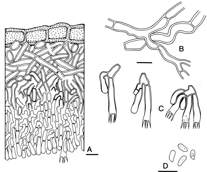
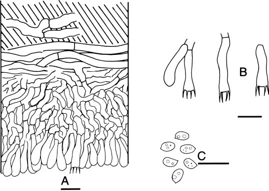
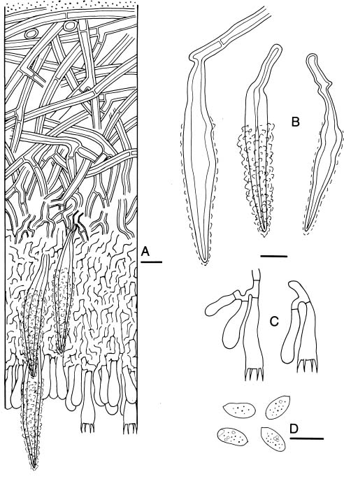
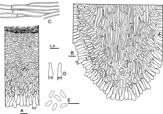
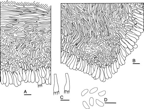
Basidiomata of Phanerochaete are resupinate. Hymenial surfaces are usually smooth, but tuberculate. Grandinioid or odontioid surfaces occur in some species. Phanerochaete is microscopically characterized by the monomitic hyphal system, generative hyphae with mostly simple septae, clavate basidia, and normally thin-walled nonamyloid and acyanophilic basidiospores. Cystidia, principally lepto- or lamprocystidia, are present in many species. Phanerochaete species possess holocoenocytic nuclear behavior, i.e. a pluri- or multi-nucleate condition occurs in the primary and secondary mycelia (Boidin and Lanquetin, 1984). Phanerochaete species are lignicolous, saprobic, and cause a uniform white rot in wood. Phanerochaete chrysosporium Burds. has been extensively studied as a microorganism used in the pulp and paper industry (Kirk and Chang, 1990). More than ninety species of Phanerochaete are known (Burdsall, 1985; Parmasto, 1997; Hjortstam, 1997; Wu, 1998). Thus it is the largest genus of the Corticiaceae s.l.

Before this study, thirty-one species of Phanerochaete has been recorded from Taiwan. Lin and Chen (1990) reported four species of Phanerochaete from Taiwan, viz. P. alba S.H. Lin & Z.C. Chen, P. commixtoides S.H. Lin & Z.C. Chen, P. globosa S.H. Lin & Z.C. Chen, and P. sordida (P. Karst.) J. Erikss. & Ryvarden (as P. cremea (Bres.) Parmasto). Wu (1990) added thirteen species to

the Taiwan flora, viz. P. aculeata Hallenb., P. affinis (Burt) Parmasto, P. albida Sheng H. Wu, P. brunnea Sheng H. Wu, P. ericina (Bourdot) J. Erikss. & Ryvarden, P. flavidoalba (Cooke) Rattan (as Phlebiopsis flavidoalba (Cooke) Hjortstam), Phanerochaete himalayensis (Dhingra) Sheng H. Wu, P. intertexta Sheng H. Wu, P. leptoderma Sheng H. Wu, P. parmastoi Sheng H. Wu, P. subglobosa Sheng H. Wu, P. taiwaniana Sheng H. Wu, and P. viticola (Schwein.:Fr.) Parmasto. Maekawa (1992) reported P. sanguinea (Fr.:Fr.) Pouzar from Lan-Yu, an island ca. 80 km east of the southern end of Taiwan. Wu (1995) described from Taiwan a new species, P. stereoides Sheng H. Wu, which has characteristic brown subicular hyphae. Wu (1997) further reported three new records: P. carnosa (Burt) Parmasto, P. filamentosa (Berk. & M.A. Curtis) Burdsall, and P. gigantea (Fr.:Fr.) S.S. Rattan et al. Wu (1998) proposed nine new species from Taiwan: P. argillacea Sheng H. Wu, P. capitata Sheng H. Wu, P. eburnea Sheng H. Wu, P. flavidogrisea Sheng H. Wu, P. fulva Sheng H. Wu, P. hyphocystidiata Sheng H. Wu, P. reflexa Sheng H. Wu, P. rubescens Sheng H. Wu, and P. suballantoidea Sheng H. Wu. In total thirty-seven species of Phanerochaete have been reported from Taiwan, including six new species described herein.

Materials and Methods

Materials for this study were collected from Taiwan during 1991-1996. All studied specimens and cultures are deposited at the herbarium of the National Museum of Natural Science Taiwan (TNM). Some isotypes will be distributed to BPI and K.

Descriptions of basidiomata were based on dried specimens. Free-hand, thin sections of basidiomata were prepared for microscopic studies. For observations and measurements of microscopic characters, 5% KOH was used as a mounting medium to ensure rehydration. Melzer’s reagent (IKI) was employed to detect amyloidity and dextrinoidity. Cotton blue in lactic acid (CB) was used as a mounting medium to determine cyanophily.

1Fax: +886-4-3258684; E-mail: shwu@nmns1.nmns.edu.tw


Botanical Bulletin of Academia Sinica, Vol. 41, 2000

Taxonomy

Phanerochaete angustocystidiata Sheng H. Wu, sp. nov. (Figures 1, 7A)

Basidiocarpus effusus, membranaceus, 100-200 µm crassus; superficies hymenialis plana. Systema hypharum monomiticum; hyphae efibulatae. Lamprocystidia subulata. Basidia clavata, 18-28 × 4.5-5.5 µm., 4 sterigmatibus. Basidiosporae cylindricae, laeves, tenuitunicatae, 6.3-8 × 2.3-3 µm, IKI-, CB-.

Cultural descriptions and species codes follow Nobles (1965) with amendments by Boidin and Lanquetin (1983). Minor modifications to Nobles’ code system have been made by other mycologists. Nobles’ code as detailed by Nakasone (1990) is adopted in this study. Nuclear staining of mycelia for P. odontoidea and P. subodontoidea were made with Giemsa according to Boidin (1958). Other practical details adopted in the cultural study were previously described (Wu, 1996).

Figure 1. Phanerochaete angustocystidiata (holotype). A, Basidiome section with substratum (cross hatched) at top; B, Hyphae from basal layer; C, Lamprocystidia; D, Basidia; E, Basidiospores. Scale bars = 10 µm.


Wu — New Phanerochaete from Taiwan

This new species resembles Phanerochaete leptoderma Sheng H. Wu, but is distinguished from it in having a distinct basal layer and narrower basidiospores (2.9-3.3 µm wide in P. leptoderma, according to Wu (1990).

Phanerochaete canolutea Sheng H. Wu, sp. nov. (Figures 2, 7B)

Basidiocarpus effusus, subceraceus, 50-150 µm crassus; superficies hymenialis plana. Systema hypharum monomiticum; hyphae efibulatae. Cystidia destituta. Basidia clavata, 15-25 × 4-5 µm, 4 sterigmatibus. Basidiosporae anguste ellipsoideae, laeves, tenuitunicatae, 4.7-6.3 × 2.5-3 µm, IKI-, CB-.

Basidiome resupinate, effuse, adnate, subceraceous, 50-150 µm thick in section. Hymenial surface grayish yellow, smooth, extensively cracked; margin thinning, concolorous or paler, byssoid-filamentous. Hyphal system monomitic; hyphae simple-septate. Subiculum composed of a medullary layer, with fairly dense texture; hyphae colorless, 3-8 µm diam., with 0.5-1.5 µm thick walls. Hymenium thickening, with dense texture; hyphae mainly vertical, colorless, thick-walled. Cystidia lacking. Basidia clavate, 15-25 × 4-5 µm, thick-walled except the apical parts, 4-sterigmate. Basidiospores narrowly ellipsoid, adaxially slightly concave, smooth, thin-walled, 4.7-6.3 × 2.5-3 µm, IKI-, CB-.

Etymology. From canus (= gray) + luteus (= yellow), referring to the color of the hymenial surface.

Holotype. TAIWAN. TAIPEI HSIEN: Yangmingshan, alt. 600 m, on branch of angiosperm, 20 Nov 1992, Wu 9211-105 (TNM).

Cultural description (mycelium derived from woody substratum of the holotype). 1 wk growth: Colony radius 40 mm. Advancing zone even. Mat white. Aerial mycelium pellicular. Advancing hyphae colorless, usually simple-septate, occasionally with single, double, or multiple clamp connections, 2.5-7.5 µm diam., thin-walled. 2 wk growth: Plates covered. 6 wk growth: Mat white, not discoloring in KOH. Aerial mycelium downy-felty. Hyphal system monomitic. Hyphae usually simple-septate, occasionally with single, double, or multiple clamp connections, colorless, 2.5-7.5 µm diam., thin-walled. Cuboid crystals present. No distinct odor. Not fruiting.

Oxidase reactions. TAA: -, 0; -, 0. GAA: (+), 20; ++, 35-40. TYA: -, 20; -, 30-35.

Species code. 2a. 5. 7. 32. 36. 38. 42. 54.

Both Phanerochaete canolutea and P. avellanea (Bres.) J. Erikss. & Hjortstam lack cystidia and have similar-sized basidiospores. However, several features separate these two species. First, the subiculum of P. avellanea is composed of a very thin basal layer (Burdsall, 1985; Eriksson et al., 1981), while that of P. canolutea is composed of a medullary layer. Secondly, basidia of P. canolutea are shorter (15-25 µm) than those of P. avellanea ((25-)30-40 µm (Burdsall, 1985)); see also the ba

Basidiome resupinate, effuse, adnate, membranaceous, 100-200 µm thick in section. Hymenial surface ivory color or light cream, smooth, cracked; margin rather determinate, white, slightly filamentous. Hyphal system monomitic; hyphae simple-septate. Subiculum composed of a basal layer and a medullary layer. Basal layer up to ca. 50 µm thick, with compact texture; medullary layer with loose texture. Subicular hyphae colorless, 2.5-5 µm diam., thin-walled near subhymenium, ± thick-walled near substratum, occasionally encrusted, horizontal and glued together in basal layer, ± vertical towards subhymenium. Hymenium thickening, with dense texture; hyphae colorless, thin-walled. Lamprocystidia numerous, immersed or projecting, cylindrical, subulate, conical when small, colorless, 30-70 × 5-9 µm (with encrustation). Basidia clavate, 18-28 × 4.5-5.5 µm, 4-sterigmate. Basidiospores cylindrical, adaxially slightly concave or flattened, smooth, thin-walled, 6.3-8 × 2.3-3 µm, IKI-, CB-.

Etymology. From angustus (narrow) + cystidium, referring to the shape of the cystidia.

Holotype. TAIWAN. NANTOU HSIEN: Yushan National Park, Nanhsi Forest Road, alt. 1,800 m, on branch of angiosperm, 13 Jun 1996, Wu 9606-39 (TNM; isotypes: BPI, K).

Additional specimens examined. TAIWAN. NANTOU HSIEN: Yushan National Park, Nanhsi Forest Road, alt. 1,850 m, on branch of angiosperm, 13 Oct 1993, Wu 9310-18 (TNM); Tungpu, alt. 1,300 m, on branch of angiosperm, 8 Oct 1992, Wu 9210-100 (TNM), Wu 9210-106 (TNM); alt. 1,350 m, on branch of angiosperm, 23 Nov 1993, Wu 9311-27 (TNM), Wu 9311-29 (TNM), Wu 9311-31 (TNM), Wu 9311-32 (TNM). Hoshe, alt. 750 m, on branch of angiosperm, Wu 9405-10 (TNM), Wu 9405-14 (TNM). HUALIEN HSIEN: Hsienpaotai, alt. 850 m, on branch of angiosperm, 6 Dec 1991, Wu 911206-22 (TNM), Wu 911206-55 (TNM).

Cultural description (combined from polysporous mycelia of Wu 9210-100 and Wu 9606-39, and mycelium derived from woody substratum of Wu 9310-18). 1 wk growth: Colony radius 30-65 mm. Advancing zone fairly even. Mat white. Aerial mycelium pellicular or almost absent near inoculum, pellicular or downy towards growth margins. Advancing hyphae colorless, usually simple-septate, occasionally with single, double, or multiple clamp connections, 2-5 µm diam., thin-walled. 2 wk growth: Plates covered. 6 wk growth: Mat white, not discoloring in KOH. Aerial mycelium pellicular or almost absent, occasionally zonate. Hyphal system monomitic. Hyphae mostly simple-septate, rarely with single, double, or multiple clamp connections, colorless, 1.5-6.5 µm diam., thin-walled, occasionally shortly septate, sometimes with adhered crystals. Odor fruity or fragrant. Not fruiting.

Oxidase reactions. TAA: -, 0; -, 0. GAA: (+), 0; (+), 0. TYA: - (yellow), 58-75; - (brownish yellow), 90+.

Species code. (2a). 5. 7. 32. 36. 38. 42. 50. 54.


Botanical Bulletin of Academia Sinica, Vol. 41, 2000

Figure 2. Phanerochaete canolutea (holotype). A, Basidiome section with substratum (woody cells) at top; B, Subicular hyphae; C, Basidia; D, Basidiospores. Scale bars=10 µm.

Figure 3. Phanerochaete ginnsii (holotype). A, Basidiome section with substratum (hatched) at top; B-C, Basidia; C, Basidiospores. Scale bars = 10 µm.


Wu — New Phanerochaete from Taiwan

Phanerochaete laxa Sheng H. Wu, sp. nov. (Figures 4, 7D)

Basidiocarpus effusus, membranaceus, 100-250 µm crassus; superficies hymenialis plana. Systema hypharum monomiticum; hyphae efibulatae. Lamprocystidia subulata. Basidia clavata, 20-30 × 6-7 µm, 4 sterigmatibus. Basidiosporae ellipsoideae, laeves, tenuitunicatae, 8-10 × 4-5 µm, IKI-, CB-.

Basidiome resupinate, effuse, adnate, membranaceous, 100-250 µm thick in section. Hymenial surface ivory-colored, slightly darkening in KOH, smooth, cracked; margin thinning, white, shortly filamentous. Hyphal system monomitic; hyphae simple-septate. Subiculum composed of a medullary layer, with loose texture; hyphae ± horizontal towards substratum, ± vertical towards subhymenium, colorless, 2-5 µm diam., with ca. 1 µm thick walls. Hymenium thickening, with dense texture; hyphae colorless, slightly thick-walled. Lamprocystidia numerous, immersed or projecting, subulate, apical half encrusted, colorless or slightly yellow, 50-120 × 10-22 µm (with encrustation), with 1.5-4.5 µm thick walls. Basidia clavate, 20-30 × 6-7 µm, 4-sterigmate. Basidiospores ellipsoid, adaxially slightly concave, smooth, thin-walled, 8-10 × 4-5 µm, IKI-, CB-.

Etymology. From laxus (= loose), referring to the texture of the subiculum.

Holotype. TAIWAN. NANTOU HSIEN: Yushan National Park, Tungpu, alt. 1,250 m, on culm of Miscanthus floridulus, 23 Nov 1993, Wu 9311-17 (TNM; isotype: BPI).

Cultural description (mycelium derived from woody substratum of the holotype). 1 wk growth: Colony radius 45 mm. Advancing zone even. Mat white. Aerial mycelium pellicular near inoculum, downy towards growth margins. Advancing hyphae colorless, usually simple-septate, with occasional single, double, or multiple clamp connections, 2.5-7.5 µm diam., thin-walled. 2 wk growth: Plates covered. 6 wk growth: Mat white, not discoloring in KOH. Aerial mycelium pellicular, occasionally floccose, felty towards plate margins. Hyphal system monomitic; hyphae usually simple-septate, occasionally with single, double, or multiple clamp connections. Aerial hyphae colorless, 1.5-5 µm diam., usually thin-walled, occasionally thick-walled. Submerged hyphae colorless, 2-8 µm diam., usually thin-walled, occasionally thick-walled. Crystals present. No distinct odor. Not fruiting.

Oxidase reactions. TAA: -, 0; -, 0. GAA: -, 23-26; -, 57-65. TYA: -, 25-27; +, 50-60.

Species code. 2b. 5. 7. 32. 36. 38. 42. 54.

This new species resembles Phanerochaete arenata (P.H.B. Talbot) Jülich, but the latter has narrower basidiospores (3-3.5 µm, according to Burdsall (1985)).

Phanerochaete odontoidea Sheng H. Wu, sp. nov. (Figures 5, 7E)

Basidiocarpus effusus, ceraceus, 50-200 µm crassus; superficies hymenialis odontoidea. Systema hypharum monomiticum; hyphae efibulatae. Cystidia destituta. Ba

sidium illustrated from the type specimen in Eriksson et al., 1981). Further, P. canolutea is distinct in having thick-walled basidia. Moreover, basidiospores of P. canolutea are slightly shorter (4.7-6.3µm) than the lectotype specimen of P. avellanea (5.5-6.5(-7) µm, according to Wu (1990: as Efibula avellanea)).

Phanerochaete ginnsii Sheng H. Wu, sp. nov. (Figures 3, 7C)

Basidiocarpus effusus, subceraceus, 40-80 µm crassus; superficies hymenialis plana. Systema hypharum monomiticum; hyphae efibulatae. Cystidia destituta. Basidia subclavata, 17-25 × 5-5.5 µm, 4 sterigmatibus. Basidiosporae late ellipsoideae, laeves, tenuitunicatae, 4-5 × 2.7-3 µm, IKI-, CB-.

Basidioma resupinate, effuse, adnate, subceraceous, 40-80 µm thick in section. Hymenial surface cream-colored, smooth, cracked; margin thinning, paler, byssoid-filamentous. Hyphal system monomitic; hyphae simple-septate. Subiculum composed of a basal layer, with dense texture; hyphae horizontal, colorless, 2.5-5 µm diam., thin- or slightly thick-walled. Hymenium distinctly thickening, with dense texture; hyphae mainly vertical, colorless, thin-walled. Cystidia lacking. Basidia subclavate, 17-25 × 5-5.5 µm, 4-sterigmate. Basidiospores broadly ellipsoid, adaxially flattened, smooth, thin-walled, usually guttulate, 4-5 × 2.7-3 µm, IKI-, CB-.

Etymology. Named in honor of Dr. James H. Ginns, who has made significant contributions to systematics of the corticioid fungi.

Holotype. TAIWAN. CHIAYI HSIEN: Yushan National Park, Nanhsi Forest Road, alt. 1,800 m, on branch of angiosperm, 6 Oct 1992, Wu 9210-22 (TNM; isotype: BPI).

Cultural description (mycelium derived from woody substratum of the holotype). 1 wk growth: Colony radius 53-60 mm. Advancing zone even. Mat white. Aerial mycelium absent. Advancing hyphae colorless, usually simple-septate, occasionally with single, double, or multiple clamp connections, 2.5-7.5 µm diam., thin-walled. 2 wk growth: Plates covered. 6 wk growth: Mat white, turning brownish yellow in KOH. Aerial mycelium pellicular, with ± reticulate appearance. Hyphal system monomitic; hyphae usually simple-septate, occasionally with single, double, or multiple clamp connections. Aerial hyphae colorless, 2.5-6.5 µm diam., thin- to thick-walled. Submerged hyphae colorless, 2-5 µm diam., thin- to slightly thick-walled. Cuboid crystals present. Odor slightly fruity. Not fruiting.

Oxidase reactions. TAA: -, 0; -, 0. GAA: -, 27; ++, 60. TYA: - (slightly yellow), 90+; - (slightly yellow), 90+.

Species code. 2a. 5. 7. 32. 36. 37. 38. 42. 50. 54.

This new species resembles Phanerochaete parvispora Sheng H. Wu & Losi, but the latter hassmaller basidiospores (3.2-4.2 × 2.2-2.8 µm, according to Wu and Losi, 1995).


Botanical Bulletin of Academia Sinica, Vol. 41, 2000

Figure 4. Phanerochaete laxa (holotype). A, Basidiome section; B, Lamprocystidia; C, Basidia; D, Basidiospores. Scale bars = 10 µm.


Wu — New Phanerochaete from Taiwan

Figure 5. Phanerochaete odontoidea (holotype). A, Basidiome section; B, Aculeus section; C, Subicular hyphae; D, Basidia; E, Basidiospores. Scale bars = 10 µm.

sidia clavata, 14-18 × 4.5-5.5 µm, 4 sterigmatibus. Basidiosporae cylindricae vel anguste cylindricae, laeves, tenuitunicatae, 6-7.2 × 2.6-3 µm, IKI-, CB-.

Basidiome resupinate, effuse, adnate, ceraceous, 50-200 µm thick in section (aculei excluded). Hymenial surface pale brown, odontioid, cracked; margin fairly determinate, paler to whitish. Aculei conical to subulate, usually separate, 2-4 per mm, 100-250 µm wide, up to 700 µm long. Hyphal system monomitic; hyphae simple-septate. Subiculum fairly uniform, a basal layer of compact texture; hyphae colorless, mainly horizontal, 4-6 µm diam., with ca. 1 µm thick walls. Hymenium thickening, with compact texture; hyphae mainly vertical, colorless, slightly thick-walled. Trama of aculei of compact texture, some with crystal masses near apex; hyphae mainly vertical, other aspects similar to those of subiculum. Cystidia lacking. Basidia clavate, 14-18 × 4.5-5.5 µm, 4-sterigmate. Basidiospores cylindrical or narrowly ellipsoid, smooth, thin-walled, 6-7.2 × 2.6-3 µm, IKI-, CB-.

Etymology. From odontoideus (= odontoid), referring to the hymenial surface.

Holotype. TAIWAN. ILAN HSIEN: Fushan Botanical Garden, alt. 600 m, on branch of angiosperm, 7 Aug 1991, Wu 910807-11 (TNM; isotypes: BPI, K).

Additional specimens examined. TAIWAN. NANTOU HSIEN: Yushan National Park, Nanhsi Forest Road, alt. 1,850 m, on branch of angiosperm, 13 Oct 1993, Wu 9310-8 (TNM). Tungpu, alt. 1,300 m, on branch of angiosperm, 23 Nov 1993, Wu 9311-46 (TNM).

Cultural description (mycelium derived from woody substratum of Wu 9310-8). 1 wk growth: Colony radius 70-80 mm. Advancing zone fairly even. Mat white. Aerial mycelium pellicular. Advancing hyphae colorless, simple-septate, 2.5-7 µm diam., thin-walled. 2 wk growth: Plates covered. 6 wk growth: Mat pale yellow, not discoloring in KOH. Aerial mycelium slightly pellicular. Hyphal system monomitic. Hyphae colorless, simple-septate, 1-10 µm diam., thin- or slightly thick-walled, occasionally encrusted with fine crystals. Cubical crystals present. No distinct odor. Not fruiting.

Oxidase reactions. TAA: +++, 20; ++++, 30-35. GAA: ++++, tr; ++++, tr. TYA: - (brownish yellow), 90+; - (brownish yellow), 90+.

Cytology. Mycelium derived from woody substratum is multinucleate.

Species code. 2a. 6. 7. 32. 36. 38. 42. 54.


Botanical Bulletin of Academia Sinica, Vol. 41, 2000

50-250 µm wide, up to 700 µm long. Hyphal system monomitic; hyphae simple-septate. Subiculum fairly uniform, composed of a basal layer, with compact texture; hyphae colorless, mainly horizontal, 3-6 µm diam., with ca. 1 µm thick walls. Hymenium thickening, with compact texture; hyphae mainly vertical, colorless, slightly thick-walled. Trama with compact texture, sometimes with crystal masses close to apices; hyphae mainly vertical, similar to those of subiculum. Cystidia lacking. Basidia clavate, 16-20 × 5-6 µm, 4-sterigmate. Basidiospores narrowly ellipsoid, smooth, thin-walled, 6.5-8.5 × 3-3.7 µm, IKI-, CB-.

Etymology. From sub (= somewhat, not completely) + odontoidea, referring to the resemblance of this new species to Phanerochaete odontoidea.

Holotype. TAIWAN. NANTOU HSIEN: Yushan National Park, Leleku, alt. 1,450 m, 13 Apr 1994, CWN 00776 (TNM; isotype: K).

Additional specimen examined. TAIWAN. TAICHUNG HSIEN: Chiapaotai, alt. 850 m, on branch of angiosperm, 6 Dec 1996, Wu 911206-38 (TNM).

Phanerochaete odontoidea is very similar to P. subodontoidea: both species have odontioid hymenial surface, compact subiculum, and lack cystidia. Multinucleate condition of secondary mycelia reported for both species in this paper, indicates a holocoenocytic nuclear behavior which supports their placement in Phanerochaete. Phanerochaete odontoidea has narrower basidiospores (2.6-3 µm) than P. subodontoidea (3-3.7 µm).

Phanerochaete subodontoidea Sheng H. Wu, sp. nov. (Figures 6, 7F)

Basidiocarpus effusus, ceraceus, 50-170 µm crassus; superficies hymenialis odontoidea. Systema hypharum monomiticum; hyphae efibulatae. Cystidia destituta. Basidia clavata, 16-20 × 5-6 µm, 4 sterigmatibus. Basidiosporae anguste cylindricae, laeves, tenuitunicatae, 6.5-8.5 × 3-3.7 µm, IKI-, CB-.

Basidioma resupinate, effuse, adnate, ceraceous, 50-170 µm thick in section (aculei excluded). Hymenial surface pale brown, odontioid, occasionally cracked; margin fairly determinate or thinning, paler brown or concolorous. Aculei conical to subulate, usually separate, 2-4 per mm,

Figure 6. Phanerochaete subodontoidea (holotype). A, Basidiome section with substratum (hatched) at top; B, Aculeus section; C, Basidia; D, Basidiospores. Scale bars = 10 µm.


Wu — New Phanerochaete from Taiwan

Figure 7. Cultures after 6 wk of growth on 1.5% MEA at 25°C. A, Phanerochaete angustocystidiata; B, P. canolutea; C, P. ginnsii; D, P. laxa; E, P. odontoidea; F, P. subodontoidea.

Cultural description (polysporous mycelium of the holotype). 1 wk growth: Colony radius 55-67 mm. Advancing zone uneven. Mat white. Aerial mycelium slightly pellicular. Advancing hyphae colorless, simple-septate, 2.5-5.5 µm diam., thin-walled. 2 wk growth: Plates covered. 6 wk growth: Mat yellow, not discoloring in KOH. Aerial mycelium slightly pellicular. Hyphal system monomitic. Hyphae colorless, or occasionally yellow from the yellow aerial solid mat lining the plate margin, simple-septate, 1-7 µm diam., thin- or slightly thick-walled, occasionally encrusted with crystals. Crystals present. No distinct odor. Not fruiting.

Oxidase reactions. TAA: +++, 30; ++++, 30-35. GAA: ++++, tr; ++++, tr. TYA: - (yellow), (90); - (brownish yellow), 90+.

Cytology. Both monosporous and polysporous mycelium are multinucleate.

Species code. 2a. 6. 7. 32. 36. 38. 42. 54.

Acknowledgments. This study was supported by the National Science Council of ROC (No. NSC 85-2311-B-178-001) and the National Agriculture Council of ROC (No. 88-AST-1.2-FCD-01(3)2).

Literature Cited

Boidin, J. 1958. Essai biotaxonomique sur les Hydnés résupinés et les Corticiés. Rev. Mycol. (Paris) Mém. Hors-Sér. 6: 1-388.

Boidin, J. and P. Lanquetin. 1983. Basidiomycètes Aphyllophoralés épithéloïdes étalés. Mycotaxon 16: 461499.

Boidin, J. and P. Lanquetin. 1984. Répertoire des données utiles pour effectuer les tests d’intercompatibilité chez les Basidiomycètes. I. Introduction. Crypt. Mycol. 5: 33-45.

Burdsall, H.H., Jr. 1985. A contribution to the taxonomy of the genus Phanerochaete (Corticiaceae, Aphyllophorales). Mycol. Mem. 10: 1-165.

Eriksson, J., K. Hjortstam, and L. Ryvarden. 1981. The Corticiaceae of North Europe 6. Phlebia - Sarcodontia. Fungiflora, Oslo, pp. 1048-1276.

Hjortstam, K. 1997. A checklist to genera and species of corticioid fungi (Basidiomycotina, Aphyllophorales). Windahlia 23: 1-54.

Jülich. W. 1981. Higher taxa of Basidiomycetes. Biblioth. Mycol. 85: 1-485.

Kirk, T.K. and H.M. Chang (eds.). 1990. Biotechnology in Pulp and Paper Manufacture. Applications and Fundamentals Investigations. Butterworth-Heinemann, Boston.


Botanical Bulletin of Academia Sinica, Vol. 41, 2000

Lin, S.H. and Z.C. Chen. 1990. The Corticiaceae and the resupinate Hydnaceae of Taiwan. Taiwania 35: 69111.

Maekawa, N. 1992. Corticiaceous fungi (Aphyllophorales) collected in Lan-Yu Island, Taiwan. Rept. Tottori Mycol. Inst. 30: 21-25.

Nakasone, K.K. 1990. Cultural studies and identification of wood-inhabiting Corticiaceae and selected Hymenomycetes from North America. Mycol. Mem. 15: 1-412.

Nobles, M.K. 1965. Identification of cultures of woodinhabiting Hymenomycetes. Can. J. Bot. 43: 10971139.

Parmasto, E. 1986. On the origin of the Hymenomycetes (what are corticioid fungi?). Windahlia 16: 3-19.

Parmasto, E. 1997. Cortbase [vers. 1.2, 1998] — a nomenclatural taxabase of corticoid fungi (Hymenomycetes). Mycotaxon 61: 467-471.

Wu, S.H. 1990. The Corticiaceae (Basidiomycetes) subfamilies Phlebioideae, Phanerochaetoideae and Hyphodermoideae in Taiwan. Acta Bot. Fennica 142: 1123.

Wu, S.H. 1995. A study of the genus Phanerochaete (Aphyllophorales) with brown subicular hyphae. Mycotaxon 54: 163-172.

Wu, S.H. 1996. Studies on Gloeocystidiellum sensu lato (Basidiomycotina) in Taiwan. Mycotaxon 58: 1-68.

Wu, S.H. 1997. Notes on resupinate Basidiomycetes of Taiwan IV. Fung. Sci. 12: 99-103.

Wu, S.H. 1998. Nine new species of Phanerochaete from Taiwan. Mycol. Res. 102: 1126-1132.

Wu, S.H. and C. Losi. 1995. Phanerochaete parvispora sp. nov. (Aphyllophorales) from Venetian Lagoon, Italy. Mycotaxon 55: 543-545.

»OÆW²£Åãµ·µßÄݤ»·sºØ

§dÁnµØ

°ê¥ß¦ÛµM¬ì¾Ç³Õª«À]´Óª«¾Ç²Õ

¥»¤å³ø¾É»OÆW²£Åãµ·µßÄݤ»·sºØ¡A§Y P. angustocystidiata, P. canolutea, P. ginnsii, P. laxa, P. odontoidea¡A¥H¤Î P. subodontoidea¡C³o¨Ç¼Ð¥»±Ä©ó 1991 ¦Ü 1996 ¦~¡C¤å¤¤´£¨Ñ¤»ºØ¤§§ÎºA´y­z¡BÅã·L¯S¼x¤§°wµ§´y¹Ï¥H¤Î°ö¾i¬ã¨s¡C

ÃöÁäµü¡G¥Ö´ßµß¬ì¡F·sºØ¡FÅãµ·µßÄÝ¡F»OÆW¡F¤ÀÃþ¾Ç¡C