Bot. Bull. Acad. Sin. (2000) 41: 251-255

Chen and Lin — Three dematiaceous hyphomycetes new to Taiwan

New records from Taiwan of three interesting dematiaceous hyphomycetes

Jin-Liang Chen1 and Weir-Sen Lin

Department of Hospital and Health Care Administration, Chia-Nan College of Pharmacy and Science, Tainan, Taiwan 717, Republic of China

(Received September 29, 1999; Accepted January 7, 2000)

Abstract. Three dematiaceous hyphomycetes, Hyphodiscosia jaipurensis, Scutisporus brunneus, and Pithomyces terricola, were isolated in pure culture for the first time in Taiwan. All three species are described and illustrated.

Keywords: Hyphodiscosia jaipurensis; Hyphomycetes; Pithomyces terricola; Scutisporus brunneus; Taiwan; Taxonomy.

Introduction

During our study of hyphomycetes on rotting litter in Taiwan, three interesting species, Hyphodiscosia jaipurensis Lodha & Chandra Reddy, Scutisporus brunneus K. Ando & Tubaki and Pithomyces terricola (Manohara Chary & Ramarao) P.M. Kirk, were isolated. They were collected from different sources: H. jaipurensis and P. terricola were isolated from rotten leaves, while S. brunneus was from rotten stems. These three species have also been reported from other countries (Ellis, 1976; Kuthubutheen and Nawawi, 1994; Matsushima, 1975, 1981, 1989, 1993; Nawawi, 1985; Tubaki, 1965), but they are described and illustrated here as new records in Taiwan. The Taiwanese isolates have unique and important characteristics which enable them to be specifically identified. Hyphodiscosia jaipurensis has polyblastic, denticulate conidiogenous cells and 1-septate, somewhat curved-cylindrical, hyaline conidia with two slender appendages (setulae). Scutisporus brunneus has polyblastic, denticulate, proliferating sympodially conidiogenous cells and 4-celled butterfly-shaped conidia with 4-appendages. Pithomyces terricola has integrated, monoblastic conidiogenous cells and fusiform to ellipsoidal, 3-5-septate, warty (verrucose), brown conidia.

Materials and Methods

Samples of rotten litters were collected in Taiwan during 1995-1996. Collections were incubated in moist chambers (plastic boxes, 30 × 20 × 12 cm, with three layers of moistened paper) for fungal sporulation. Pure culture was established by inoculating a single spore or spores onto 3% water agar using a sterile glass microneedle. A piece of agar containing isolated spores was transferred to oat

meal agar (OMA) slants or plates under a stereomicroscope. Details of fungal characteristics and conidiogenesis were studied, measured, described, illustrated and photographed with an Olympus light microscope (BX50). Both live cultures and dried specimens were deposited in the Herbarium of the Chen-fungus-Collection (Herb. CFC).

Taxonomy

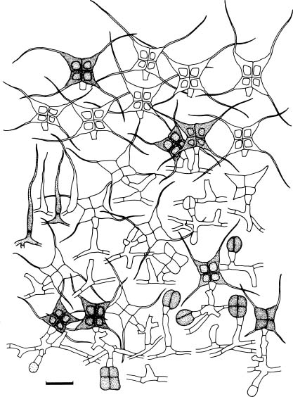
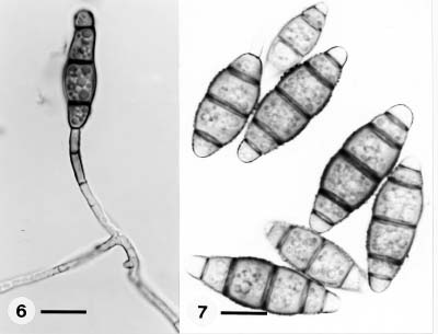
Hyphodiscosia jaipurensis Lodha & Chandra Reddy, 1974, Trans. Br. Mycol. Soc. 62: 418-421. (Figure 1)

Colonies on oat meal agar effuse, low, thin, white to pale brown or pale reddish purple; reverse pale brown to middle brown or reddish purple. Mycelium mostly immersed, partly superficial, composed of branched, septate, smooth or roughened, hyaline to pale brown 0.6-2.4 µm wide hyphae. Conidiophores macronematous, mononematous, terminal or lateral, simple, cylindrical or clavate, usually 1-2-septate, erect or flexuous, smooth to roughened or verrucose at the base, pale brown to reddish purple, 17.2-52.0 × 3.2-6.4 µm, sympodial proliferating at the apex. Conidiogenous cells polyblastic, integrated, denticulate. Conidia solitary, cylindrical, often slightly curved, rounded at the apex, truncate at the base, 1-septate, smooth, hyaline, 15.0-20.0 × 3.2-5.1 µm, usually with two slender, tapering curved setulae on the same side of the conidium, setulae up to 17.2 µm long.

Specimens examined. On a rotten leaf, Chungpu, Taiwan, 31 Aug 1995, Herb. CFC-3 (dried culture).

Notes. Lodha and Chandra Reddy (1974) established the genus Hyphodiscosia which include one species, H. jaipurensis Lodha and Chandra Reddy. It was found on dead wood in India. Hyphodiscosia is defined as having integrated, terminal, polyblastic, clavate, denticulate conidiogenous cells and cylindrical, 1-septat, truncate at the apex, conico-truncate at the base, hyaline, smooth conidia with two delicate setulae. Later, four species, H.

1Corresponding author. Tel: 06-266-4911 ext. 284; Fax: 06-266-7322; E-mail: ccl51911@ms29.hinet.net


Botanical Bulletin of Academia Sinica, Vol. 41, 2000

europaea Holubova-Jechova and Borowska, H. mirabilis Melnik and Sutton, H. radicicola Ts. Watan., and H. queenslandica T. Matsushima were added to the genus. H. europaea was isolated from dead bark and branches of Abies alba in Czechoslovakia. H. mirabilis was isolated from dead bark of Alnus incana in U.S.S.R. H. radicicola was isolated from roots of Fragaria chiloensis var. ananassa in Japan. H. queenslandica was isolated from dead leaf of Cryptocarya mackinnoniana in Queensland. Characteristics of Codinaea triseptata Matsushima (Matsushima, 1981) are close to H. jaipurensis, but the former species has polyphialidic conidiogenous cells and 3-septate conidia. Hyphodiscocioides tambopataensis Matsushima (Matsushima, 1993) is similar to H. jaipurensis, but the former species was described as having monoblastic, annellidic conidiogenous cells and 1-celled, smooth, pale brown conidia with two short setulae. The conidiophores of the Taiwanese isolate (up to 52 µm) are conspicuously shorter than the Indian isolate (up to 150 µm) which was collected from dead bark and was described by Ellis (1976). However, the conidia of the Japanese isolate (up to 26.5 µm) which was collected from bark of Diospyroris kakii and the Indian isolate (up to 25 µm) are longer than the Taiwanese isolate (up to 20 µm). The setulae of the three isolates are almost identical in length (Ellis, 1976; Matsushima, 1975).

Scutisporus brunneus K. Ando & Tubaki, 1985. Trans. Mycol. Soc. Japan 26: 151 (Figures 2, 4, 5)

Colonies on oat meal agar growing slowly, 10-12 mm diameter in 14 days at 25°C, somewhat floccose, velutinous, greyish brown to dark olive or dark green when sporulating. Mycelium partly superficial, partly immersed composed of branched, septate, smooth to roughened or verrucose, hyaline to subhyaline or pale brown, 0.6-2.0 µm wide hyphae. Conidiophores macronematous, mononematous, simple or branched, erect or flexuous, mostly lateral, rarely terminal, aseptate or septate, smooth or roughened, hyaline or subhyaline, 4.0-37.6 × 1.8-2.6 µm. Conidiogenous cells polyblastic, integrated, terminal, intercalary, cylindrical or sympodial proliferating, denticulate. Conidia solitary, dry, flattened, butterfly-shaped (with cross-septa, 4-cells, 4-appendages and a basal cell) olivaceous brown or olive, smooth, main body 9.6-12.8 × 9.6-16.0 µm, appendages subulate, smooth, long, slender, 8.0-27.2 µm long, 0.5-0.6 µm wide, hyaline or subhyaline; basal cell cuneiform or conical, truncate at the base, 2.4-5.2 × 2.0-3.0 µm. Setulae tapering to a point, smooth, brown paler towards the apex, 12.4-50.0 × 1.4-2.8 µm.

Specimens examined. On a rotten stem, Lantan, Chiayi City, Taiwan, 25 Oct 1996, Herb. CFC-4 (dried culture).

Figure 1. Hyphodiscosia jaipurensis. Conidiophores, conidia and conidiogenesis. Scale = 10 µm.

Figure 2. Scutisporus brunneus. Conidiophores, conidia and conidiogenesis. Scale = 10 µm.


Chen and Lin — Three dematiaceous hyphomycetes new to Taiwan

Notes. Ando and Tubaki (1985) erected the genus Scutisporus with S. brunneus as the type species isolated from fallen needles of Pinus densiflora in Japan. S. brunneus has since been recorded in several locations (Kuthubutheen and Nawawi, 1994; Matsushima, 1989; Nawawi, 1985; Tubaki, 1965). To date, it is the only one species reported in the genus Scutisporus. The Taiwanese isolate from rotten stem has shorter conidiophores (up to 37.6 mm) compared to the Malaysian isolate (up to 60 µm) from decaying twigs (IMI 355834) which was described by Kuthubutheen and Nawawi (1994). Also, the conidial dimensions of the Taiwanese isolate are slightly wider than those from Malaysia and Japan. Moreover, the appendage length of the Malaysian isolate (up to 25 µm) and the Japanese isolate (up to 20 µm) are somewhat shorter than the Taiwanese isolate (up to 27 µm) (Kuthubutheen and Nawawi, 1994; Matsushima, 1975).

Pithomyces terricola (Manohara Chary & Ramarao) P.M. Kirk, 1983, Trans. Br. mycol. Soc. 80: 462. (Figures 3, 6, 7)

Colonies effuse, plane to floccose, pale grey brown, grey brown to dark brown; reverse brown to dark brown. Mycelium partly superficial, partly immersed, composed of branched, septate, smooth to roughened or verrucose, subhyaline to brown hyphae, 0.8-5.6 µm wide. Conidiophores micronematous or semimacronematous, simple or branched, erect or flexuous, smooth to roughened or veruculose, subhyaline to brown, 1.8-5.4 µm wide. Conidiogenous cells monoblastic, mostly terminal, rarely intercalary, integrated, sometimes proliferous percurrent. Conidia solitary fusiform to broadly fusiform

Figure 3. Pithomyces terricola. Conidiophores, conidia and conidiogenesis. Scale = 10 µm.

Figures 4-5. Scutisporus brunneus. Figure 4. Conidiophore with developing conidium. Figure 5. Butterfly-shaped conidia. Figures 4-5. Scales = 10 µm.


Botanical Bulletin of Academia Sinica, Vol. 41, 2000

Figures 6-7. Pithomyces terricola. Figure 6. Conidiophore with developing conidium. Figure 7. Fusiform and verrucose conidia, mostly 3-5 septate. Figures 6-7. Scales = 10 µm.

or broadly ellipsoidal, mostly 5-septate, occasionally 3 or 4-septate, usually verruculose to verrucose, mid brown to brown, paler and roughened or verruculose at the both ends, 18.8-40.8 × 5.2-12.2 µm, often constricted at the septa, occasionally retaining the part of the conidiophore at the base.

Specimens examined. On a rotting leaf, Chiaokengtzu, Tainan Pref., Taiwan, 20 Oct 1995, Herb. CFC-5 (dried culture).

Notes. In 1983, Stemphyliomma terricola Manohara Chary and Ramarao was accommodated as a basionym of Pithomyces terricola (Manohara Chary and Ramarao) P.M. Kirk by Kirk. P. terricola is similar to other Pithomyces species, but the former species was described as having monoblastic, sometimes proliferous percurrent conidiogenous cells and longer, fusiform, 3-5 septate, roughened to verrucose, mid brown to brown conidia. The Taiwanese isolate has smaller conidia (18.8-40.8 × 5.2 -12.2 µm) compared to the type species and other collections (Ellis, 1976; Kirk, 1983; Matsushima, 1981,1993). Ellis' collection (Ellis, 1976) from pond mud in India has conidia up to 60 µm long × 17 µm wide, and Matsushima's collection has conidia up to 55 µm long × 14-16.5 µm wide (Matsushima, 1981, 1993).

Acknowledgements. We are grateful for the support of the National Science Council, ROC., by grant (NSC-85-2311-B-041-001 and NSC-86-2314-B-041-008) to J.L. Chen.

Literature Cited

Ando, K. and K. Tubaki. 1985. Three new hyphomycetes from Japan: Anthopsis microspora, Scutisporus brunneus, and Titaeella capnophila. Trans. Mycol. Soc. Japan 26: 151-160.

Ellis, M.B. 1976. More Dematiaceous Hyphomycetes. CAB International Mycological Institute: Kew, England.

Lodha, B.C. and K.R. Chandra Reddy. 1974. Hyphodiscosia gen. nov. from India. Trans. Br. mycol. Soc. 62: 418-421.

Kirk, P.M. 1983. New or interesting microfungi IX. Dematiaceous hyphomycetes from Esher Common. Trans. Br. Mycol. Soc. 80: 449-467.

Kuthubutheen, A.J. and A. Nawawi. 1994. Henicospora longissima sp. nov., Obeliospora triappendiculata sp. nov., Paraulocladium fabisporum sp. nov. and other hyphomycetes from Malaysia. Mycol. Res. 98: 677-685.

Matsushima, T. 1975. Icones Microfungorum a Matsushima Lectorum. Matsushima, Kobe, Japan.

Matsushima, T. 1981. Matsushima Mycological Memoirs No.2. Matsushima, Kobe, Japan.

Matsushima, T. 1989. Matsushima Mycological Memoirs No.6. Matsushima, Kobe, Japan.

Matsushima, T. 1993. Matsushima Mycological Memoirs No.7. Matsushima, Kobe, Japan.

Nawawi, A. 1985. Aquatic hyphomycetes and other water-borne fungi from Malaysia. Malay. Nature J. 39: 75-134.

Tubaki, K. 1965. Short note on aquatic spora in East New Guinea. Trans. Mycol. Soc. Japan 6: 11-16.


Chen and Lin — Three dematiaceous hyphomycetes new to Taiwan

¤TºØ¥xÆW²£¤£§¹¥þµß·s°O¿ýºØ

³¯«°ãSªL¬°´Ë

¨p¥ß¹Å«nÃIJz¾Ç°|Âå°ÈºÞ²z¨t

¥»¤å¤¤°£´y­z¤TºØ¥xÆW²£µ·ÌUºõ¤£§¹¥þµß¡A¥]¬A Hyphodiscosia jaipurensis Lodha & Chandra Reddy, Scutisporus brunneus K. Ando & Tubaki »P Pithomyces terricola (Manohara Chary & Ramarao) P.M. Kirk ¥~¡A¥ç±N¦¹¤TºØ¤£§¹¥þµß¦C¬°¥xÆW·s°O¿ýºØ¡C

ÃöÁäµü¡G ¤£§¹¥þµß¡F·s°O¿ýºØ (Hyphodiscosia jaipurensis, Scutisporus brunneus, Pithomyces terricola)¡F¥xÆW¡F¤ÀÃþ¾Ç¡C