Bot. Bull. Acad. Sin. (2000) 41: 283-291

Kuo-Huang et al. — Sclereids in Nymphoides and Nuphar

Ultrastructural study on the formation of sclereids in the

floating leaves of Nymphoides coreana and Nuphar schimadai

Ling-Long Kuo-Huang1,2, Su-Hwa Chen1, and Shiang-Jiuun Chen1

1 Department of Botany, National Taiwan University, Taipei, Taiwan, Republic of China

(Received December 29, 1999; Accepted April 14, 2000)

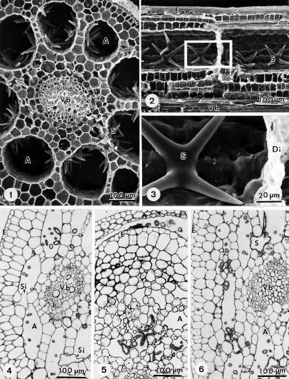
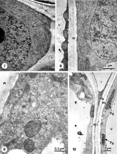
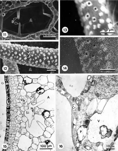
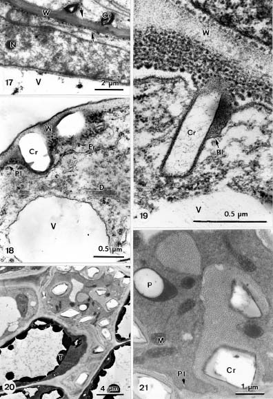
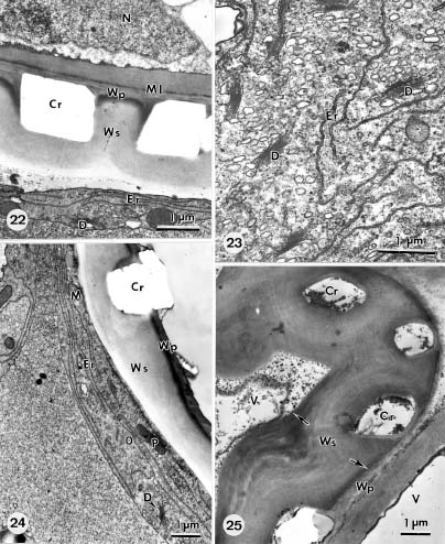
Abstract. The formation of star-shaped sclereids in the floating leaves of Nymphoides coreana and Nuphar schimadai was studied microscopically. These foliar sclereids were associated with the aerenchyma and found as the form of idioblast. The outer surface of mature sclereids was smooth in Nymphoides, but with many prismatic calcium oxalate crystals in Nuphar. However, the early morphogenesis of these two kinds of sclereids was similar. The sclereid initials were distinguished from the neighboring cells by their distinctly large nucleus. The expanding sclereid initials were constrained by the neighboring cells. Crystal formation in young sclereids of Nuphar started near the cessation of sclereid expansion. The crystals were bounded by crystal sheath and located in crystal chambers between the primary cell wall and plasma membrane. Calcium antimonate precipitates were found, especially on the crystal sheaths as well as between the plasma membrane and the primary cell walls. The crystal chambers have a paracrystalline appearance connected with the crystal sheath and the plasma membrane. After formation of crystals, the secondary wall was deposited and then the crystals became embedded between the primary and secondary walls. The possible functions of the foliage sclereids and the plans for further investigation are discussed.

Keywords: Calcium antimonate precipitates; Calcium oxalate crystals; Cell wall; Foliar sclereids; Nuphar schimadai; Nymphoides coreana.

Introduction

Sclereids can be found in many different parts of plant tissues and organs. They may serve various protective, strengthening, and other functions. For example, sclereids may form bands around seeds or cover roots or stems, possibly for strengthening purposes (Harris, 1983). Individual sclereids, called idioblasts, may cause tissues to be coarse and gritty and less palatable to insects (Mouseth, 1988). Such sclereids are frequently observed in the mesophyll of leaves of many plant genera, e.g., Borinia (Foster, 1955), Nymphaea (Gaudet, 1960; Kuo-Huang, 1992), and others (Rao and Das, 1979). The foliar sclereids of Olea europaea may also function as optical fibers to improve the light microenvironment within the mesophyll (Karabourniotis et al., 1994). The formation of sclereids can be observed during normal development of tissues and organs. However, differentiation of sclereids also can be induced artificially. For example, sclereids differentiated in the pith of Arabidopsis thaliana following repeated cutting of developing inflorescences (Lev Yadun, 1994), and a continuous pericyclic sclereid band developed in regenerated bark of Cinnamomum cassia, three years after the bark was removed (Hong and Tong,

1997). Various types of sclereids are found among different species, and sclereids in different tissues and organs have been used for plant classification (Rao and Banerjee, 1979; Villaron-Franceschinelli and Yamamoto, 1993).

The occurrence of sclereids in vascular plants has been studied mostly in terrestrial species, and rarely in aquatic species (Chiang and Huang, 1984; Kuo-Huang, 1990). In a preliminary study, we found many sclereids in the petiolated leaves of Nymphoides coreana (Menyanthaceae) and Nuphar schimadai (Nymphaeaceae). Both are members of the nymphaeid macrophytes (Smits et al., 1989). When comparing the foliar sclereids of these two aquatic species we found that their outer surfaces were quite different, which compelled us to investigate further. Results of light and electron microscopy are presented in this paper.

Materials and Methods

The Nymphoides coreana (L.) Hara (Menyanthaceae) and Nuphar schimadai Hayata (Nymphaeaceae) plants were grown in ponds located on the campus of National Taiwan University. Floating leaves in various stages were collected, and their petioles and laminae were cut into small pieces. The specimens were routinely processed through fixation (2.5-5% glutaraldehyde and 3-4% paraformaldehyde in 0.1 M sodium cacodylate buffer),

2 Corresponding author. Tel: (02) 2363-0231 ext. 2364; Fax: (02) 2391-8940; E-mail: linglong@ccms.ntu.edu.tw


Botanical Bulletin of Academia Sinica, Vol. 41, 2000

postfixation (1% OsO4), and dehydration (acetone series). For subcellular localization of calcium, some specimens of Nuphar schimadai were fixed in 2.5% glutaraldehyde-4% paraformaldehyde in 0.1 M phosphate buffer (pH 7.6) with 2% (w/v) potassium antimonate and 0.1% tannic acid in the same buffer (Slocum and Roux, 1982). All treated specimens were then embedded in Spurr's resin under vacuum. One-micron sections were stained with 1% toluidine blue in borax buffer for light microscope observation. Thin sections for electron microscopy were cut with the Ultracut E, using a diamond knife. The thin sections were stained with uranyl acetate and lead citrate and examined by TEM, using the Hitachi H-600 microscope at 75 kV.

Specimens for SEM observation were dried with a Hitachi Critical Point Dryer (HCP-1), coated with IB-2 ion coater, and examined with the Hitachi S-550 microscope. Specimens for X-ray microanalysis were quenched in freon 22 (cooled by liq. N2) for 20 sec. and immersed in liq. N2 for 1 h before being transferred into a freeze-dryer for two days. The freeze-dried specimens were coated with carbon and the Jeol JXA-733 EPMA at 25kV was used for X-ray microanalysis.

Results

As with most other aquatic angiosperms, the amount of vascular tissue in floating leaves of Nymphoides coreana and Nuphar schimadai was limited while aeren


Figures 1-6. Nymphoides coreana. 1: SEM photograph of the cross section of petiole showing the air-canals surrounding vascular bundle and many star-shaped sclereids bordering the air-canals; 2, 3: Longitudinal section of petiole showing the air-canal separated by the transverse diaphragm and the mature sclereids with smooth outer surface; 4: LM photograph of the cross section of young petiole showing the sclereid initials in the young lacunate parenchyma; 5, 6: Cross sections of mature petiole showing the sclereids with bending or intercellular growth. Key to lettering: A, Air canal; Di, Diaphragm; E, Epidermis; Vb, Vascular bundle; S, Sclereid; Si, Sclereid initial.


Kuo-Huang et al. — Sclereids in Nymphoides and Nuphar

chyma tissue was well developed. In the petioles of these two species, 6-10 air-canals separated by the transverse diaphragms were found (Figures 1-3). The foliar sclereids were mostly observed bordering the air-canals and in the highly lacunate spongy mesophyll of both species.

Sclereids in Nymphoides coreana

In the petioles of Nymphoides coreana, sclereid initials were first found in the layer of cells bordering the air canals (Figures 4-6), especially at the petiole-rhizome and the petiole-lamina junctions. In the lamina, the first laminar sclereid initials were found in the spongy mesophyll near the midribs; however, the sclereid initials in the lamina appeared later than in the petiole, i.e., when initials in the petiole had started developing into young sclereids.

The sclereid initials were generally detected sporadically in young, lacunate parenchyma. They were distinguished from the neighboring cells by their large nucleus (Figure 7). Nevertheless, there were no predictable patterns of sclereid initials bordering the air-canals. In young sclereids, large vacuoles and thin dense cytoplasm with numerous organelles were observed (Figures 7, 8). When growth of the sclereids was hindered by surrounding cells, bending or intercellular growth of the sclereids were observed (Figures 4-6). Usually, the initials would expand into an air-canal and sprawl into adjacent air-canals or into large intercellular spaces. Such expansions resulted in star-shaped sclereids.

Toward the end of growth and expansion, the secondary cell wall started being laid down between the primary


Figures 7-10. Nymphoides coreana. 7-8: TEM photographs of the sclereid initials showing the large nucleus and dense cytoplasm with numerous organelles; 9: TEM photograph of the sclereids during the formation of secondary cell wall showing numerous vesicle-producing dictyosomes in the cytoplasm; 10: TEM photograph of the mature leaf showing the sclereid (right cell) containing plastids with thylakoid membranes and plastoglobuli, however, smaller than those in the neighboring parenchyma-tous cell (left cell). Key to lettering: D, Dictyosome; I, Intercellular space; M, Mitochondrium; Ml, Middle lamella; N, Nucleus; P, Plastids; Pg, Plastoglobulus; V, Vacuole; W, Cell wall; Wp, Primary wall; Ws, Secondary wall.


Botanical Bulletin of Academia Sinica, Vol. 41, 2000

wall and the plasma membrane. As the wall increased in thickness, the cytoplasm was confined to a thin layer around the cell periphery and formed numerous vesicle-producing dictyosomes (Figure 9). The plastids in mature sclereids were associated with thylakoid membranes and plastoglobuli (Figure 10); in general, they were smaller than those of neighboring parenchymatous cells. The outer surface of mature sclereid was smooth (Figure 3). In the cell walls of sclereids, primary and secondary wall layers were clearly observed in mature cell walls (Figure 10).

Sclereids in Nuphar schimadai

The sequence of formation and the early morphogenesis of foliar sclereids in petioles and laminae of Nuphar schimadai were similar to those of foliar sclereids of Nymphoides coreana. However, the formation of sclereid cell walls of these two species was different. On outer

surfaces of the mature sclereids of Nuphar schimadai, many prismatic crystals were found. These crystals were distributed densely, but irregularly, on all surfaces of the cell wall (Figures 11, 12). They were found on the wall that was in contact with neighboring cells (Figures 17, 22, 25) as well as on surfaces exposed to the air-canals. The crystals contained substantial amounts of calcium (Figures 13, 14). All crystals found in Nuphar were insoluble in acetic acid. But they were soluble in hydrochloric acid, without forming bubbles, suggesting that they were composed of calcium oxalate.

Ramifications of the initial cell of sclereids were similar to those observed in Nymphoides coreana (Figure 15). At cessation of apical intrusive growth of the sclereids, clusters of crystal chambers formed sporadically, and almost simultaneously, between the primary cell wall and the plasma membrane (Figure 16). The young sclereid contained a thin primary cell wall and an organelle-rich


Figures 11-16. Nuphar schimadai. 11: SEM photograph of the cross section of petiole showing an air-canal and many star-shaped sclereids bordering the air-canal; 12: A branch of the mature sclereid showing many prismatic crystals irregularly on the outer surface; 13,14: SEM photograph and X-ray map localizing the calcium electron transition energy, indicating the high calcium content of crystals; 15: LM photograph of the cross section of young leaf lamena showing the sclereid initials in the young lacunate parenchyma with ramification and intercellular growth; 16: TEM photograph of the sclereid initial showing the organelle-rich cytoplasm and many crystal chambers formed sporadically and almost simultaneously between the primary cell wall and the plasma membrane. Key to lettering: A, Air canal; Cr, Crystal; E, Epidermis; M, Mitochondrium; N, Nucleus; P, Plastids; V, Vacuole; Vb, Vascular bundle; S, Sclereid; Si, Sclereid initial.


Kuo-Huang et al. — Sclereids in Nymphoides and Nuphar

Figures 17-21. Nuphar schimadai. 17-18: TEM photographs of the crystal-forming sclereid showing the calcium antimonate precipitates accumulated on the crystal sheaths and between the plasma membrane and the primary cell walls; 19: TEM photograph of the crystal-forming sclereid showing crystals were bounded by crystal sheath that appeared to be connected with the plasma membrane; 20, 21: TEM photographs of the crystal-forming sclereid in a paradermal sectioning plane showing an ordered substructure with a paracrystalline appearance. Key to lettering: Cr, Crystal; D, Dictyosome; Er, Endoplasmic reticulum; M, Mitochondrium; N, Nucleus; P, Plastids; Pl, Plasmalemma; V, Vacuole; S, Sclereid; Si, Sclereid initial; T, Tannin; W, Cell wall.


Botanical Bulletin of Academia Sinica, Vol. 41, 2000

cytoplasm. The plastids were found to contain mostly small starch grains and small laminar stacks (Figure 16).

As an individual crystal developed, it underwent periclinal and anticlinal growth and formed a prismatic crystal, pressing the plasma membrane into the cytoplasm. The shape of the developing crystals could be recognized in electron-transparent areas (Figure 16), where the crystals were dissolved or had dislodged from the thin sec

tions during staining or sectioning. The crystals were bounded by a crystal sheath that appeared to be connected with the plasma membrane (Figures 17-19). Calcium antimonate precipitates were found especially on crystal sheaths as well as between the plasma membrane and the primary cell walls. In a different paradermal sectioning plane, parts of crystal chambers were transversely sectioned, and an ordered substructure with a

Figures 22-25. Nuphar schimadai. 22-24: TEM photographs of the sclereids during the formation of secondary cell wall showing numerous vesicle-producing dictyosomes in the cytoplasm. The cytoplasm condensed and formed numerous vesicle-producing dictyosomes and many a long and rough endoplasmic reticulum parallel to the plasma membrane; 25: TEM photograph of mature sclereid showing the crystals embedded in the thick layered secondary wall. Key to lettering: Cr, Crystal; D, Dictyosome; Er, Endoplasmic reticulum; M, Mitochondrium; N, Nucleus; O, Oil drop; P, Plastids; Pl, Plasmalemma; Ml, Middle lamella; V, Vacuole; Wp, Primary wall; Ws, Secondary wall.


Kuo-Huang et al. — Sclereids in Nymphoides and Nuphar

paracrystalline appearance was discernible (Figures 20, 21). There appeared to be a close association between the ordered substructure, the crystal sheath, and the plasma membrane.

After the formation of crystals, the secondary cell wall was deposited. As the wall thickened, the cytoplasm condensed and formed numerous vesicle-producing dictyosomes and many a long and rough endoplasmic reticulum, parallel to the plasma membrane (Figures 22-24). The thickening secondary cell wall encroached on the crystal sheath and started growing around the crystals. During the process, the crystals on the wall exposed to intercellular spaces became protruded and covered with thin primary cell walls. Wall thickening continued until the stratified and thickened secondary wall of the sclereids was deposited around the crystals (Figure 25).

Discussion

Sclereids are generally initiated in fundamental parenchyma (Mauseth, 1988). In Camellia, a terrestrial plant, the foliar sclereids originate and develop simultaneously (Boyd et al., 1982). However, in tissues of petioles and laminae of floating leaves of aquatic species of Nymphaea (Gaudet, 1960; Chiang and Huang, 1984; Kuo-Huang, 1992), Nuphar and Nymphoides, a different differentiation pattern is found. In petioles of these aquatic plants, sclereid initials are first observed at the petiole-rhizome and petiole-lamina junctions. Sclereid initials in the lamina appeared later. The foliar sclereids of aquatic plants may function by helping stiffening of air channels in the petiole and strengthening the joints between rhizome, petiole, and lamina, while maintaining their flexibility and air circulation in air channels in water (Guertler, 1905; Mevi-Schutz and Grosse, 1988).

As observed in other studies (Gaudet, 1960; Harris, 1983; Kuo-Huang, 1992), distinguishable features among sclereid initial, large nucleus and prominent nucleolus, were found in both Nymphoides coreana and Nuphar schimadai. The enlarging sclereids are constrained by neighboring cells. Foliar sclereids with an angular transverse shape indicate that expansion and ramification of young sclereids depended on the amount of available space. It has been thought that secondary cell walls are deposited only after cells have attained their final size and shape; but, Juniper et al. (1981) reported that wall deposition can begin even while the cell is still elongating. In our plants, secondary cell wall was begun laid down near the end of the expanding stage of the sclereid. In general, lignification begins at the central parts of cells, in the region of the middle lamella or the primary wall, which is the most heavily lignified area. The secondary wall becomes lignified last. These findings also indicate that the secondary cell wall was being initiated near the center of the cell, while the tips of the cell were approaching their destination.

For many cellular processes the presence of free calcium in the cytoplasm is considered an important devel

opmental regulator (Borowitzka, 1984; Bush, 1993). Various functions have been attributed to the formation of its crystal idioblasts. The reversible biological calcification with oxalic acid, an end product of metabolism, may also imply an effort to maintain an ionic equilibrium in the cells (Franceschi and Horner, 1980). The formation of calcium crystals is a complex process. Factors which control oxalate synthesis and cellular calcium uptake and mobility may affect crystal induction (Borchert, 1986). In leaves, calcium oxalate crystals are generally present in the central vacuoles as crystal idioblasts, e.g., in Phaseolus (Kuo-Huang and Zindler-Frank, 1998), Morus (Wu and Kuo-Huang, 1997), and Lemna (Franceschi, 1987). In Phaseolus and Morus, crystal growth is accompanied by thickening of secondary cell walls which make contact with the crystals and grow around them. However, in Nuphar schimadai and some species of Nymphaea (Gandet, 1960; Kuo-Huang, 1992) the calcium crystals are deposited between the primary and secondary cell walls of the sclereids. In plants, calcium is an essential apoplastic nutrient and serves as a structural component of the cell wall (Bush, 1993). When sclereids of Nuphar and Nymphaea form their crystals in the cell wall, calcium antimonate precipitates are accumulated, especially on the crystal sheaths (Figure 17; Kuo-Huang and Chen, 1999). The presence of an ordered substructure in crystal chamber connected with the crystal sheath and plasma membrane seems to be closely associated with crystal development. However, the mechanism of calcium crystal initiation in cell walls is still unknown.

Smits et al. (1990, 1995) have compared seeds of species of Nymphaea, Nuphar, and Numphoides. Their germination experiments demonstrated that factors influencing dormancy and germination of the seeds were similar for Nymphaea and Nuphar, but different for Nymphoides. We found that, concerning the occurrence of calcium crystals in the wall, foliar sclereids of Nymphaea and Nuphar are also different from those of Nymphoides. The presence or absence of calcium crystals in the plant body is an important character for understanding evolutionary relationships among plant species (Franceschi and Horner, 1980). Obviously, the different morphology of foliar sclereids between Nymphoides coreana and Nuphar schimadai has taxonomical significance; however, we think its ecological function and evolutionary implications are even more important. Studies of the individuals or species that are growing in calm water or moving water should provide further information on the function of foliar sclereids of aquatic plants. In progress are investigations of sclereid and calcium crystal formation in germinating seeds of these taxa.

Acknowledgements. This research was financially supported by the National Science Council of the Republic of China. The help of Miss Ching-Yen Lin of the Electron Microscope Laboratory of College of Science of National Taiwan University with preparing photographs is acknowledged.


Botanical Bulletin of Academia Sinica, Vol. 41, 2000

ment of crystal-forming sclereids in Nymphaea tetragona. Taiwania 37: 104-114.

Kuo-Huang, L.L. and S.J. Chen. 1999. Subcellular localization of calcium in the crystal-forming sclereids of Nymphaea tetragona Georgi. Taiwania 44: 520-528.

Kuo-Huang, L.L. and E. Zindler-Frank. 1998. Structure of crystal cells and influences of leaf development on crystal cell development and vice versa in Phaseolus vulgaris (Leguminosae). Bot. Acta 111: 337-345.

Lev Yadun, S. 1994. Induction of sclereid differentiation in the pith of Arabidopsis thaliana (L.) Heynh. J. Exp. Bot. 45: 11845-1849.

Mevi-Schutz, J. and W. Grosse. 1988. The importance of water vapor for the circulating air flow through Nelumbo nucifera. J. Exp. Bot. 39: 1231-1236.

Mauseth, J.D. 1988. Plant Anatomy. 1st. edn. The Benjamin/Cummings Pub. Com. Inc.

Rao, T.A. and B.C. Banerjee. 1979. On foliar sclereid in the Nymphaeaceae and their use in familial classification. Proc. Indian. Acad. Sci. 88: 413-422.

Rao, T.A. and S. Das. 1979. Leaf sclereids - occ Srence and distribution in the angiosperms. Bot. Notiser. 132: 319-324.

Slocum, R.D. and S.J. Roux. 1982. An improved method for the subcellular localization of calcium using a modification of the antimonate precipitation technique. J. Histochem. Cytochem. 30: 617-629.

Smits, A.J.M., P.H. Van Avesaath, and G. Van Der Velde. 1990. Germination requirements and seed banks of some nymphaeid macrophytes Nymphaea alba L. Nuphar lutea L. Sm. and Nymphoides peltata Gmel. O. Kuntze. Freshwater Bio. 24: 315-326.

Smits, A.J.M., R. Van Ruremonde, and G. Van Der Velde. 1989. Seed dispersal of three Nymphaeid macrophytes. Aqua. Bot. 35: 167-180.

Smits, A.J.M., G.H.W. Schmitz, G. Van Der Velde, and L.A.C.J. Voesenek. 1995. Influence of ethanol and ethylene on the seed germination of three water plants. Freshwater Bio. 34: 39-46.

Villaron-Franceschinelli, E. and K. Yamamoto. 1993. Taxonomic use of leaf anatomical characters in the genus Simarouba aublet (Simaroubaceae). Flora (Jena) 188: 117-124.

Wu, C.C. and L.L. Kuo-Huang. 1997. Calcium crystals in the leaves of some species of Moraceae. Bot. Bull. Acad. Sin. 38: 97-104.

Literature Cited

Borchert, R. 1986. Calcium acetate induces calcium uptake and formation of calcium oxalate crystals in isolated leaflets of Gleditsia triacanthos L. Planta 168: 571-578.

Borowitzka, K.A. 1984. Calcification in aquatic plants. Plant Cell Envir. 7: 457-466.

Boyd, D.W., W.M. Harris, and L.E. Murry. 1982. Sclereid development in Camellia japonica petioles. Am. J. Bot. 69: 339-347.

Bush, D.S. 1993. Regulation of cytosolic calcium in plants. Plant Physiol. 103: 7-13.

Chiang, S.H.T. and L.L. Huang. 1984. The formation of foliar sclereids in Nymphaea. Yushania 1: 33-52.

Foster, A.S. 1955. Structure and ontogeny of the terminal sclereids in the leaf of Mouriria huberi Cogn. Am. J. Bot. 42: 551-560.

Franceschi, V.R. 1987. Oxalic acid metabolism and calcium oxalate formation in Lemna minor L. Plant Cell Envir. 10: 397-406.

Franceschi, V.R. and H.T. Horner. 1980. Calcium oxalate crystals in plants. Bot. Rev. 46: 361-427.

Gaudet, J. 1960. Ontogeny of the foliar sclereids in Nymphaea odorata. Am. J. Bot. 47: 525-532.

Guertler, F. 1905. Ueber interzellulare Haarbildungen, insbesondere ueber die sognannten inneren Haare der Nymphaeaceen und Menyanthoideen. Inaugural Dissertation, Friedrich-Wilhelms Universitaet, Berlin.

Harris, W.M. 1983. On the development of macrosclereids in seed coats of Pisum sativum. Am. J. Bot. 70: 1528-1535.

Hong, L. and C.Y. Tong. 1997. Bark regeneration of Cinnamomum cassia Persl after ringing. J. Plant Resour. Envir. 6: 1-7.

Juniper, B.E., J.R. Lowton, and P.J. Harris. 1981. Cellular organelles and cell-wall formation in fibres from the flowering stem of Lolium temulentum L. New Phytol. 89: 609-619.

Karabourniotis, G., N. Papastergiou, E. Kabanopoulou, and C. Fasseas. 1994. Foliar sclereids of Olea europaea may function as optical fibres. Can. J. Bot. 72: 330-336.

Kuo-Huang, L.L. 1990. Calcium oxalate crystals in the leaves of Nelumbo nucifera and Nymphaea tetragona. Taiwania 35: 178-190.

Kuo-Huang, L.L. 1992. Ultrastructural study on the develop


Kuo-Huang et al. — Sclereids in Nymphoides and Nuphar

¤p×cµæ»P¥xÆWµÓ½´¯óº}¯B¸­¤º«p¾À²Ó­M§Î¦¨¤§¶W·L¬ã¨s

¶À¬ÂÄn¡@³¯²QµØ¡@³¯­»§g

°ê¥ß¥xÆW¤j¾Ç´Óª«¾Ç¬ã¨s©Ò

¥H¥ú¾Ç»P¹q¤lÅã·LÃè§Þ³NÆ[¹î¤p×cµæ»P¥xÆWµÓ½´¯óº}¯B¸­¤º¬Pª¬«p¾À²Ó­M¤§§Î¦¨¹Lµ{¡C³o¨Ç¸­ ³¡«p¾À²Ó­M¥H²§§Î²Ó­M¤è¦¡¦s¦b©ó³q®ð²Õ´¡C¤p×cµæ¦¨¼ô«p¾À²Ó­Mªº¥~ªí¥­·Æ¡A¦ý¥xÆWµÓ½´¯ó¦¨¼ô «p¾À²Ó­Mªº¥~ªí«h¥iÆ[¹î¨ì³\¦h¦h­±Å骺¯ó»Ä¶t´¹Åé¡AµM¦Ó¤p×cµæ»P¥xÆWµÓ½´¯ó¸­³¡«p¾À²Ó­M¦­´Á ªº§ÎºAµo¥ÍÃþ¦ü¡C«p¾À©l·½²Ó­M¥D­n¥H¨ä¨ã¤jªº²Ó­M®Ö¦Ó»P¾Fªñ¤@¯ëÁ¡¾À²Ó­M°Ï¤À¡A¥B©µ¦ù¥ç¨ü¨ä ­­¨î¡C¥xÆWµÓ½´¯ó«p¾À²Ó­M¤§¶t´¹Å骺§Î¦¨¤j¬ùµo¥Í©ó«p¾À²Ó­M¤£¦A¦æ¤ÀªK©µ¦ù¥Íªø¡A¯ó»Ä¶t´¹Åé ¥Ñ´¹ÅéÀT½¤¥]³ò¡A¦Ó¦ì©óªì¥Í²Ó­M¾À»P²Ó­M½¤¤§¶¡ªºµ²´¹µÄ¤¤¡A´¹ÅéÀT½¤»P²Ó­M½¤¬Û³s±µ¡C«p¾À©l ·½²Ó­M¤º¡A·L²ÓªºµJ¾O»Ä¶tªº¨I¾ýª«±K¶°¦a°ï¿n©ó´¹ÅéÀT½¤¤W¤Î¤À§G©ó²Ó­M½¤»P²Ó­M¾À¤§¶¡¡Cµ²´¹ µÄÅã¥Ü´¹®æª¬ºc³y¡A¨ä»P´¹ÅéÀT½¤¤Î²Ó­M½¤¬Û³s±µ¡C«p¾À²Ó­M¤§²Ó­M¾À¤ºªº´¹Åé§Î¦¨«á¡A§Y¶i¦æ¦¸ ¥Í²Ó­M¾Àªº°ï¿n¡A¨Ã±N´¹Åé¥]®I©óªì¥Í»P¦¸¥Í²Ó­M¾À¤§¶¡¡C¥»¤å¥ç°Q½×¸­³¡«p¾À²Ó­M¥i¯àªº¥\¯à»P ¶i¤@¨B±´°Qªº¥DÃD¡C

ÃöÁäµü¡G ¤p×cµæ¡F¥xÆWµÓ½´¯ó¡F¸­³¡«p¾À²Ó­M¡F²Ó­M¾À¡F¯ó»Ä¶t´¹Åé¡FµJ¾O»Ä¶t¨I¾ý¡C