Bot. Bull. Acad. Sin. (2001) 42: 115-121

Wu et al. — Identification of Chlorella spp. isolates

Identification of Chlorella spp. isolates using ribosomal DNA sequences

Hsiuan-Lin Wu, Ruey-Shyang Hseu and Liang-Ping Lin*

Graduate Institute of Agricultural Chemistry, National Taiwan University, Taipei, Taiwan 106, Republic of China

(Received May 24, 2000; Accepted October 31, 2000)

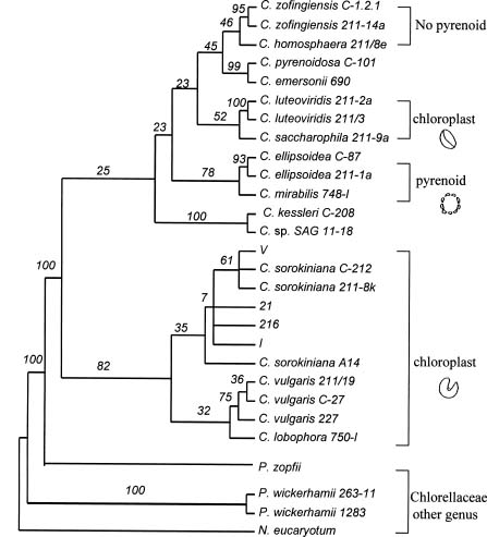
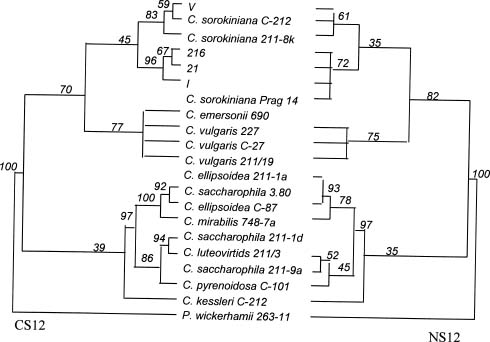
Abstract. Members of the Chlorella species are very simple unicellular algae, easy to cultivate and widely used in various physiological studies. Their morphological and physiological characteristics, however, normally change with the environment, making species identification difficult. To elucidate the relationship between various strains of Chlorella, this investigation analyzed the nuclear-encoded and chloroplast-encoded small-subunit rDNA sequences of four strains of Chlorella using PCR techniques. These strains were isolated from different rivers and ponds in Taiwan and Indonesia, and then compared and identified using stock strains of Chlorella spp. from the culture collection centers, and published DNA sequence data from Genbank. Experimental results attributed the isolated strains mainly to C. sorokiniana, a common species of green algae which grows in freshwater ecosystems at around 36°C. In addition, phylogenetic analysis of nuclear-encoded and chloroplast-encoded small-subunit rDNA sequences from spherical green algae of the genera Chlorella revealed the sequences to closely resemble each other. Further analyses indicated that Chlorella spp. 216 was close to Chlorella spp. 21 and I. Generally, the chloroplast data sets supported the lineages more than the nuclear data sets did. Strains 21 and 216 were closer to I. Comparisons with some of the morphological and biochemical data indicated that the phylogenetic analysis of rDNA sequences was in line with results obtained by conventional methods.

Keywords: Chlorella; Chloroplast; Nuclear; PCR; Phylogeny; Small subunit.

Introduction

The simple and common green algae of the genus Chlorella (Beijerinck, 1890) are placed below the order Chlorococcales and family Chlorellaceae (Hoek et al., 1995). Reproduction is asexual and achieved by producing non-motile autospores. Species of this genus are widespread in fresh water and in the sea, air, and soil. Warburg (1919) discovered that pure cultures of these fast-growing micro-organisms can be used as the ideal experimental materials for research on photosynthesis, nitrate reduction, physiology and biochemistry. Recently, Chlorella have been extensively studied and employed in various practical applications in agriculture and biotechnology. Chlorella are also used as protein-rich foods for sewage oxidation (Kessler, 1982). However, their cells do not exhibit characteristics that differentiate them from the morphological properties which are typically the basis of a classical taxonomic treatment of other algae (Shihira and Krauss, 1965).

Although the traditional taxonomic characteristics of Chlorella spp. indicate that morphological, biochemical and physiological properties are used in its identification, the cell size and shape are variable and largely depend on varying age, nutrition, and environmental factors (Fott and Novakova, 1969; Komárek and Fott, 1983). Additionally, certain biochemical and physiological characteristics are

not species specific (Kessler, 1982; Kessler, 1984; Kalina and Puncocharova, 1987). Therefore, classifying unknown isolated samples can be difficult.

The high number of copies and the inclusion of conserved and variable regions by evolution of ribosomal RNA genes (rDNA) have made possible a new method of species identification, classification and phylogenetic relationship determination (Mullis et al., 1986; Mullis and Faloona, 1987; Saiki et al., 1988). Algae cells have chloroplast genes just like higher plants, meaning cells have two varieties of SSU rDNA, nuclear-encoded and chloroplast-encoded. These characteristics have been considered for algae taxonomy and phylogenetic relationships (Huss and Sogin, 1990; Wilcox et al., 1992; Steinkötter et al., 1994; Schreiner, 1995). Meanwhile, Krienitz et al. (1996) compared the morphology and nuclear encoded SSU rDNA of green algae and found them closely related.

Recently, biochemical, physiological, and ultrastructural characters, together with molecular phylogeny based on the complete 18sRNA sequence, have led to the proposal that only four species should be kept in the genus Chlorella: C. vulgaris, C. lobophora, C. sorokiniana, C. kessleri (Huss et al., 1999).

Phylogenetic analysis of the order Chlamydomonadales revealed that this set of chloroplast data exhibited stronger support for comparable lineages than the set of nuclear data (Buchheim et al., 1996). Therefore, in this study field isolates from rivers in Indonesia and Taiwan are investigated, and the main observed cells were spherical,

*Corresponding author. Fax: 886-2-23626455; E-mail: m046@ccms.ntu.edu.tw


Botanical Bulletin of Academia Sinica, Vol. 42, 2001

Table 1. Source of the Chlorella spp.

Strain number Location Ecological environment

pH Temp (°C)

21 Wonokromo, Jagir, Surabaya, Indonesia 7.9 30

216 Wonokromo, Jagir, Surabaya, Indonesia 7.5 32

I Ching-lung His, Ching-lung Village, Taimali Town, Taitung, Taiwan 7.2 27

V Alun-alun, Malany, Indonesia 7.9 27


Table 2. Chlorellaceae rDNA sequence from GenBank (EMBL).

Chlorellaceae species Source of SSU rDNA

Nuclear Chloroplast

Nanochlorum eucaryotum

Strain Mainz 1 X06425

Prototheca wickerhamii

Strain 263-11 X56098 X74309

Strain Pore 1283 X56099

Prototheca zopfii

Strain SAG 263-1a X63519

Chlorella species

C. ellipsoidea

Strain 211-1a X63520

Strain IAM C87 D13324 X12742

C. emersonii

Strain NIES 690 AJ242761 (NS12) AJ242751 (CS12) AJ242747 (CS34)

C. homosphaera

Strain CCAP 211/8e X73996

C. kessleri

Strain SAG 211-11g X65099

Strain SAG 211-11h D11346

Strain IAM C-208 AJ242765 (NS12) AJ387750 (CS12) AJ242769 (CS12)

C. lobophora

Strain Andreyeva 750-I X63504

C. luteoviridis

Strain CCAP 211/3 AJ242758 (NS12) AJ242767 (CS12) AJ242768 (CS34)

Strain SAG 211-2a X73998

C. mirabilis

Strain Andreyeva 748-I X74000 X65100

C. pyrenoidosa

Strain IAM C-101 AJ242762 (NS12) AJ242752 (CS12) AJ242749 (CS34)

C. protothecoidea

Strain 211-7a X65688

C. saccharophila

Strain 3.80 D11348

Strain 211-1d D11349

Strain SAG 211-9a X63505

Chlorella sp. 21 AJ242760 (NS12) AJ242754 (CS12) AJ387748 (CS34)

Chlorella sp. 216 AJ242759 (NS12) AJ387753 (CS12) AJ387749 (CS34)

Chlorella sp. I AJ242764 (NS12) AJ238891 (CS12) AJ387755 (CS34)

Chlorella sp. V AJ242766 (NS12) AJ387759 (CS12) AJ387752 (CS34)

C. sorokiniana

Strain 211-8k X62441 X65689

Strain Prag A14 X74001

Strain IAM C-210 AJ242763 (NS12) AJ387756 (CS12) AJ387751 (CS34)

C. vulgaris

Strain 211-11b X16579

Strain 211-1e D11347

Strain IAM 211/19 AJ242755 (NS12) AJ242754 (CS12) AJ242771 (CS34)

Strain NIES 227 AJ242756 (NS12) AJ242750 (CS12) AJ242770 (CS34)

Strain IAM C-27 AJ242757 (NS12) AJ242753 (CS12) AJ242748 (CS34)

C. zofingiensis

Strain SAG 211-14a X74004

Strain Bethesda C-1.2.1 X74005

Chlorella sp. SAG 11-18 X74006


Wu et al. — Identification of Chlorella spp. isolates

reproduced with autospores, and had no flagella. These basic characteristics suggest that isolates are primary and belong to Chlorella. Partial SSU rDNA data from nuclear regions are compared with the chloroplast of all isolates herein, and various other conventional identification methods are also examined.

Materials and Methods

Taxon Selection

Field samples of the investigated microalgae were collected from selected rivers and ponds in Taiwan and Indonesia. Table 1 lists some physicochemical and ecological data from these environments. Meanwhile, Table 2 lists the nuclear and chloroplast encoded SSU rDNA sequence data (accession numbers are given in parentheses) taken from the GenBank/EMBL databases and integrated into the analyses, including Chlorella, Prototheca, and Nanochlorum, three genera of Chlorellaceae.

Nuclear and Chloroplast Sequence Data

Both nuclear and chloroplast sequence data were obtained from the PCR amplifications using genomic DNA. Cells were cultivated in an autotrophic media at 25°C under continuous illumination. They were then harvested by centrifugation (10,000 rpm for 30 min) and ground in porcelain vessels with liquid nitrogen. Total cellular DNA was extracted from cell samples, as described by Hseu et al. (1996a). Table 3 illustrates PCR primers used for amplifying and sequencing the partial SSU segment. PCR amplification reactions were performed as described by Hseu et al. (1996a,b). For DNA sequence analysis, an ABI PRISM 377-96 DNA-sequencer (Perkin-Elmer, CA, USA), was used with ABI PRISM BigDye. Meanwhile, a terminator cycler sequencing ready reaction kit (PE Applied Biosystem, USA) was also employed.

Data Analyses

The alignment of nuclear and chloroplast sequences was conducted using SeqApp 1.9 (Gilbert, 1992). Phylogenetic analyses were performed via the SSU data set using the parsimony method with the program PAUP, Version 4.0 (Swofford, 1998). Bootstrap values from 100 repeated samplings were calculated for each set of data. All tree relationships were rooted through the outgroup method.

Conventional Methods

Isolated cell samples were observed under Nikon Eclipse E600 optical microscopes with a phase contrast device. Meanwhile, thermophilic ability was tested by cells grown on agar slants at elevated temperature. Finally, starch hydrolysis abilities were assessed for all isolates on agar plates containing suitable quantities of soluble starch.

Results

Structures of the Sequence Data

Table 4 summarizes the primary structure of the two independent data sets. This table also compares variable and informative sites, indicating that the chloroplast data are more variable than the nuclear data in terms of number and percentage of both variable and informative sites.

Phylogenetic Analysis of Nuclear Data

According to the primers designed by White et al. (1990), the SSU sequence from the GenBank/EMBL database was separated into several regions, namely the NS12, NS34, NS56, and NS78 regions. Different regions in parsimony analysis exhibit the same tendency (data not shown), so the NS12 region was employed herein to ana

Table 3. Primers used in PCR reaction.

Primer Source Primer sequence Product size (bp)

NS1 Nuclear SSU 5'-GTAGTCATATGCTTGTCTC-3'

NS2 Nuclear SSU 5'-GGCTGCTGGCACCAGACTTGC-3' 550

CS1 Chloroplast SSU 5'-CGGCTGATTAGCTTGTTGG-3'

CS2 Chloroplast SSU 5'-GAGTGCTTTCGCCTTTGG-3' 500

CS3 Chloroplast SSU 5'-AAGGCCAAAGCACTCTGC-3'

CS4 Chloroplast SSU 5'-TTCCTCCGGCTTATCACC-3' 450-500

Table 4. Nucleotide sequence variation in the nuclear SSU NS12 region and in the chloroplast SSU CS14 region in Chlorellaceae.

Category of comparison Nuclear Data Chloroplast Data

Positions aligned 446 854

Chlorellaceae

Total variable sites 129 (28.9%) 380 (44.5%)

Phylogenetically informative sites 41 (9.2%) 164 (19.2%)

Informative / variable 31.8% 43.1%

Chlorella spp.

Total variable sites 73 (16.4%) 243 (28.5%)

Phylogenetically informative sites 23 (5.2%) 132 (15.5%)

Informative / variable 31.7% 54.4%


Botanical Bulletin of Academia Sinica, Vol. 42, 2001

lyze the isolates from the field samples. Parsimony analysis of the nuclear data set resulted in an efficient method for identification of strains. This analytical result illustrated the taxonomic congruence between the morphological characteristics (Figure 1). The confidence of the Chlorella spp. cluster was higher, up to 100%, and four strains were closely associated with the C. sorokiniana, C. vulgaris, and C. lobophora clusters. Furthermore, strains I, 21, and 216 were closer to C. sorokiniana Prag A14 while strain V was closer to C. sorokiniana C-212 and C. sorokiniana 211-8k.

Phylogenetic Analysis of Chloroplast Data

Parsimony analysis of the chloroplast SSU CS12, CS34 region has demonstrated sufficient congruence with morphological characters, as using P. wickerhamii as the outgroup, the four isolated strains were most closely related to C. sorokiniana (Figure 2). Furthermore, strains 21, 216 and I were closer to each other, in the middle degree, and strains 21 and 216 were most closely related to each other. Strain V was also more closely related to C. sorokiniana C-212 and C. sorokiniana 211-8k, from the phylogenetic analysis of nuclear data, so the chloroplast data were more robust across methods of phylogenetic reconstruction than the nuclear data (Figure 3).

Some Data from Applying Traditional Methods

Table 5 summarizes the results of microscopic observations of field isolates. The cells are spherical, cup-shaped chloroplasts with pyrenoids making it extremely difficult to differentiate them from one another. Under field conditions the algae propagated mostly via the two and four autospore formation.

At the upper temperature range, the growth experiment showed the four strains were all grown thermophilic, to 36°C. This finding also indicated that all isolates could be placed in the C. sorokiniana species with similar results in the molecular investigation. Starch hydrolysis testing revealed that strain V tested negative for starch hydrolysis ability, while strains 21, 216 and I all possessed proven starch hydrolysis ability coinciding with the results for C. sorokiniana Prag A14 (Table 5).

Discussion

Parsimony analyses in nuclear-encoded SSU rDNA, Chlorella spp. exhibited 90-100% similarity in the NS12 region. Meanwhile, Chlorella spp. cluster confidence

Figure 1. Phylogenetic relationships inferred by Chlorella spp. nuclear SSU NS12 nucleotide sequence data. The tree produced by use of a heuristic search in PAUP 4.0 from cladistically informative characters. Tree length = 355; consistency index (CI) = 0.6347; retention index (RI) = 0.6899. Values above branches are confidence levels estimated by 100 bootstrap replicates.

Figure 2. Phylogenetic relationships between CS12 and CS34 nucleotide sequences data.

Table 5. Morphology characters, growth limited temperature and starch hydrolysis of 4 Chlorella spp.

Species Cell form Pyrenoid Chloroplast form Cell size (µm) Temp. limit Thermophily Starch hydrolysis

C. sp. 21 Spherical + Cup-shaped 3~6 ¡Ü 38°C + +

C. sp. 216 Spherical + Cup-shaped 3~6 ¡Ü 38°C + +

C. sp. I Spherical + Cup-shaped 3~6 ¡Ü 38°C + +

C. sp. V Spherical + Cup-shaped 3~6 ¡Ü 41°C + -


Wu et al. — Identification of Chlorella spp. isolates

gene, a situation similar to that found in prokaryotes (Cedergren et al., 1988; Peer et al., 1990). Chloroplast and mitochondria are the places for processing energy change, and they produce more freeradicals, leading to genetic variation.

Nuclear data were more prominent in Chlorella gene levels than the chloroplast data. However, combining chloroplast CS12 with CS34 regions improves phylogenetic analysis in Chlorella genus levels (data not shown), making the analysis similar to that of the nuclear NS12 region. Perhaps the chloroplast gene was more variable among the species, making this region more suitable for species analysis. Combining the two regions into a longer sequence, enhances its phylogenetic analysis (Buchheim et al., 1996).

The molecular classification was examined using conventional methods. Microscopic observation of four isolates revealed significant similarities to one another, and also that they were congruent with the species C. sorokiniana. Elevated growth temperature also indicated a similar observation. According to Kessler (1982) and Huss et al. (1999), C. sorokiniana is the only species of Chlorella capable of growing above 36°C. Notably, strain V had the highest growth temperature. Kessler (1982) reported that C. sorokiniana 211-8k and C. sorokiniana Prag A14 exhibited differing starch hydrolysis ability. Therefore, in this study, we examined their biochemical characteristics. We suspect that strains 21, 216 and I possessed this ability as did C. sorokiniana Prag A14. Meanwhile, strain V did not perform the same as C. sorokiniana 211-8k. This molecular method of ribosomal DNA sequences was employed not only to reveal unknown strains, but also to reveal strain relationships through phylogenetic analysis. The process applied herein could be applied to classifying other unknown species of green algae.

Acknowledgements. The authors would like to thank Taiwan Chlorella Industry Manufacturing Company (Taipei, Taiwan) for assistance in isolating Chlorella strain I, and also PT. Sunchlorella Indonesia Manufacturing Corporation (Jawa Timur, Indonesia) for cooperation in isolating Chlorella strains 21, 216 and V.

Literature Cited

Beijerinck, M.W. 1890. Culturversuche mit Zoochlorellen, Lichenen-goniden und anderen niederen Algen. Bot. Ztg. 45: 726-739, 741-753, 757-767, 781-784.

Buchheim, M.A., C. Lemieux, C. Otis, R.R. Gutell, R.L. Chapman, and M. Turmel. 1996. Phylogeny of the Chlamydomonadales (Chlorophyceae): A comparison of ribosomal RNA gene sequences from the nucleus and the chloroplast. Mol. Phyl. Evol. 5: 391-402.

Cedergren, R., M.W. Gray, and D. Sankoff. 1988. The evolutionary relationships among known life forms. J. Mol. Evol. 28: 98-112.

Fott, B. and M. Novakova. 1969. A monograph of the genus Chlorella. In B. Fott (ed.), The Freshwater Species, Stud

Figure 3. Phylogenetic relationships between NS12 and CS12 nucleotide sequences data.

reached 100%, indicating that Chlorella genes had a unique sequence in this region. This feature could be used to distinguish them from non-Chlorella species. Four isolates (strains 21, 216, I, V) were located on the C. sorokiniana lineage, and their similarity, up to 98%, was sufficient to consider them the same species. For phylogenetic analyses in this investigation, the morphological characteristics were compared and the same result was reached. Algae with cup-shaped chloroplast were clustered together, and other characteristics, such as the presence of pyrenoid, were also consistent with this finding (Figure 1). Phylogenetic analysis values on non-cup-shaped chloroplast algae were not high, and perhaps taxonomic characters here were more complicated. Chlorella ellipsoidea and C. saccharophila were ellipsoid-shaped algae. Although C. ellipsoidea and C. mirabilis shared a similar pyrenoid structure, C. saccharophila and C. luteoviridis shared a similar chloroplast structure. These characteristics made phylogenetic analyses more difficult.

Parsimony analyses of chloroplast-encoded SSU rDNA CS12 and CS34 regions was conducted herein. Apparently, these four isolates more closely resembled the species C. sorokiniana, rather than C. vulgaris. Chloroplasts with cup-shaped species are all clustered together. Strains 21, 216 and I were relatively close to one another, and strain V, C. sorokiniana C-212 and C. sorokiniana 211-8k were also relatively close to one another. Meanwhile, strains 21 and 216 were the most closely related species, and they had been isolated from the same environment. Comparing nuclear-SSU NS12 region data with chloroplast SSU CS12 region data revealed that outgroup to belong to C. wickerhamii. Phylogenetic analysis results share the same tendency, and in unknown species analysis, chloroplast SSU data had results clearer than the conventional method (Figure 3).

In primary structure analysis (Table 4), chloroplast data had more variable regions than nuclear data. This phenomenon could increase the difficulty of getting phylogenetic results. The changeable chloroplast gene may exist because its replication was less compact than the nuclear


Botanical Bulletin of Academia Sinica, Vol. 42, 2001

ies in Phycology. Academia, Praha, pp. 10-74.

Gilbert, D.G. 1992. SeqApp: A Biosequence Editor and Analysis Application. Version 1.9. Writen using MacApp, 1985-1991 Apple Computer, Inc.

Hoek, C., D.G. Mann, and H.M. Johns. 1995. Algae _ An Introduction to Phycology. Cambridge University Press.

Hseu, R.S., H.H. Wang, H.F. Wang, and J.M. Moncalvo. 1996a. Differentiation and grouping of isolates of the Ganoderma lucidum complex by random amplified polymorphic DNA-PCR compared with grouping on the basis of internal transcribed spacer sequences. Appl. Environ. Microbiol. 62: 1354-1363.

Hseu, R.S., J.M. Moncalvo, H.F. Wang, and H.H. Wang. 1996b. Application of PCR-amplified DNA to differentiate the Ganoderma isolates. J. Chinese Agri. Chem. Soc. 34: 129-143.

Huss, V.A.R. and M.L. Sogin. 1990. Phylogenetic position of some Chlorella species within the Chlorococcales based upon complete small-subunit ribosomal RNA sequences. J. Mol. Evol. 31: 432-442.

Huss, V.A.R., C. Frank, E.C. Hartmann, M. Hirmer, A. Kloboucek, B.M. Seidel, P. Wenzeler, and E. Kessler. 1999. Biochemical taxonomy and molecular phylogeny of the genus Chlorella sensu lato (Chlorophyta). J. Phycol. 35: 587-598.

Kalina, T. and M. Puncocharova. 1987. Taxonomy of the subfamily Scotiellocystoideae Fott 1976 (Chlorellaceae, Chlorophyceae). Arch. Hydrobiol. Suppl. 73.4 (Algological Studies) 45: 437-521.

Kessler, E. 1982. Chemotaxonomy in the Chlorococcales. In F.E. Round and D.J. Chapman (eds.), Progress in Phycological Research, Vol. 1. Elsevier, Amsterdam, pp. 111-135.

Kessler, E. 1984. A general view on the contribution of chemotaxonomy to the systematic of green algae. Systematics Association, Special vol. 17, Academic Press, London, pp. 391-407.

Komárek, J. and B. Fott. 1983. Das Phytoplankton des Suesswassers. Systematik und Biologie. 7. Teil, 1 Haelfte. Chlorophyceae (Gruenalgen). Ordnung Chlorococcales, In G. Huber-Pestalozii, (ed.), Die Binnengewasser 16. E. Schweizerbart'sche Verlagsbuchhandlung (Naegele & Obermiller), Stuttgart, pp. 1-1044.

Krienitz, L., V.A.R. Huss, and C. Hümmer. 1996. Picoplanktonic Choricystis species (Chlorococcales,

Chlorophyta) and problems surrounding the morphologically similar `Nannochloris-like algae'. Phycologia 35: 332-341.

Mullis, K.B., F.A. Faloona, S.J. Scharf, R.K. Saiki, G.T. Horn, and H.A. Erlich. 1986. Specific enzymatic amplification of DNA in vitro: the polymerase chain reaction. Cold Spring Harbor Symo. Quant. Biol. 51: 263-273.

Mullis, K.B. and F.A. Faloona. 1987. Specific synthesis of DNA in vitro via a polymerase-catalyzed chain reaction. Methods Enzymol. 155: 335-350.

Peer, Y.V., J.M. Neefs, and R. Wachter. 1990. Small ribosomal subunit RNA sequences, evolutionary relationships among different life forms, and mitochondrial origins. J. Mol. Evol. 30: 463-476.

Saiki, R.K., D.H. Gelfand, and S. Toffel. 1988. Primer-directed enzymatic amplification of DNA with a thermostable DNA polymerase. Science 239: 487-491.

Schreiner, M., M. Geisert, M. Oed, J. Arendes , U. Gungerich, H.J. Breter, K. Stuber, and D. Weinblum. 1995. Phylogenetic relationship of the green alga Nanochlorum eucaryotum deduced from its chloroplast rRNA sequences. J. Mol. Evol. 40: 428-442.

Shihira, I. and R.W. Krauss. 1965. Chlorella. Physiology and Taxonomy of Forty-One Isolates. University of Maryland, College Park, Maryland, pp. 1-92.

Steinkötter, J., D. Bhattacharya, I. Semmelroth, C. Bibeau, and M. Melkonian.1994. Prasinophytes from independent lineages within the Chlorophyta: evidence from ribosomal RNA sequence comparisons. J. Phycol. 30: 340-345.

Swofford, D.L. 1998. PAUP: Phylogenetic Analysis Using Parsimony. Version 4.01. Champaign: Illinois Natural History Survey.

Warburg, O. 1919. Uber die Geschwindigkeit der Kohlensàurezersetzung in lebenden Zellen. Biochem. Z. 100: 20-270.

White, T.J., T. Bruns, S. Lee, and J. Taylor. 1990. Amplification and Direct sequencing of fungal ribosomal RNA genes for phylogenetics. PCR Protocols: A Guide to Methods and Applications, Copyright 1990 by Academic Press, Inc., pp. 315-322.

Wilcox, L.W., L.A. Lewis, P.A. Furest, and G.L. Floyd. 1992. Assessing the relationships of autosporic and zoosporic Chlorococcalean green algae with 18S rDNA sequence data. J. Phyol. 28: 381-386.


Wu et al. — Identification of Chlorella spp. isolates

§Q¥Î®Ö¿}Åé®Ö»Ä§Ç¦CŲ©w¤p²yĦ¤§¤ÀÂ÷®è

§d«ÅÀM ³\·ç²» ªL¨}¥­

°ê¥ß¥xÆW¤j¾Ç¹A·~¤Æ¾Ç¬ã¨s©Ò

¤p²yĦ Chlorella spp. ¬°¥Ø«e³Q¼sªxÀ³¥Îªº·LĦ¤§¤@¡A¥Ñ©ó Chlorella spp. ¬°ºc³y²³æ¤§³æ²Ó­MĦÃþ¡A §@¬°ºØŲ©w¨Ì¾Úªº§ÎºA¯S¼x±`¦]¥ÍºAÀô¹Ò§ïÅܦӲ§¡A³y¦¨Å²©w¤Wªº§xÃø¡C¬°ÁA¸Ñ Chlorella spp. ºØ¶¡ªº¿Ë½tÃö«Y¡A¥»¬ã¨s¥H¤À¤l¥Íª«§Þ³N¤¤ªº»E¦X ú@³sÂê¤ÏÀ³©ñ¤j¯S©w°Ï°ì¡A¤ÀªR¥|®è¤À§O±q¥xÆW©M¦L¥§©Ò¤ÀÂ÷¤§ ¤p²yĦ²Ó­M®Ö¡A¤Î¸­ºñÅ餺ªº®Ö¿}Å餧¤p¦¸Åé°ò¦]§Ç¦C¡A»P°ò¦]¸ê®Æ®w¤wµoªíªº°ò¦]§Ç¦C¥[¥H¤ñ¹ï¡A¥H¶i ¦æ¤p²yĦÄÝĦ®è Chlorella spp. ªº¤ÀÃþŲ©w¡A¨Ã»P«O¦s¤¤¤ßĦºØ¡A¤ñ¸û¤ÀÃþµ²ªG¤¬¬ÛÅçÃÒ¡Cµ²ªGÅã¥Ü¨Ñ¸Õ ªº¥|®è¤p²yĦ§¡±µªñ©ó±`¨£ªººñĦ Chlorella sorokiniana¡A¦¹¬°¤@¥i¥Íªø©ó 35°C ¤ô°ì¤¤ªº­@°ª·Å¤p²yĦ¡A ¤ÀªRµ²ªG§óÅã¥ÜĦ®è 216 §ó±µªñ©óĦ®è 21 ©M I¡C¸­ºñÅé SSU rDNA °ò¦]§Ç¦C¤ñ²Ó­M®ÖªÌ§ó²M·¡ªº¤ÀªR ¥X¥|®è¤p²yĦªº¿Ë½tÃö«Y¡CĦ®è 21 »P 216 ¸òĦ®è I ¸û¬°±µªñ¡C±q§ÎºA»P¥Í¤Æ©Ê½è¤ñ¸ûÅçÃÒ«á¡AÅã¥Ü¦¹ rDNA ¤ÀÃþµ²ªG»P¶Ç²Î¤ÀÃþ¤èªk©Ò±oµ²ªG¡A§e±j¯Pªº¬ÛÃö©Ê¡C

ÃöÁäµü¡G ¤p²yĦ¡F®Ö¿}Å餧¤p¦¸Åé¡F²Ó­M®Ö¡F¸­ºñÅé¡F»E¦X ²¡³sÂê¤ÏÀ³¡F¨t²Îµo¥Í¾Ç¡C