Bot. Bull. Acad. Sin (2001) 42: 215-222

Chiang and Lin — Diphyscium of Taiwan

Taxonomic revision and cladistic analysis of Diphyscium (Family Diphysciaceae) of Taiwan

Tzen-Yuh Chiangl,* and Shang-Hsiung Lin2

lDepartment of Biology, National Cheng-Kung University, Tainan 701, Taiwan

2Department of Biology, Tunghai University, Taichung 407, Taiwan

(Received April 2, 1999; Accepted September 7, 1999)

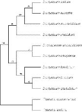
Abstract. Six species of Diphyscium, with three new additions to the moss flora of Taiwan, are revised taxonomically, including D. chiapense Norris ssp. unipapillosum (Deguchi) T. Y. Chiang and S. H. Lin, stat. nov. Diagnosable keys are provided. In order to test Norris' and Deguchi et al.'s classifications, cladistic analyses were carried out on eleven morphological traits scored from nine Asiatic species of Diphyscium. Two equally parsimonious trees rooted at the Theriotia species were recovered by PAUP. The reconstructed phylogeny, which suggested a close relationship between the taxa with single papillose lamina cells and the species with pluripapillose lamina cells, invalidated Norris' hypothesis. The parsimony tree agrees with Deguchi et al.'s classification, in which three groups were classified based on cell-wall papillosity. Based on the inferred phylogeny, the "orifice" stomata may have evolved more than once in Diphyscium, while a single "secondary loss" event occurred in the group of D. involutum, D. rupestre, D. mucronifolium and D. suzukii.

Keywords: Cladistics; Diphyscium; D. chiapense ssp. unipapillosum; Orifice stomata; Papillosity; Phylogeny; Taiwan; Taxonomy.

Introduction

Diphyscium is a conspicuous moss characterized by its obliquely ovoid capsule with dorsiventral differentiation and a narrow mouth as well as strongly differential, bristled perichaetial leaves. Diphyscium and Theriotia constitute the Diphysciaceae. Diphyscium is distinguished from the latter by the well differentiated laminate portion of the leaves, waxy anticlinal walls in the dorsal side of the capsules, and the absence of chlorophyllosd cells in the leaves (cf. Deguchi, 1984a). Phylogenetically, Diphysciaceae are closely related to the Buxbaumiaceae, sharing common structure and ontogeny of the capsules (Crum and Anderson, 1981). The reduced gametophytes and elongated setae in Buxbaumiaceae, however, suggest an independent lineage from the Diphysciaceae. Despite the remarkable differences of the sporophytic structure, the gametophytes of Diphyscium in some cases [e.g., D. pocsii (Bizot) Zand.] resemble the Pottiaceae. Deguchi (1975, 1984b) and Norris (1981) utilized the "collared" axillary hairs to differentiate the Diphysciaceae from the Pottiaceae.

About twenty-three species have been recognized, with a distribution ranging from the tropics to the temperate (van der Wijk et al., 1967; Crum and Anderson, 1981;

Crosby et al., 1992; Crosby and Magill, 1994; Deguchi et al., 1997). Deguchi et al. (1997) distinguished three groups of Diphyscium based on the papillosity of their leaf-cell surface: 1) species with smooth leaf cells (e.g., D. involutum), 2) species with unipapillose-mamillose leaf cells (e.g., D. chiapense), and 3) species with pluripapillose-mamillose leaf cells (e.g., D. fulvifolium). In contrast, Norris (1981) recognized two groups, with D. foliosum and D. involutum being representative. Diphyscium chiapense was regarded as a member of the D. involutum group according to his classification. Although Deguchi et al. (1997) treated D. chiapense and its relatives as a new group, the phylogenetic relationships among the three groups were not further discussed and therefore have remained unknown.

Interestingly, although the plants have discernible morphological characteristics, little research has been carried out on the east Asiatic (i.e. As 2; van der Wijk et al., 1967) taxa except for the Japanese species (Iwatsuki, 1976; Deguchi, 1983, 1984b; Noguchi, 1987; Deguchi et al., 1997). For example, the floristic survey of the Chinese Diphyscium has been poorly documented, with only four species and one variety known (cf. Redfearn et al., 1996). The tiny plants, with pottiaceous appearance when the sporophytes are absent, may have caused it to be overlooked by collectors.

The first record of this genus in Taiwan is from 1935, with the collections of Diphyscium fulvifolium and D. formosicum (= D. involutum) (Horikawa, 1935a, 1935b).

*Corresponding author. Tel: +886-6-2757575 ext. 65525; Fax: +886-6-2742583; E-mail: tychiang@mail.ncku.edu.tw


Botanical Bulletin of Academia Sinica, Vol. 42, 2001

Wang and Lin (1974a, 1974b) reported two new species, D. macrophyllum and D. rotundatifohum, which were later synonymized to D. granulosum and D. fulvifolium, respectively (Iwatsuki, 1976). Subsequently the above species have been revised in taxonomic treatments (Chuang, 1973) or described by floristic surveys (Lin, 1981,1984; Lin and Tsai, 1984; Lai, 1986).

We studied the taxonomy of Diphyscium in Taiwan based on both our own collection and herbaria specimens. Six species, including three newly found taxa, are reported in this paper. We analyzed the phylogenetic relationships between the species belonging to the three groups of Deguchi et al.'s (1997) classification cladistically and tested the validity of Norris' (1981) hypothesis. Based on our reconstructed phylogeny, the morphological evolution of leaf-cell papillosity and stomata is discussed.

Materials and Methods

Materials were obtained both from field collection and loans from herbaria (HAST, HIRO, NMNS, TNS, TUNGH). Specimens were examined with stereo and light microscopes. Six species were identified and distinguished based on the diagnosable characters. In total, 11 characters were examined and scored to character states. Based on the previous taxonomy, Theriotia lorifolia and T. kashmiretisis were chosen as outgroups and were used for character polarity (Deguchi, 1984a). In order to gain more insight into the phylogeny and morphological evolution, D. suzukii (a Japanese species), D. mucronifolium (a species of Java) and D. chiapense (a Mexican species) were also included in the analysis. The characters included in this analysis and the polarization of character states are as follows (Table 1):

1. Chlorophyllose cells in cross section of costa: present (0) and absent (1). The chlorophyllose cells are only present in Theriolia.

2. Thickness of lamina: multistratose (0) and bistratose (1). Theriolia species have multistratose lamina; while Diphyscium species have bistratose lamina (cf. Noguchi, 1987).

3. Waxy anticlinal walls in exothecial cells: absent (0) and present (1). The waxy anticlinal walls are differentiated on the dorsal side of the capsules of Diphyscium only.

4. Cell-wall papillosity: smooth (0), single-papillose (1), and pluri-papillose (2).

5. Stomata: differentiated at apophysis (0), differentiated at orifice (1), and absent (2). Most species, both ingroup and outgroup, have differentiated stomata either at apophysis (e.g., D. foliosum) or at orifice (e.g., D. chiapense). Stomata are absent in D. involutum and its relatives.

6. Subula of inner perichaetial leaves: smooth (0), and scabrous (1).

7. Upper portion of perichaetial leaves: entire (0), shallowly bifurcate (1), and deeply bifurcate (2). State 1 exists in D. fulvifolium only; state 2 exists in D. foliosum and D. granulosum.

8. Margins on inner perichaetial leaves: cilia (0), dissected (1), and entire (2). The appendages are differentiated in outgroups and most ingroup taxa except for D. chiapense.

9. Leaf-shape: linear with a short-ovate sheathing base (0), ovate-lanceolate (1), and linear without sheathing base (2). State 2 exists in most Diphyscium, except for D. granulosum (state 1).

10. Leaf apex: acute (0), apiculate (1), and obtuse (2).

11. Costa: excurrent (0), reaching apex (1), ending below apex (2).

Cladistic Analysis

Cladistic analyses of the morphological characters were carried out using the exhaustive searches of the PAUP computer software (Version, 3. 1. 1, Swofford, 1993). Most characters were treated as ordered, except for characters 5 and 10. All characters were unweighted.

Confidence of clades was tested using the complete-and-partial (CP) bootstrap technique to correct the bias (conservativeness) of the standard bootstrap approach (Li

Table 1. Distribution and polarization of characters and character states for Diphyscium and Theriofia in cladistic analysis.

Taxa\Characters 1 2 3 4 5 6 7 8 9 10 11

Outgroups:

Therlotia kashmirensis 0 0 0 0 0 0 0 0 0 0 0

Therlotia lorifolia 0 0 0 0 0 0 0 0 0 0 0

Ingroups:

Diphyscium suzukii 1 1 1 0 2 0 0 0 2 1 2

Diphyscium rupestre 1 1 1 0 2 0 0 1 2 1 2

Diphyscium mucronifolium 1 1 1 0 2 0 0 1 2 1 0

Diphyscium involutum 1 1 1 0 2 0 0 1 2 0 1

Diphyscium chiapense 1 1 1 1 1 1 0 2 2 0 2

ssp. unipapillosum 1 1 1 1 1 0 0 2 2 0 2

Diphyscium fulvifolium 1 1 1 2 1 1 1 0 2 0 1

Diphyscium foliosum 1 1 1 2 0 1 2 0 2 2 2

Diphyscium granulosum 1 1 1 2 0 1 2 0 1 2 2


Chiang and Lin — Diphyscium of Taiwan

and Zharkikh, 1995). The fit of character data to phylogenetic hypotheses was evaluated using the consistency index (CI) (Kluge and Farris, 1969) and the retention index (RI) (Farris, 1989). The statistical significance of CI was determined according to Klassen et al. (1991).

Results and Discussion

Taxonomic Treatments

Six taxa of Diphyscium are revised, including three new additions to the moss flora of Taiwan. Diagnostic keys are provided.

Keys to the Species of Diphyscium in Taiwan

1. Laminal cells smooth 2

2. Leaf apex apiculate D. rupestre

2. Leaf apex acute D. involutum

1. Laminal cells mamillose or papillose 3

3. Laminal cells unipapillose D. chiapense ssp. unipapillosum

3. Laminal cells pluri-papillose 4

4. Upper portion of inner perichaetial leaves shallowly bifurcate, stomata distributing at orifice D. fulvifolium

4. Upper portion of inner perichaetial leaves deeply bifurcate, stomata distributed at apophysis 5

5. Leaves linear D. foliosum

5. Leaves ovate-lanceolate D. granulosum

DIPHYSCIUM Mohr, 1803.

1. Diphyscium chiapense Norris ssp. unipapillosum (Deguchi) T. Y. Chiang and S. H. Lin, stat. nov. Figure 1

Basionym. Diphyscium unipapillosum Deguchi, J. Jap. Bot. 59: 97, f. 1. 1984.—TYPE: Japan, Pref. Kochi: Kamiyashiki, Nakainura-shi; 380 m alt., Deguchi 24901 (holotype in HIRO; isotype in TNS and Kochi University).

Syn. Diphyscium buckii Tan, Bryologist 93: 429, f. 1-5. 1990.

Taxonomic notes. This taxon was first described as D. unipapillosum (Deguchi, 1984b). Deguchi et al. (1997) synonymized both D. unipapillosum and D. buckii, a species described from the Philippines (Tan, 1990), with Diphyscium chiapense based on the uni-mamillose-papillose (both dorsal and ventral sides) lamina cells. Nevertheless, they pointed out that D. chiapense has papillose subula of the perichaetial leaves, while the subulae of the two other taxa are smooth on the surface. They considered the papillosity of the subula as local expressions and summarized a conspecific relationship between the three taxa. Our own observation shows that materials collected in Taiwan also have smooth subula, as do D. unipapillosum and D. buckii. The smooth subula there

fore appears to be stable in east Asian populations, from Japan to the Philippines, although few materials have been examined. From the view of phytogeography, the disjunct distribution between D. chiapense (Mexican populations) and D. unipapillosum (Asiatic populations) provides an opportunity for differentiation across geographical regions. Furthermore, the papillosity of subulae seems quite conserved in most Diphyscium species with no known polymorphism between populations. Based on the shared uni-mamillose-papillose lamina cells, which suggest a common origin, as well as the differentiation of the papillosity of subulae, we recognize a subspecies status for D. unipapillosum.

Diagnosable characters. This taxon is characterized by uni-papillose laminal cells, differentiated stomata distributed at orifice of the capsules, smooth subula, and the absence of appendages on margins of inner perichaetial leaves.

Illustrations. Deguchi (1984): f. 1-2 (as D. unpapillosum); Tan (1990): f. 1-5 (as D. buckii); Deguchi et al. (1997): f. 1.

Figure 1. Diphyscium chiapense Norris ssp. unipapillosum (Deguchi) T. Y. Chiang and S. H. Lin (drawn from Chiang 25316). A-C, Leaves (×32); D-E, Perichaetial leaves (×32); F, Antheridia (×71); G, Apical cells of leaf (×303); H, Median laminal cells (×303); I, Cells from basal part of leaf (×303); J-N, Parts of cross-sections of leaf (×303).


Botanical Bulletin of Academia Sinica, Vol. 42, 2001

Specimen examined. NANTOU HSIEN: Mt. Lu-lin, 2,600 m alt., Chiang 25316 (MO).

Distribution. Japan, Philippines and Taiwan (new record).

2. Diphyscium foliosum (Hedw.) Mohr, Ind. Mus. PI. Crypt.: 3. 1803. Figure 2

Taxonomic notes. Diphyscium foliosum is closely related to D. granulosum, sharing pluripapillose leaf cells and a bifurcate apex of perichaetial leaves. The lingulate leaves distinguish D. foliosum from the latter species, which has ovate-lanceolate leaves. Diphyscium foliosum is a new addition to the moss flora of Taiwan.

Diagnosable characters. Leaves are strongly curled and contorted when dry, and are erect-spreading when moist. This species is characterized by the lingulate leaves, scabrous subula, deeply bifurcate upper portion of the perichaetial leaves, and pluri-papillose lamina cells.

Illustrations. Iwatsuki & Mizutani (1972) f. 32; Noguchi (1976): f. 14h; Crum and Anderson (1981): f. 615; Noguchi (1987): f. 5A.

Specimen examined. KAOHSIUNG HSIEN: Mt. Chitou, ca. 2,700 m, Chiang 11465 (HAST)

Distribution. N. and C. America, Europe, Africa, Japan and Taiwan (new record).

3. Diphyscium fulvifolium Mtt., Trans. Linn. Soc. Bot. 2, 3: 143. 1891. Syn. Diphyscium rotundatifolium Wang & Lin, Ann. Mssouri Bot. Gard. 61: 526. 1974.—TYPE: Taiwan. Nantou Hsien, Luku Hsiang, Chitou, 1,150-1,750 m alt., 12 Nov 1971, Lin s. n. (holotype, TUNGH no. 1267!).

Taxonomic notes. Diphyscium fulvifolium is one of the most common species of this genus in Taiwan and is also highly polymorphic in leaf shape. Wang and Lin (1974b) reported D. rotundatifolium based on the obtuse to rounded leaf apices and uni-mamillose leaf cells. However, the description of the mamillosity of the leaf-cell surface was later found to be erroneous and was corrected to pluri-papillose (Iwatsuki, 1976). Iwatsuki (1976) further showed the variation in range of the leaf apices in both D. fulvifolium and D. rotundatifolium and reached the conspecificity conclusion. This species is generally distributed in the middle elevations of mountains in Taiwan.

Diphyscium fulvifolium is closely related to both D. foliosum and D. granulosum, sharing the pluri-papillose leaf cells. The shallowly bifurcate upper portion of the perichaetial leaves distinguishes it from the latter two species, in which the perichaetial leaves are deeply bifurcate.

Diagnosable characters. Diphyscium fulvifolium is characterized by pluri-papillose lamina cells, scabrous subulae, and shallowly bifurcate upper portion of the perichaetial leaves.

Illustrations. Iwatsuki & Mizutani (1972): Pl. 2, 23; Noguchi (1976): f. 14f; Wang and Lin (1974b): f. 1-34 (as D. rotundatifolium); Iwatsuki (1976): f. VI; Noguchi (1987): f. 4.

Specimens examined. TAICHUNG HSIEN: Anmashan, ca. 1,650 m, Lin 4020, Wang 0042 (identified as D. perminutum) (TNM).

Distribution. Japan, Korea, China, Taiwan, and the Philippines.

4. Diphyscium granulosum Chen, Rep. Spec. Nov. Regn. Veg. 58: 34. 7. 1955. Syn. Diphyscium macrophyllum Wang & Lin, Hikobia 7: 21-24. 1974.—TYPE: Ilan Co., Tatung Hsiang, Mt. Taiping, ca. 2,000 m alt., 7 Jul 1972, Lin s. n. (holotype, TUNGH, no. 2804!).

Taxonomic notes. Diphyscium granulosum is phylogenetically related to D. foliosum in sharing the scabrous subulae and pluri-papillose lamina cells. Ovate-lanceolate leaves distinguish D. granulosum from the latter. This species is distributed in the middle elevations of mountain areas in Taiwan.

Figure 2. Diphyscium foliosum (Hedw.) Mohr (drawn from Chiang 11465). A-B, Leaves (×32); C, Perichaetial leaf (×32); D, Shoulder part of perichaetial leaf (×71); E, Surface of subula (×71); F, Antheridia (×71); G, Apical cells of leaf (×303); H-I, Marginal cells of leaf (×303); J, Cells of basal part of leaf (×303); K, Part of Cross-section of leaf (×303).


Chiang and Lin — Diphyscium of Taiwan

Diagnosable characters. Diphyscium granulosum is characterized by incurred leaves, both when dry and moist. Leaves are broadest at the base, tapering to an acute apex.

Illustrations. Wang and Lin (1974a): f. 1-25; Iwatsuki (1976): f. V.

Specimens examined. ILAN HSIEN: Mt. Taiping, ca. 2,200 m alt., Lin 2804b (TNM); KAOHSIUNG HSIEN: Hsiangyang, ca. 2,500 m alt., Lin 1133 (TNM).

Distribution. China and Taiwan.

5. Diphyscium involutum Mitt., J. Linn. Soc. Bot. Suppi. 1: 149. 1859. Syn. Diphyscium formosicum Horikawa, Bot. Mag. Tokyo 49: 677. 1935.

Taxonomic notes. Diphyscium involutum is closely related to D. suzukii and D. rupestre, sharing the smooth lamina cells and subulae, and the absence of stomata. Narrowly-spathulate leaves and acute apices distinguish it from the latter two species. This species also shares dissected appendages on perichaetial leaves with D. rupestre.

Diagnosable characters. Diphyscium involutum is characterized by spathulate leaves with costa reaching to the apex.

Illustrations. Horikawa (1935): f. 31 (as D. formosicum); Bartram (1939): Pl. 28, f. 491; Iwatsuki and Mizutani (1972): f. 32; Noguchi (1987): f. 6.

Specimens examined. NANTOU HSIEN: Funghuangku, Lin 11 (TNM). YUNLIN HSIEN: Kukeng, 1,400 m alt., 21 Feb 1991, Lin s. n. (TNM); KAOHSIUNG HSIEN: Mt. Nanfeng, Wang 1098 (TNM).

Distribution. Japan, Taiwan, the Philippines, India, Sri Lanka.

6. Diphyscium rupestre Doz. et Molk., Pl. Jungh. 3: 340. 1854. Figure 3

Taxonomic notes. Diphyscium rupestre was described only from tropical Asia. This species is related to D. mucronifolium Mtt. and D. suzukii Iwats. phylogenetically. The three species, belonging to the group with smooth lamina cells, also share the apiculate leaf apices that distinguish them from D. involutum. The cilia appendages on the perichaetial leaves distinguish D. suzukii from the other two species, which in contrast have dissected apices on the appendages. The excurrent costa distinguishes D. mucronifolium from D. rupestre. Diphyscium rupestre is reported for the first time in Taiwan and in the As2 area as well.

Diagnosable characters. Diphyscium rupestre is characterized by its three-celled margins, smooth lamina cells, and apiculate leaf apices.

Illustrations. Fleischer (1922): f. 248; Bartram (1939): pl. 28. f. 492.

Specimens examined. TAINAN HSIEN: Mt. Tadon, ca. 600 m alt., Chiang 5547, Lin 5547 (TNM; MO).

Distribution. Philippines, Malesia, and Taiwan (new record).

Figure 3. Diphyscium rupestre Doz. & Molk. (drawn from Chiang 5547). A-B, Leaves (×32); C-E, Perichaetial leaves (×32); F-G, Leaf apices (×72); H, Archegonia (×72); I-J, Cross-sections of leaf margin (×303); K, Cross-section of costa (×303); L, Marginal cells of leaf (×303); M, Appendages of pedchaetial leaf (×303); N, Cells of basal part of leaf (×303); O, Rhizoid (×303).

Phylogeny Reconstruction

In order to test Norris' (1981) classification, cladistic analysis was conducted on the morphological characters based on the parsimony assumption using PAUP. Two equally parsimonious trees (Figure 4) were recovered with 22 steps, a consistency index (CI) of 0.818 (p< 0.05), and a retention index (RI) of 0.840, rooted at Theriotia kashimirensis and T. lorifolia. Diphyscium is differentiated from Theriotia by four morphological characters (1-3, 11). The monophyly of Diphyscium is significantly supported, with a bootstrap value of 100%. Three groups can be identified: the group with smooth lamina cells [D. involutum, (D. mucronifolium, (D. rupestre, D. suzukii))], the group with pluripapillose lamina cells [D. fulvifolium, (D. granulosum, D. foliosum)] and the group with single-


Botanical Bulletin of Academia Sinica, Vol. 42, 2001

papillose lamina cells (D. chiapense, D. chiapense ssp. unipapillosum). Both of the groups with pluripapillose lamina cells and single-papillose lamina cells were significantly supported, with a bootstrap value of 85% and 71%, respectively. This pluripapillose celled group is also characterized by scabrous subulae and bifurcate perichaetial leaves. Within this group, a close relationship between D. foliosum and D. granulosum was suggested with a bootstrap of 97%. In contrast, although the monophyly of the group with smooth lamina cells was suggested, it was not significantly supported (bootstrap value = 65%). Within this clade a group of three species (D. suzukii, D. rupestre, and D. mucronifolium) sharing apiculate leaf apices was supported significantly (bootstrap value = 83%).

Three groups were thus identified based on the parsimony tree, as Deguchi et al. (1997) proposed. Accordingly, the group with single-papillose lamina cells is closely related to the one with pluri-papillose lamina cells. The monophyletic group consisting of the above two groups was significantly supported, with a bootstrap of 73%. The cladistic analysis suggests the invalidity of Norris' classification, which grouped the taxa with single-papillose lamina cells (including D. chiapense and D.

chiapense ssp. unipapillosum) and the taxa with smooth lamina cells (including D. involutum and its relatives) together. Norris (1981) suggested two origins of the papillosity on lamina cells from the primitive "smooth" state, which led to the single-papilla and multi-papillae respectively. Obviously, this multiple evolution hypothesis was not supported by our data.

Deguchi (1984) and Deguchi et al. (1997) suggested the use of stomata in taxonomy of Diphyscium. They claimed a correlation between smooth lamina cells and the absence of stomata. According to the inferred phylogeny, the absence of stomata in Diphyscium was a secondary loss and occurred once to the group of D. involutum, D. rupestre, D. mucronifolium and D. suzukii. In contrast, the stomata differentiated at the orifice of capsules were interpreted as a derived state compared to the "apophysis" stomata that occur in Theriolia and most other Diphyscium (cf. Deguchi, 1984b). Such "orifice" stomata, distributed in D. chiapense, D. chiapense ssp. unipapillosum, and D. fulvifolium, however, did not characterize a monophyletic group (see Figure 4). Multiple origins explain the parallel evolution of the orifice stomata, which is possibly ascribed to natural selection or developmental constraints (cf. Chiang, 1995).

Conclusion

In this study we identified six species of Diphyscium distributed in Taiwan. Three taxa, D. chiapense ssp. unipapillosum stat. nov., D. foliosum, and D. rupestre, are new additions to the moss flora of this island. Diphyscium rupestre is centered in tropical Asia. The population in Taiwan represents the most northern limit of the distribution. Cladistic analysis was conducted on nine species of Diphyscium in order to test Norris' classification. Three taxonomic groups proposed by Deguchi et al. (1997) were supported by the reconstructed phylogeny. In contrast, Norris' hypothesis was invalidated. The close phylogenetic relationship between the taxa with single-papillose lamina cells and the taxa with pluripapillose lamina cells indicates a single origin of cell wall papillosity in Diphyscium. Based on the cladogram, the "orifice" stomata may have evolved in multiple events, while a single secondary loss occurred in the monophyletic group of D. involutum and its relatives.

Acknowledgements. We are indebted to Prof. H. Deguchi for sending us his galley proof manuscript and the type specimens of D. unipapillosum. We thank Dr. Higuchi for the loan of the type specimen of Diphyscium unipapillosum. We are grateful to two anonymous reviewers for their constructive comments.

Literature Cited

Bartram, E.B. 1939. Mosses of the Philippines. Philippine J. Sci. 68: 1-437.

Chiang, T.Y. 1995. Phylogeny and morphological evolution of Macrothamnium M. Fleisch. and related taxa (Bryopsida: Hypnaceae). Bot. Bull. Acad. Sin. 36: 143-153.

Figure 4. Parsimony phylogeny of Diphyscium, rooted at Theriotia kashmirensis and T. lorifolia, recovered by PAUP based on morphological data. Numbers at nodes indicate the bootstrap values.


Chiang and Lin — Diphyscium of Taiwan

Chuang, C.C. 1973. A moss flora of Taiwan exclusive of essentially plurocarpous families. J. Hattori Bot. Lab. 37: 419-509.

Crosby, M.R. and R.E. Magill. 1994. Index of mosses, 1990-1992. Monographs in Systematic Botany from the Missouri Botanical Garden, Vol. 50. Missouri Botanical Garden, St. Louis.

Crosby, M.R., R.E. Magill, and C.R. Bauer. 1992. Index of Mosses. Missouri Botanical Garden, St. Louis.

Crum, H.A. and L.E. Anderson. 1981. Mosses of Eastern North America. Vol. 2. Columbia University Press, New York.

Deguchi, H. 1975. Male inflorescence of Theriotia lorifolia and its Diphyscium type paraphyses. Proc. Bryol. Soc. Japan 1: 120-122.

Deguchi, H. 1983. Mosses of Diphysciaceae. Shida to Koke, special issue 1983: 27-33. (in Japanese)

Deguchi, H. 1984a. Study on Theriotia kashmirensis (Diphysciaceae, Musci). Bull. Natl. Sci. Mus. Ser. B. Tokyo 10: 143-152.

Deguchi, H. 1984b. Diphyscium unipapillosum, sp. nov. (Diphysciaceae, Musci) from Japan. J. Jap. Bot. 59: 97-103.

Deguchi, H., J. Ueno, and T. Yamaguchi. 1997. Taxonomic notes on Diphyscium species with unipapillose leaf cells. J. Hattori Bot. Lab. 82: 99-104.

Farris, J. S. 1989. The retention index and homoplasy excess. Syst. Zool. 38: 406-407.

Horikawa, Y. 1935a. Contributions to the bryological flora of eastern Asia (II). J. Jap. Bot. 11: 499-508.

Horikawa, Y. 1935b. Symbolae Florae Bryophytae Orientali-Asiae et Mscronesiae VII. Bot. Mag. Tokyo 49: 671-678, 735-738.

Iwatsuki, Z. 1976. Bryological miscellanies, XXI-XXII. J. Hattori Bot. Lab. 40: 139-149.

Iwatsuki, Z. and M. Mzutani. 1972. Colored Illustrations of Biyophytes of Japan. Hoikusha Publislfing Co., Osaka.

Klassen, G.J., R.D. Mooi, and A. Locke. 1991. Consistency in

dices and random data. Syst. Zool. 40: 446-457.

Kluge, A.G. and J.S. Farris. 1969. Quantitative phyletics and the evolution of anurans. Syst. Zool. 18: 1-32.

Lai, M.J. 1986. Investigation of Bryophytes and Lichens in Kenting National Park. Kenting National Park, Hengchun, Taiwan. (in Chinese)

Li, W.H. and A. Zharkikh. 1995. Statistical tests of DNA phylogenies. Syst. Biol. 44: 49-63.

Lin, S.H. 1981. Exsiccatae of the bryophytes of Taiwan. Bryologist 84: 359-362.

Lin, S.H. 1984. Notes on some Formosan moss collections at the Taiwan Museum. Bryologist 87: 343-348.

Lin, S.H. and C.C. Tsai. 1984. Notes on a preliminary survey of the high mountain bryophytes and lichens in Taiwan. J. Taiwan Mus. 37: 81-100.

Noguchi, A. 1976. Handbook of Japanese Mosses. Tokyo.

Noguchi, A. 1987. Illustrated Moss Flora of Japan. Part 1. Hattori Botanical Laboratory, Nichinan.

Norris, D.N. 1981. Diphyscium chiapense, a new species from southern Mexico. Bryologist 58: 375-378.

Redfearn, Jr.P.L., B.C. Tan, and S. He. 1996. A newly updated and annotated checklist of Chinese mosses. J. Hattori Bot. Lab. 79: 163-357.

Swofford, D.L. 1993. PAUP: Phylogenetic Analysis Using Parsimony, Version 3. 1. 1. Computer program distributed by the Illinois Natural History Survey, Champaign, Illinois.

Tan, B.C. 1990. Six new taxa of Malesian mosses. Bryologist 93: 429-437.

Van der Wijk, R., W.D. Margardant, and P.A. Florschutz. 1967. Index Muscorum. Utrecht.

Wang, C.K. and S.H. Lin. 1974a. A new species of Diphyscium from Taiwan. Hikobia 7: 21-24.

Wang, C.K. and S.H. Lin. 1974b. Diphyscium rotundatifolium (Musci), a new species from Taiwan. Ann. Missouri Bot. Gard. 61: 526-529.


Botanical Bulletin of Academia Sinica, Vol. 42, 2001

¥xÆW²£µuÀVÄöÄÝ´Óª«ªº¤ÀÃþ­q¥¿¤Î¿Ë½t¤ÀªR¬ã¨s

½±Âí¦t1¡@ªLµ½¶¯2

1°ê¥ß¦¨¥\¤j¾Ç¥Íª«¾Ç¨t

2¨p¥ßªF®ü¤j¾Ç¥Íª«¾Ç¨t

¥»¬ã¨s°w¹ï¥xÆW²£¤»ºØµuÀVÄö´Óª«¶i¦æ¤ÀÃþ­q¥¿¡A¨ä¤¤¥]§t³æ¬ÐµuÀVÄö¡]·s¦ì¶¥¡^¡A¨Ã´£¨Ñ¤@ À˯Áªí¡A¬°´ú¸Õ Norris ¤Î Deguchi et al. ªº¤ÀÃþ°²»¡¡A¥»¬ã¨s°w¹ï¤EºØ¤À¥¬©ó¨È¬wªºµuÀVÄö®Ú¾Ú¤Q¤@ ­Ó§ÎºA¯S¼x¶i¦æ¿Ë½t¤ÀªR¡C¥H«p¸­Äö¬°¥~¸s¡A PAUP Ų©w¥X¨â­Óµ¥Â²¬ùªº¿Ë½t¾ð¡A¥Ñ­««Øªº¿Ë½tÅã²{ ¸­²Ó­M¨ã³æ¬Ð¤Î¦h¬Ðªº¤ÀÃþ¸s³Ì¬°ªñ½t¡A¦Ó¤£¤ä«ù Norris ªº°²»¡¡A¬Û¹ï¦a¡A Deguchi et al. ¨Ì¾Ú²Ó­M¾Àªº¯S©Ê©Ò´£¥Xªº¤T¤ÀÃþ¸s³QÅãµÛªº¤ä«ù¡C¨Ì¾Ú­««Øªºªº¿Ë½t¡A¤À¥¬©ó­Mã·¤f½tªº®ð¤Õ¦b¥»Äݬ°¦h ¦¸ºt¤Æ¡A¦Ó¦b¨÷¸­µuÀVÄö¤Î¨äªñ½tºØ¤¤«h¬°³æ¤@ªº¤G¦¸®ø¥¢¨Æ¥ó¡C

ÃöÁäµü¡G ¤Àª[¤ÀÃþ¾Ç¡FµuÀVÄö¡F³æ¬ÐµuÀVÄö¡F¤f½t®ð¤Õ¡F¬Ð¡F¿Ë½t¡F¥xÆW¡F¤ÀÃþ¡C