Bot. Bull. Acad. Sin. (2001) 42: 273-279

Lin et al. — Centromere position in maize

RFLP mapping of the centromere of chromosomes 1, 6 and 9 by B-A translocations in maize

Bor-Yaw Lin*, S.-J. Chang, and H.-M. Lin

Institute of Molecular Biology, National Chung Hsing University, Taichung 402, Taiwan, Republic of China

(Received October 16, 2000; Accepted June 27, 2001)

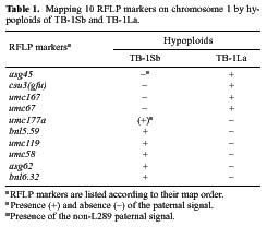
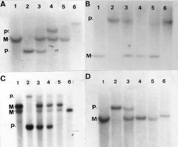
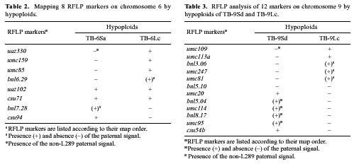
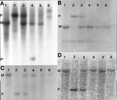
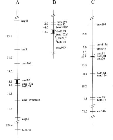
Abstract. The centromere of chromosomes 1, 6 and 9 are physically mapped by the hypoploids of the six most proximal B-A translocations. The hypoploids are deficient for a paternal chromosome arm and, as a result, lose the paternal signal of those RFLP markers located on the missing chromosome arm. Of those markers missing from the hypoploids, the two most proximal ones on each arm of a chromosome define the physical location of the centromere. Analysis of 10 RFLP markers on chromosome 1, 8 on chromosome 6 and 12 on chromosome 9, maps the first centromere to the umc67-umc177a region, the sixth centromere to the bnl6.29-bnl7.28 region, and the ninth centromere to the bnl5.10-umc20 region, an interval of about 3.3, 3 and 0.5 map units, respectively. Other interesting observations are that the A-B chromosome of five of the six B-A translocations is associated with anomalous signals not originated from the paternal parent, suggesting probable presence of chromosome rearrangement(s).

Keywords: B-A translocation; Centromere; Deletion mapping; Hypoploid; RFLP marker.

Introduction

The centromere is a distinct cytological region—the primary constriction—on metaphase chromosomes. Its primary function is to align chromosomes at the metaphase plate and then to separate them at anaphase during cell division. It exerts these functions through the attachment of its kinetochore with microtubules, to promote chromosome movement by the kinetochore motor (Hyman and Mitchison, 1991). Despite its significant biological function, the centromeric location is rarely determined and has been mapped only in a few species due to lack of a proper phenotype for genetic analysis. This stumbling block was circumvented in yeast (Clarke, 1990) and recently in Arabidopsis (Copenhaver et al., 1999) using tetrad analysis to define the centromere position at the molecular level.

In plants, the centromere has been mapped with a telocentric or an isochromosome. The telocentric chromosome is derived from a normal homolog, usually a univalent, by a misdivision of the centromere; semicolon and one end of this telocentric chromosome is the centromere. An isochromosome is structurally similar to a telocentric. It is a metacentric chromosome, carrying two identical chromosome arms. Any genetic marker located at or near the terminal position on a telocentric will be either most proximal or distal to the centromere. After comparing with the genetic map of the normal homolog, the centromeric position can be determined to be located between two closely

linked genetic markers. Centromeres have been mapped this way in wheat (Sears, 1969), cotton (Endrizzi and Kohel, 1966), rice (Singh et al., 1996) and tomato (Frary et al., 1996). Telosomes associated with chromosomes 4 and 5 have been isolated in maize (Rhoades, 1933, 1936 and 1940; Muzumdar et al., 1997) and used to map the centromere of chromosome 4 (Schneerman et al., 1998).

In addition to telocentric chromosomes, a maize centromere has been well localized by hypoploids generated from B-10 translocations. Beckett (1973) induced a translocation between the B chromosome and the short arm of chromosome 10 (10S). He used the translocation to synthesize hypoploids carrying the terminal deficiency of 10S to map physically the 10th centromere to be proximal to y9 on 10S. Subsequently, Lin (1974) created 38 translocations between the B chromosome and the long arm of chromosome 10 (10L) and observed that the hypoploids derived from two most proximal translocations have a breakpoint proximal to du1 on 10L (Lin, 1974 and 1979). Thus, the 10th centromere is located in the du1-y9 region which covers two map units.

This paper utilizes a similar approach to map the centromere of chromosomes 1, 6, and 9 on RFLP map. The centromere was mapped to a region of 3.3, 3, and 0.5 map units, respectively.

Materials and Methods

Plant Material

L289 and the B-A translocation-carrying L289 stocks are the same as previously described (Lin and Chou, 1997). Three translocations (TB-1Sb, TB-1La and TB-6Sa), iso

*Corresponding author. Fax: (04) 2287-4879; E-mail: bylin@dragon.nchu.edu.tw


Botanical Bulletin of Academia Sinica, Vol. 42, 2001

lated by Roman (1947), Roman and Ullstrup (1951), and three others (TB-6Lc, TB-9Sd, and TB-9Lc), isolated by Beckett (1978), were used for generation of hypoploids, which have a structure similar to the four previous B-A translocations described previously (Lin and Chou, 1997). They are carried by L289 and have a breakpoint on the B long arm and the second breakpoint on one of the following chromosome arms: the short and the long arm of chromosome 1 (TB-1Sb and TB-1La, respectively), the short and the long arm of chromosome 6 (TB-6Sa and TB-6Lc, respectively), and the short and the long arm of chromosome 9 (TB-9Sd and TB-9Lc, respectively). B73, an inbred, was used in this study as the maternal parent for production of hypoploids. W22, also an inbred, carried several original B-A translocations, which were subsequently transferred to L289 by continuous backcrosses. It was used in most analyses to exclude the possibility that the non-L289 signals were of W22 by origin.

Synthesis of Hypoploids

Procedures for production of hypoploids have been published (Lin and Chou, 1997), Briefly, hypoploids were synthesized by pollinating B73 with B-A translocation-carrying L289. The hypoploid progeny from the cross were identified by their large kernel size and small plant size for TB-1Sb and TB-1La and only small plant size for TB-9Lc. All six hypoploids exhibited 50% pollen sterility (for details, see Lin and Chou, 1997). Their identity was further substantiated by the absense of the paternal signal of RFLPs located on the respective deleted chromosome arm (see Results).

Strategy for RFLP Mapping of Centromere

Hypoploids of the most proximal B-A translocations on both arms of chromosomes 1, 6, and 9 were used to map the centromeric position. The mapping strategy is based on the fact that a centromere is located between the breakpoints of the two most proximal translocations: one on the short arm and the other on the long arm of the same chromosome. The two RFLP markers closest to, but distal to, the translocation breakpoints on both arms define the map position of the centromere.

The marker position in relation to translocation breakpoints is determined by the terminal deficiencies generated by B-A translocations. Like the B chromosome, one of the two B-A translocation chromosomes, termed B-A, undergoes nondisjunction at the second pollen mitosis. This process generates two sperm: one with two B-As (hyperploid) and the other without any B-A (hypoploid). Upon fertilization with an egg carrying the normal chromosome complement, the latter sperm results in a hypoploid embryo deficient for the paternal copy of the chromosome arm distal to the translocation breakpoint. In other words, the hypoploid embryo associated with the terminally deficient chromosome, termed A-B, was employed to generate DNA for RFLP analysis. For simplification of the RFLP analysis, each parent of the hypoploid progeny is in two different inbred backgrounds: the ma

ternal parent is B73 and the paternal parent, L289. The RFLP analysis is done by Southern hybridization of the hypoploid DNA probed with each marker whose position in reference to the breakpoint is determined by presence (proximal) or absence (distal) of the paternal signal.

RFLP Markers

Sources of maize RFLP markers used in this study are the same as those previously published (Lin et al., 1997).

Genomic DNA Isolation, Restriction Digestion of Genomic DNA, Blotting onto Filters, Probe Preparation, Hybridization and Washing

These protocols have been previously published (Lin et al., 1997). At least, two hypoploid plants of each translocation were used for genomic DNA preparation. The autoradiograms were prepared by exposure of the hybridized membrane either to X-ray film (Kodak) or by scanning the membrane with Phosphoimager (BAS 1000, FUJIX).

Results

Table 1 gives results of mapping 10 RFLP markers on chromosome 1 with hypoploids of TB-1Sb and TB-1La. Four markers (asg45, csu3, umc167 and umc67) exhibit no paternal signal on the hypoploid of TB-1Sb , and six others (umc177a, bnl5.59, umc119, umc58, asg62, and bnl 6.32; Figure 1A) give no paternal signal on the hypoploid of TB-1La. None of the 10 markers have the paternal signal on the hypoploids of both translocations, indicating that they are not located in the region delimited by the breakpoints of the two translocations. The paternal signal of one marker (umc67) deserves special attention; it is absent on the hypoploids of TB-1Sb but present as a very faint diffused band on the hypoploid of TB-1La (Figure 1B). Thus, the locus is located on the short arm of chromosome 1, and the data place the centromere in the umc67-umc177a region (Figure 3), an interval of 3.3 map units in Neuffer et al. map (1997).


Lin et al. — Centromere position in maize

Figure 1. A-D, Four representative analyses of mapping RFLP loci by hypoploids of four B-A translocations. The B-A translocations used are TB-1Sb, TB-1La (A and B), TB-9Sd and TB-9Lc (C and D). Probes used are umc177a (A), umc67 (B), umc20 (C) and bnl5.10 (D). The genomic DNA was digested with either EcoRV (A and B) or EcoRI (C and D). M, maternal signal; P, paternal L289 signal; P´, paternal non-L289 signal; p, duplicate paternal L289 signal. A, A non-L289 signal of umc177a is present on lane 4 (P´). Lane 1, B73; lane 2, L289; lane 3, hybrid between B73 and L289; lane 4, hypoploid of TB-1Sb; lane 5, hypoploid of TB-1La; lane 6, W22. B, The paternal signal (P) of umc67 is absent on the hypoploid of TB-1Sb but present as a faint band on the hypoploid of TB-1La. Lane 1, B73; lane 2, L289; lane 3, hybrid between B73 and L289; lane 4, hypoploid of TB-1Sb; lane 5, hypoploid of TB-1La; lane 6, W22. C, The paternal signal of umc20 is present on the hypoploid of TB-9Sd but absent on the hypoploid of TB-9Lc. Lane 1, B73; lane 2, L289; lane 3, hybrid between B73 and L289; lane 4, hypoploid of TB-9Sd; lane 5, hypoploid of TB-9Lc; lane 6, W22. D, The paternal signal of bnl5.10 is absent on the hypoploid of TB-9Sd and TB-9Lc. Lane 1, B73; lane 2, L289; lane 3, hybrid between B73 and L289; lane 4, hypoploid of TB-9Sd; lane 5, hypoploid of TB-9Lc; lane 6, W22.

Table 2 shows the result of RFLP analysis of eight markers on chromosome 6 with hypoploids of TB-6Sa and TB-6Lc. Four markers (uaz330, umc159, umc85 and bnl6.29) show no paternal signal on the hypoploids of TB-6Sa, and two others (bnl7.28 and csu94) do not have the paternal signal of the hypoploids of TB-6Lc. The last two markers (uaz102 and csu71) possess the paternal signal on the hypoploids of both TB-6Sa and TB-6Lc, indicating their location, in conjunction with that of the centromere, in the region between the two breakpoints. This result places the centromere in the bnl6.29-bnl7.28 region (Figure 3), the length of which is either 5.1 map units according to the map of Matz et al. (1995) or 3 map units based on the map of Neuffer et al. (1997). The data provides no information on the centromeric position in relation to that of uaz102 or csu71 loci.

Table 3 depicts the results of mapping 12 RFLP markers with hypoploids of TB-9Sd and TB-9Lc. The paternal signal of five markers (umc109, umc113a, bnl3.06, umc247, and

umc81) is absent on the hypoploid DNA of TB-9Sd, and that of six others (umc20, bnl5.04, umc114, bnl8.17, umc95, and csu54b; Figure 1C and 2A) is not present on the hypoploid of TB-9Lc. The last marker (bnl5.10) has no paternal signal on the hypoploid of TB-9Sd or TB-9Lc (Figures 1D, 2B, 2C and 2D), and none of the markers have the paternal signal on the hypoploids of both translocations, implying all twelve markers are distal to the breakpoints of the two translocations. Without consideration of bnl5.10, current data place the centromere in the umc81-umc20 region, the length of which is about 1.5 map units on the Neuffer et al. map (1997) or 4 map units on the Matz et al. map (1995). Since bnl5.10 is located at the proximal end of a rearrangement on the 9-B chromosome of TB-9Lc, it was deleted by multiple breakages, which occurred during the formation of the translocation (see Discussion). Accordingly, bnl5.10 is proximal to umc81, and the centromere is located in the bnl5.10-umc20 interval (Figure 3), the length of which is


Botanical Bulletin of Academia Sinica, Vol. 42, 2001

Figure 2. A-D, Anomalous paternal signals associated with the 9-B chromosome of TB-9Sd and TB-9Lc. The hypoploid DNA was digested with EcoRI (A), EcoRV (B), HindIII (C) or KpnI (D) and hybridized with bnl5.04 (A) or bnl5.10 (B, C, and D). The paternal non-L289 signal (P´) is different from the W22 and L289 signal in A; and the paternal signal is missing in hypoploid DNAs after digested with three different enzymes in B, C, and D. Lane 1, B73; lane 2, L289; lane 3, hybrid between B73 and L289; lane 4, hypoploid of TB-9Sd; lane 5, hypoploid of TB-9Lc; lane 6, W22; M, maternal signal; P, paternal L289 signal; P´, paternal non-L289 signal.


Lin et al. — Centromere position in maize

the Neuffer et al. map (1997) is more widely used, its values are tentatively assigned to the region in this study.

During the RFLP analysis of hypoploids of five B-A translocations, unexpected results were observed. Since in this study hypoploids were produced by crossing B73 with pollen of the B-A translocation-carrying L289, they are expected to give or lack the paternal L289 signal following hybridization with RFLP markers, depending on whether the markers are proximal or distal, respectively, to the breakpoint of the relevant B-A translocations. Most markers show the expected signal pattern on hypoploids, but some do not.

The unexpected results are associated with the hypoploids of five translocations (TB-1Sb, TB-6Sa, TB-6Lc, TB-9Sd and TB-9Lc) that exhibit two different paternal signals: L289 or non-L289. For example, the hypoploid of TB-1Sb, following hybridization with umc177a, gave two signals: B73 and non-L289 (Tables 1, 2 and 3; Figure 1A). The paternal L289 signal is present on the hypoploid of neither TB-1Sb nor TB-1La, but a non-L289 signal is present on the hypoploid of TB-1Sb (P´, lane 4, Figure 1A). Of the ten markers in chromosome 1 analyzed in this study, umc177a is the only one showing the paternal non-L289 signal on the hypoploid of TB-1Sb; other markers adjacent to the locus exhibit the expected L289 signal. The non-L289 signal must be paternal by origin, since it is absent in inbred B73, the maternal parent included in the same analysis.

In addition to TB-1Sb, the non-L289 signal was also observed in the hypoploids of TB-6Sa, TB-6Lc, TB-9Sd, and TB-9Lc. Similar to the hypoploid of TB-1Sb, the hypoploid of TB-6Sa and TB-6Lc has a single marker (bnl6.29 and bnl7.28, respectively) displaying the paternal non-L289 signal (Table 2). In case of the hypoploid of TB-9Sd, four markers (bnl5.04, umc114, bnl8.17, and umc95) on 9L, show the paternal non-L289 signal (Table 2). For the hypoploid of TB-9Lc, three markers (bnl3.06, umc247, and umc81) behave similarly (Table 2). The size of the paternal non-L289 signals is variable: five are larger than the L289 signal, and five others are smaller; but none of them are the same size as those of W22 (data not shown.)

The non-L289 signals may originate from two sources, both of which are associated with the paternal parent. It may arise from the original carrier of the B-A translocations and has been retained in the present stocks by linkage drag (Zeven et al., 1983). Of the six B-A translocations used in this study, TB-1Sb, TB-1La, and TB-6Sa were isolated by Roman and Ullstrup (1951); TB-6Lc, TB-9Sd and TB-9Lc by Beckett (1978 and 1991). The original carrier of the first three translocations has not been recorded in the literature; hence, its genetic constitution can not be determined. The remaining three translocations were derived either directly from L289 or indirectly from W22 inbred. To look into the possibility of linkage drag, W22 DNA was included in most RFLP analyses, and it produced a signal distinctively different from the non-L289 (lanes 4 and 6, Figure 2A), ruling out W22 as the original source of the non-L289 signal.

Figure 3. A-C, Map position of the centromere of chromosomes 1, 6, and 9. Only markers used in this study are included. The map order and distance between markers are based on the Neuffer et al. map (1997). Markers in parenthesis are located between two flanking markers of the Neuffer et al. map, but their exact position can not be determined. A, Map of the centromere of chromosome 1; B, Map of the centromere of chromosome 6; C, Map of the centromere of chromosome 9. Solid box: the centromeric region resolved by the data of this study; aMarkers of Helenjaris's map (unpublished); bMarker of Matz et al. (1995).

0.5 map units on the Neuffer et al. (1997) map but about 9 map units on Matz et al. (1995) map. This difference originates from the different map order between bnl5.10 and umc81, which is reversed in the two maps.

Discussion

The physical position of the centromere of chromosomes 1, 6, and 9 was mapped in this study by hypoploids generated from the six most proximal B-A translocations. The RFLP analysis of 10 markers on chromosome 1, 8 on chromosome 6 and 12 on chromosome 9 places the first centromere in the umc67-umc177a region, the sixth centromere in the bnl6.29-bnl7.28 region, and the ninth centromere in the bnl5.10-umc20 region (Figure 3). The centromere was placed within an interval of 3.3 and 3 map units for chromosomes 1 and 6, respectively. It was mapped to a region of either 0.5 or 9 map units for chromosome 9 in the Neuffer et al. map (1997) and Matz et al. map (1995), respectively. This difference originates partly from the different map order between bnl5.10 and umc81: bnl5.10 is proximal to umc81 in the former but distal in the latter. The basis for the difference is not clear. Since


Botanical Bulletin of Academia Sinica, Vol. 42, 2001

The alternative explanation for appearance of the paternal non-L289 signals is presence of additional chromosome rearrangement(s)—most likely an inversion— on the A-B chromosome of the five B-A translocations. This is consistent with the fact that markers giving the paternal non-L289 signals are linked together: Four markers of this nature on 9L, spanning a region of about 19 map units, are closely linked; and three others on 9S, covering about 15 map units, are likewise linked. The affected regions are not interrupted by markers that show the L289 signal. If this supposition is valid, the proposed rearrangement(s) would be cytologically visible in the pachytene chromosomes of heterozygous translocations.

This explanation is consistent with the existence of markers lacking the paternal signal (L289 or non-L289) on the hypoploids of two B-A translocations associated with the two arms of the same chromosome. For example, the affected region on the 9-B chromosome is closely associated with bnl5.10, which is deficient of the paternal signal on the hypoploid of TB-9Sd and TB-9Lc (Table 3; Figure 1D). Since the former carries the deficiency of 9S and the latter the deficiency of 9L, the paternal signal is expected to be absent on at least one of the two hypoploids. The fact that it is absent on both hypoploids suggests the possibility of a complex rearrangement that leads to the formation of a chromosome deletion covering the bnl5.10 locus or a shifting of the signal to the same gel region as the maternal signal. The latter possibility was disproved by digestion of the hypoploid DNA with three additional polymorphic enzymes (EcoRV, HindIII and KpnI), all of which failed to produce any paternal signal—L289 or non-L289 (Figure 2B, 2C and 2D). In addition, since the locus is linked to the three other markers on 9S that give the paternal non-L289 signal—no marker giving the L289 signal is located between them—these four markers may be involved in the same rearrangement; bnl5.10 was deleted by an additional breakage before ligation.

The occurrence of additional rearrangement(s) is not unique to maize B-A translocations synthesized by X-irradiation. It also occurs in a maize stock carrying the r-X1 deletion on the long arm of chromosome 10, which induces chromosome breakages during megagametogenesis (Lin, 1987). The stock produced an aberrant chromosome 2 carrying two terminal deficiencies, one on each arm, in addition to multiple chromosome rearrangements (Lin et al., 1997). Similar observations have been reported in wheat, where chromosome breakage is invoked by an alien chromosome of Aegilops cylindrica. Chromosome breakages were observed in a portion of progeny lacking the alien chromosome (Endo, 1988). The order of some RFLP loci on the resulting deficient chromosome is not consistent with that of the normal homologs (Hohmann et al., 1994). In addition, multiple rearrangements were found associated with several terminally deficient chromosomes (Hohmann et al., 1995). It is evident that chromosome breakage, induced either by X-irradiation or by other genetic means, is frequently associated with multiple events.

Acknowledgement. This research was supported in part by Grants NSC 86-2311-B-005-016 and NSC87-2311-B-005-028 from the National Council of Sciences (Republic of China).

Literature Cited

Beckett, J.B. 1973. Tentative map positions of genes and A-B translocations on chromosome 10. Maize Genet. News Lett. 47: 145-146.

Beckett, J.B. 1978. B-A translocations in maize. J. Hered. 69: 27-36.

Beckett, J.B. 1991. Cytogenetic, genetic and plant breeding applications of B-A translocations in maize. In P.K. Gupa and T. Tsuchiya (eds.), Chromosome Englineer in Plants, Part A Elsevier Science Publishers, Amsterdam, pp. 493-529.

Clarke, L. 1990. Centromeres of budding and fission yeasts. Trends Genet. 6: 150-154.

Copenhaver, G.P., K. Nickel, T. Kuromori, M. Benito, S. Kaul, X. Lin, M. Bevan, G. Murphy, B. Harris, L.D. Parnell, W.R. McCombie, R.A. Martienssen, M. Marra, and D. Preuss. 1999. Genetic definition and sequence analysis of Arabidopsis centromeres. Science 286: 2468-2473.

Endo, T.R. 1988. Induction of chromosomal structural changes by a chromosome of Aegilops cylindrica L. in common wheat. J. Hered. 79: 366-370.

Endrizzi, J.E. and R.J. Kohel. 1966. Use of telosomes in mapping three chromosomes in cotton. Genetics 54: 535-550.

Frary, A., G.G. Presting, and S.D. Tanksley. 1996. Molecular mapping of the centromeres of tomato chromosomes 7 and 9. Mol. Gen. Genet. 250: 295-304.

Hohmann, U., T.R. Endo, K.S. Gill, and B.S. Gill. 1994. Comparison of genetic and physical maps of groups 7 chromosomes from Triticum aestivum L. Mol. Gen. Genet. 245: 644-653.

Hohmann, U., T.R. Endo, R.G. Hermann, and B.S. Gill. 1995. Characterization of deletions in common wheat induced by an Aegilops cylindrica chromosome: detection of multiple chromosome rearrangements. Theor. Appl. Genet. 91: 611-617.

Hyman, A.A. and T.J. Mitchison. 1991. Two different microtubule-based motor activities with opposite polarities. Nature 351: 206-211.

Lin, B.-Y. 1974. TB-10 breakpoints and marker genes on the long arm of chromosome 10. Maize Genet. News Lett. 48: 182-184.

Lin, B.-Y. 1979. Two new B-10 translocations involved in the control of nondisjunction of the B chromosome in maize. Genetics 92: 931-945.

Lin, B.-Y. 1987. Cytological evidence of terminal deficiencies produced by the r-X1 deletion in maize. Genome 29: 718-721.

Lin, B.-Y. and H.-P. Chou. 1997. Physical mapping of four RAPDs in the B chromosome of maize. Theor. Appl. Genet. 94: 534-538.

Lin, B.-Y., S.F. Peng, Y.-J. Chen, H.-S. Chen, and C.-F. Kao. 1997. Physical mapping of RFLP markers on four chromosome arms in maize using terminal deficiencies. Mol. Gen. Genet. 256: 509-516.


Lin et al. — Centromere position in maize

Matz, E.C., F.A. Burr, and B. Burr. 1995. Molecular map based on TxCM and COxTx recombinant inbred families. Maize Genet. News Lett. 69: 257-267.

Muzumdar, D.A., M.C. Schneerman, G.G. Doyl, and D.F. Weber. 1997. Identification of an isochromosome for the long arm of chromosome 7 in maize. Maize Genet. News Lett. 71: 67.

Neuffer, M.G., E.H. Coe, and S.R. Wessler. 1997. Maps of nuclear genes and molecular markers. In Mutants of Maize. Cold Spring Harbor Laboratory Press 1997, pp. 33-53.

Rhoades, M.M. 1933. A secondary trisome in maize. Proc. Natl. Acad. Sci. USA 19: 1031-1038.

Rhoades, M.M. 1936. A cytogenetic study of a chromosome fragment in maize. Genetics 21: 491-502.

Rhoades, M.M. 1940. Studies of a telocentric chromosome in maize, with reference to the stability of its centromere. Genetics 25: 483-520

Roman, H. 1947. Mitotic nondisjunction in the case of inter

changes involving the B-type chromosome in maize. Genetics 32: 391-409.

Roman, H. and A.J. Ullstrup. 1951. The use of A-B translocations to locate genes in maize. Agron. J. 43: 450-454.

Schneerman, M.C., W.S. Lee, G. Doyle, and D.F. Weber. 1998. RFLP mapping of the centromere of chromosome 4 in maize using isochromosomes for 4S. Theor. Appl. Genet. 96: 361-366.

Singh, K., T. Ishii, A. Parco, N. Huang, D.S. Brar, and G.S. Khush. 1996. Centromere mapping and orientation of the molecular linkage map of rice (Oryza sativa L.) Proc. Natl. Acad. Sci. USA 93: 6163-6168.

Sears, E.R. 1969. Wheat cytogenetics. Ann. Rev. Genet. 3: 451-468.

Zeven, A.C., D.R. Knott, and R. Johnson. 1983. Investigation of linkage drag in near isogenic lines of wheat by testing for seedling reaction to races of stem rust leaf rust and yellow rust. Euphytica 32: 319-327.