Bot. Bull. Acad. Sin. (2002) 43: 37-56

Tsou and Mori — Seed coat of Lecythidoideae

Seed coat anatomy and its relationship to seed dispersal in subfamily Lecythidoideae of the Lecythidaceae (The Brazil Nut Family)

Chih-Hua Tsou1 and Scott A. Mori2,*

1Institute of Botany, Academia Sinica, Taipei, Taiwan 115, Republic of China

2Nathaniel Lord Britton Curator of Botany, Institute of Systematic Botany, The New York Botanical Garden, Bronx, New York 10458-5126, USA

(Received April 19, 2001; Accepted August 31, 2001)

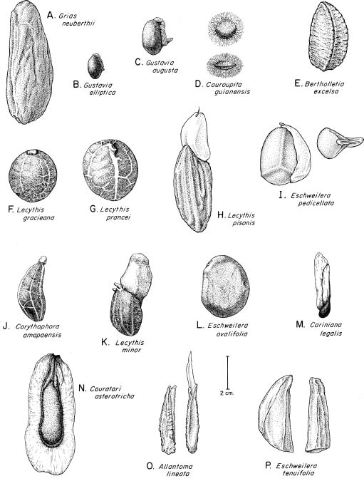
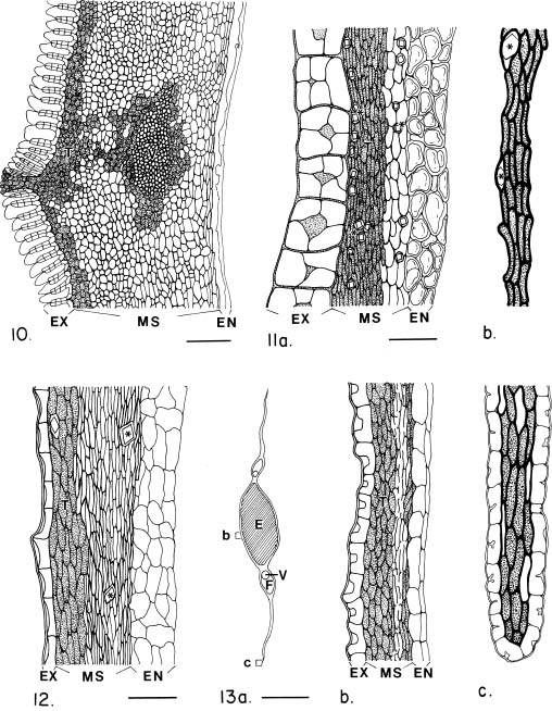
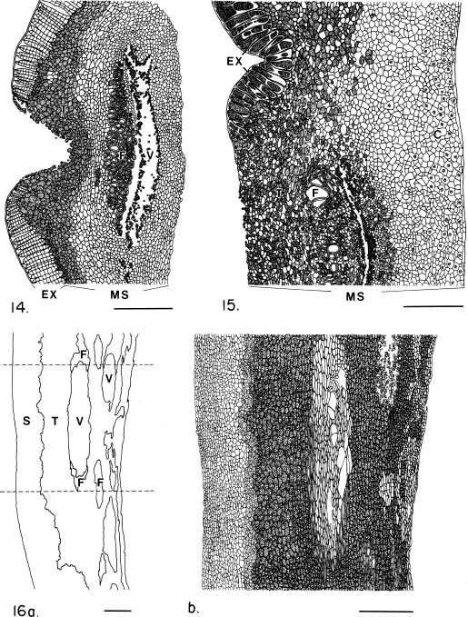
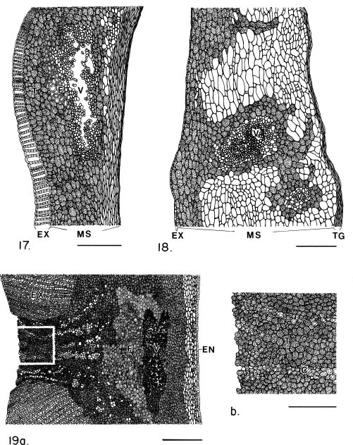
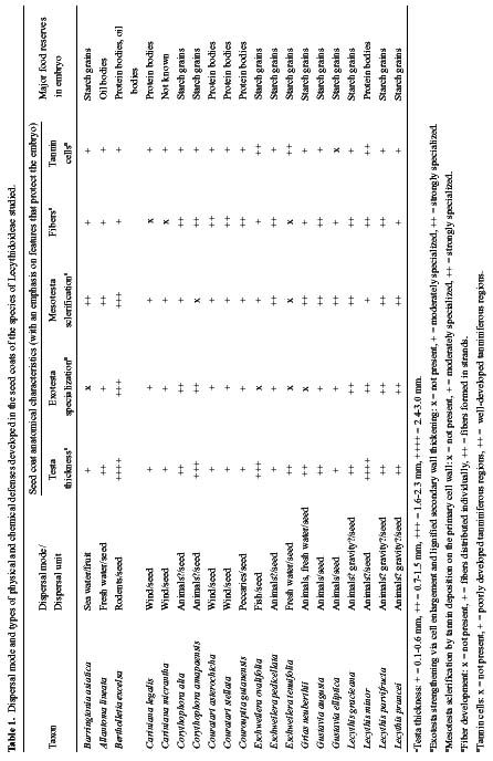
Abstract. The seed coat anatomy of representative species from all 10 Neotropical genera of Lecythidaceae subfamily Lecythidoideae and from the Paleotropical Barringtonia (Lecythidaceae subfamily Planchonioideae) was studied. The seed coat is mainly composed of the testa, which is developed through moderate or intensive multiplication of the outer integument of the ovule. The tegmen, derived from the inner integument of the ovule, is mostly crushed at seed maturity. Barringtonia and Grias, with fruits as diaspores, have an unspecialized exotesta and a poorly differentiated seed coat. In contrast, species of Lecythidoideae, with seeds as diaspores, possess well-differentiated seed coats with diversified protective mechanisms. Examples include: an expanded and lignified exotesta that serves as a water barrier and protects the embryo; an extensive area of tannin cells that provides a chemical defense against pathogens and predators; a thick and sclerotic mesotesta that protects the embryo; and large fibers surrounding and supporting the vascular bundles. In addition, other modifications of the seed coat are probably adaptations for seed dispersal by different dispersal agents. These include the exotestal hairs of Couroupita guianensis, which possibly protect the embryos as they pass through the digestive tracts of peccaries; an extension of the seed coat into a wing in species of Cariniana and Couratari , which facilitate wind dispersal; the papillate exostestal cells of Allantoma lineata, which probably promote dispersal by water; the well-developed arils of species of Corythophora and Lecythis, probably sought after by animals, which in turn disperse the seeds; and the sarcotesta of Eschweilera ovalifolia, consumed by fish, which in turn disperse the seeds. We conclude that the highly diversified anatomy and morphology of the seed coat of the Lecythidoideae has evolved from relatively homogeneous ovules in response to pathogens and predators as well as to abiotic and biotic dispersal agents.

Keywords: Brazil nut family; Lecythidaceae; Lecythidoideae; Planchonioideae; Seed coat anatomy; Seed dispersal.

Introduction

The Brazil nut family (Lecythidaceae), a pantropical family of small to very large trees, is represented by 11 genera and approximately 200 species in the New World tropics. All New World Lecythidaceae, except Asteranthos brasiliensis Desf. (Lecythidaceae subfamily Scytopetaloideae), belong to Lecythidaceae subfamily Lecythidoideae (Prance and Mori, 1979; Mori and Prance, 1990a; Morton et al., 1997, 1998). Barringtonia is an Old World member of Lecythidaceae subfamily Planchonioideae and is sister to the Lecythidoideae (Tsou, 1994).

New World Lecythidaceae are an important element of the flora at latitudes between 19° N and 25° S (Mori and Prance, 1990a). They rank as the first or second most important family of trees in terms of species and numbers of individuals in the vicinity of Belém, Brazil (Black et al., 1950;

Cain et al., 1956). They are the third most important tree family at La Fumée Mountain, French Guiana (Mori and Boom, 1987); and one of the most important in central Amazonia (Prance et al., 1976; Mori and Lepsch-Cunha, 1995; De Oliveira and Mori, 1999) and the Chocó of Colombia (Gentry, 1982). Although most common in lowland, non-flooded forests, the family is also found in reduced numbers in cloud forests, periodically flooded riverine habitats, and in savannas. Although occasionally found in disturbed areas, species of Lecythidaceae are usually not a dominant component of secondary vegetation.

Members of New World Lecythidaceae are diversified in fruit and seed morphology and are well adapted for dispersal by both abiotic (water, wind, and gravity) and biotic (fish and mammal) agents (Prance and Mori, 1978, 1979; Mori and Prance, 1990b; Mori, 2000). Fruit and seed morphology in relation to seed dispersal has been extensively studied in general (Pijl, 1982; Murray, 1986), and the role of the seed coat in protecting the embryo and as a means of controlling dormancy and germination has long been recognized (Bewley and Black, 1994). In contrast, seed

*Corresponding author. Tel: (718) 817-8629; Fax: (718) 817-8648; E-mail: smori@nybg.org


Botanical Bulletin of Academia Sinica, Vol. 43, 2002

coat adaptations to dispersal agents have not been given the attention they deserve. In this paper, we describe and discuss seed coat structure and its relationship with dispersal agents in the Lecythidoideae. Previously, Corner (1976a, 1976b) described the seed coat anatomy of Bertholletia excelsa, Couroupita guianensis, and Gustavia sp. (probably G. augusta), and Vishinskaya (1992) reviewed the seed anatomy of the family.

An understanding of fruit and seed morphology and dispersal agents of the Lecythidoideae, in particular the species studied herein, is important in interpreting seed coat anatomy. Thus, we provide a review taken from published sources (Prance and Mori, 1978, 1979, 1983; Mitchell and Mori, 1987; Mori and Prance, 1990a, 1990b) and from the field observations of the second author. Illustrations of the fruits and seeds of many Lecythidoideae can be found in Prance and Mori (1979) and Mori and Prance (1990a).

New World members of the Lecythidaceae have either indehiscent or dehiscent fruits, and abiotic and biotic dispersal agents are associated with both types of fruits. The diaspores (= dispersal unit) are either the entire fruit or the individual seeds; the seeds either possess or lack appendages (arils, wings, hairs, sarcotestas); the embryos are either chlorophyllous or achlorophyllous; and the embryo either has or lacks cotyledons. We divide the following review into species with indehiscent fruits and those with dehiscent fruits.

Species with Indehiscent Fruits

Grias (Prance and Mori, 1979) and Barringtonia (Payens, 1967) have single-seeded, indehiscent fruits that serve as the diaspore. Barringtonia asiatica is a seashore plant dispersed by sea currents as are several other species of the genus (Payens, 1967). The corky mesocarp of the fruit serves as a flotation device in much the same way that the husk of a coconut (Cocos nucifera L.) facilitates dispersal. Some other species of Barringtonia, however, have fleshy fruits and are probably dispersed by animals (e.g., B. edulis Seem., Payens, 1967). Grias neuberthii has a fleshy mesocarp surrounding a fibrous endocarp. The fruits of this species are sold in western Amazonian markets for the edible mesocarp. The endocarp, however, facilitates flotation of the fruit once the mesocarp has been removed by animals, and it therefore appears that although G. neuberthii is primarily animal dispersed, it may also be secondarily water dispersed. The most widely distributed species, G. cauliflora L., is found in Jamaica, throughout Central America, and into northwestern South America (Prance and Mori, 1979). Its fruits are so efficiently dispersed by water that they frequently wash up on beaches well beyond the known distribution of the species (Gunn and Dennis, 1976).

Most species of Gustavia (Prance and Mori, 1979) and all Couroupita (Mori and Prance, 1990a) possess multiple-seeded fruits. In these species, the seeds serve as the diaspores. In Gustavia elliptica and its close relative G. augusta (the two species studied by us), the fruits rot open

to expose fleshy, contorted funicular arils that may attract animals, which in turn may disperse the seeds after eating the arils. In other species of Gustavia, the funicle does not develop, and it is the pulp surrounding the seed that animals seek. The yellow pulp of G. speciosa (Kunth) DC. is sucked from the fruit raw or is cooked with meat and rice by humans (Romero-Castañeda, 1991). The orange pulp surrounding the seeds of G. grandibractea Croat & S. A. Mori and G. superba (Kunth) O. Berg, neither of which has developed funicles, has reportedly been eaten by mammals (Prance and Mori, 1983).

In species of Couroupita, mature fruits fall to the ground and crack open to expose a bluish-green, foul-smelling pulp, in which are embedded numerous, flattened, small seeds (usually less than 15 mm long) with a hairy testa. In the Brazilian Amazon, peccaries of an undetermined species have been reported to eat the pulp and swallow the seeds. It is presumed that the seeds pass unharmed through their digestive tracts and subsequently germinate some distance from the mother trees thereby effecting dispersal (Prance and Mori, 1979). The pulp is often fed to domestic pigs and poultry (Smith, 1999) and seeds have been reported by people living along the Amazon River to germinate in the feces of these animals (S. A. Mori, pers. obs.).

Bertholletia, with its only species B. excelsa, has evolved a very specialized dispersal system, first reported in 1910 after it was observed that agoutis (Dasyprocta spp.) gnaw open the woody fruits and remove the seeds (Huber, 1910). Subsequently, Amazonian squirrels (Sciurus spadiceus) and brown capuchin monkeys (Cebus apella) have been reported to be able to open mature fruits and other arboreal predators, e.g., the bearded saki monkey (Chiropotes satanas), several species of macaws, and the red-necked woodpecker (Campephilus rubricollis) were discovered to be capable of opening immature fruits of the Brazil nut (Peres and Baider, 1997). At maturity, the fruits drop to the ground with the seeds trapped inside because the size of the fruit opening is smaller than the size of the seeds (Mori and Prance, 1990b; Mori, 1992). The fruits are so rot resistant that after they fall to the ground they serve as homes to an assemblage of anuran and insect species (Caldwell, 1993) for many months after the seeds have been removed.

Although the fruits of Bertholletia are technically dehiscent (they have a small, inwardly falling operculum), they are functionally indehiscent because the seeds are not naturally released from the fruit. The agoutis enlarge the natural opening, remove the seeds, consume some of them, and bury the remaining ones in hidden caches for subsequent use. Those forgotten by these rodents germinate 12 to 18 months after the woody seed testa breaks down (Mori and Prance, 1990a, 1990b; Mori, 1992).

Fruits of several species of Lecythis, e.g., L. gracieana, L. lurida (Miers) S. A. Mori, and L. prancei, also fall to the ground with the seeds inside and lack an aril (Mori and Prance, 1990a). These species differ from Bertholletia in that there is no sign of a natural fruit opening and there


Tsou and Mori — Seed coat of Lecythidoideae

have been no reports of animals eating or hoarding the seeds.

Lecythis rorida (O. Berg) Miers, which grows in periodically inundated forest along the Rio Negro of Brazil, drops entire indehiscent fruits into the water (reported as L. chartacea O. Berg by Kubitzki and Ziburski, 1994) at high water level in June and July (S. A. Mori, pers. obs.). The fruits float and are frequently observed washed up on white sand beaches. The seeds, which rattle around inside the fruit at maturity, do not possess an aril. As far as we know, all species of Lecythis with indehiscent fruits do not have developed funicle/arils (S. A. Mori, pers. obs.). A morphologically similar species of non-flooded forest, Lecythis chartacea, possesses dehiscent fruits and seeds with arils. Lecythis rorida and L. chartacea are so similar in vegetative and flowering features that the former was placed in synonymy under the latter in the most recent monograph of the family (Mori and Prance, 1990a).

Species with Dehiscent Fruits

Lecythis pisonis Cambess, distributed in the Atlantic forests of eastern Brazil and in eastern Amazonia (Mori and Prance, 1990a) is a dehiscent-fruited, animal dispersed species of Lecythidaceae. The very large (25 or more cm in diameter) fruit opens while still on the tree. The seeds are attached to the fruit wall by a funicle, which, in turn, is surrounded by a fleshy, white aril reportedly sought after by the bat Phyllostomus hastatus (Greenhall, 1965). The seeds are dropped unharmed either in flight or under bat roosts.

Many species of Corythophora, Eschweilera, and Lecythis have dehiscent fruits, arillate seeds, and seeds attached to the fruit wall by funicles, and, hence, are similar to L. pisonis. It is not known, however, what animals disperse most of the other species of Lecythidaceae with this fruit/seed syndrome. Marc van Roosmalen (reported in Prance and Mori, 1978) has observed that the fleshy aril of Lecythis poiteaui O. Berg is so thoroughly harvested by the monkeys Chiropotes satanas and Ateles paniscus that he was unable to collect untouched ones despite much effort. Parrots and macaws have been reported to open the fruits of Lecythidaceae (Prance and Mori, 1979). They may be dispersal agents as well as seed predators.

At least one species of dehiscent-fruited Lecythidaceae, Eschweilera ovalifolia, is dispersed by fish (Goulding, 1980). This species grows in periodically inundated forest, mostly along the Amazon River to the west of the mouth of the Rio Negro (Mori and Prance, 1990a). The seeds, which are surrounded by a thin, white, fleshy sarcotesta, drop into the water where they are consumed by fish. The fish apparently remove the sarcotesta and pass the seeds unharmed through their digestive tracts.

Abiotic dispersal is found in at least four genera of dehiscent-fruited Lecythidoideae. All species of both Cariniana and Couratari are dispersed by the wind. Species in these genera are generally very tall trees. If shorter,

they grow in more open, wind exposed habitats such as savanna. In both genera, the fruits open to release seeds that are either winged at one end in Cariniana (Prance and Mori, 1979) or winged all around the seed in Couratari (Mori and Prance, 1990a). The seeds are relatively light and possess embryos with leaf-like cotyledons. Upon germination, eight to twelve days after dispersal in Cariniana micrantha (Imakawa and Ferraz, 1995), the persistent, leaf-like chlorophyllous cotyledons enable the seedlings to immediately photosynthesize. Although the fruit wall of C. micrantha is very thick and woody, it does not deter brown capuchin monkeys from opening the lids and eating the seeds of fruits nearly ready to dehisce. Each fruit contains an average of 18 seeds (Peres, 1991). Peres (1991) has estimated that 99.5% of the entire seed production of a population of this species in one fruiting season in central Amazonia was destroyed by these monkeys.

Water dispersal occurs in at least several dehiscent-fruited species of Lecythidoideae. Allantoma lineata occurs along rivers and small streams in central and eastern Amazonia (Mori and Prance, 1990a). At maturity, the fruits open and the seeds fall into the water, where they are carried away by currents. The seed coat has been reported to be rich in oil (Ducke, 1948), and this has been suggested to aid in flotation (Prance and Mori, 1979). A similar dispersal system is found in Eschweilera tenuifolia, which is found along the Orinoco, Negro, and Amazon rivers from the mouth of the Rio Negro eastward (Mori and Prance, 1990a). This species drops its seeds into the water at high water. In this case, it is the corky seed coat that facilitates seed dispersal, either because of its buoyant nature or by keeping water from penetrating the seeds, and there is no sign of an aril.

Embryo Types of Lecythidoideae

Three embryo types are found in species of Lecythidoideae (Prance and Mori, 1978). In Cariniana (Figure 17D in Prance and Mori, 1979), Couratari (Figure 18G in Prance and Mori, 1979), and Couroupita (Figure 17H in Prance and Mori, 1979), the embryo possesses chlorophyllous, foliaceous cotyledons. In Gustavia (Figures 15D, 16G in Prance and Mori, 1979), the embryo has achlorophyllous, fleshy cotyledons; and, in the remaining taxa, the achlorophyllous macropodial embryos lack cotyledons (Figures 17L, 18C, 18M, 18R, 19C in Prance and Mori, 1979). The seeds of species with chlorophyllous, foliaceous cotyledons carry relatively little food reserve compared with the seeds of species with achlorophyllous embryos and no cotyledons.

Materials and Methods

Fruits and seeds of most species were fixed in FAA (Formalin-Glacial Acetic Acid-70% alcohol) in the field and later transferred to 70% alcohol for long-term storage. Pieces of seed coat attached to a small piece of embryo were taken from the medium portion of the seed body.


Botanical Bulletin of Academia Sinica, Vol. 43, 2002

When seed wings or arils were present, a small piece of each was removed. These pieces were dehydrated in an ethanol-t-butyl alcohol series and embedded in paraplast. Cross sections with a thickness of 5-8 µm were obtained by sectioning with a rotary microtome, and the sections were prepared as microscope slides. Most of the slides were stained with safranin O-fast green FCF. Condensed tannin bodies always stained deep red with safranin O. Protein bodies also reacted with safranin O, but stained a lighter red. Both tannin and protein bodies could be identified by their positions in the seed, with tannins common in the seed coat and protein bodies in the endosperm and embryo. Some sections were stained with phloroglucinol to identify lignins and with iodine in potassium iodide to reveal the presence of starch grains (Gahan, 1984). Free hand sections of embryos were stained with iodine in potassium iodide and Sudan black to detect the presence of starch grains and lipid bodies, respectively. Samples obtained from herbarium specimens were softened in 2.5% NaOH overnight or longer and subsequently dehydrated and embedded as above.

Seeds examined include one species of Planchonioideae: Barringtonia asiatica (L.) Kurz (C. F. Shen, 12 Mar 1993, not vouchered) and samples from 19 species of Lecythidoideae: Allantoma lineata (Mart. ex O. Berg) Miers (J. C. Ongley and J. F. Ramos P23254), Bertholletia excelsa Humb. & Bonpl. (Freitas 610), Cariniana legalis (Mart.) Kuntze (Mori 22632), Cariniana micrantha Ducke (Krukoff 5095), Corythophora alta Knuth (Prance et al. 5094), Corythophora amapaensis Pires ex S. A. Mori & Prance (Mori 8783), Couratari asterotricha Prance (Mori 22630), Couratari stellata A. C. Sm. (Freitas 545), Couroupita guianensis Aubl. (Mori et al. 22153), Eschweilera ovalifolia (DC.) Nied. (Mori et al. 9159), Eschweilera pedicellata (Rich.) S. A. Mori (Mori 8623), Eschweilera tenuifolia (O. Berg) Miers (Prance 24357), Grias neuberthii J. F. Macbr. (Callejas and Marulandia 5897), Gustavia augusta L. (Mori and Bolten 8650), Gustavia elliptica S. A. Mori (Freitas 530), Lecythis gracieana S. A. Mori (Freitas 745), Lecythis minor Jacq. (Mori et al. 22152), Lecythis parvifructa S. A. Mori (Freitas 726), and Lecythis prancei S. A. Mori (Freitas 739). Vouchers for all specimens are archived in the herbarium of the New York Botanical Garden (NY), except that fruits of Barringtonia asiatica were collected in the Tropical Botanical Garden, Pingtong, Taiwan, and no voucher was made for this species. These collections also serve as vouchers for the species illustrated in the figures.

Terminology for seed morphology and seed coat anatomy follows Corner (1976a). The drawings of the cross sections of the seed coat in Figures 2-19 are all arranged with the exotesta to the left. If endosperm is present, it is indicated in the figures. Parenchymatous mesotestal cells are bordered with the thinnest lines; sclerotic cells (impregnated by tannins but without lignin deposition) are bordered by slightly thicker lines; fibers and exotestal cells with secondarily thickened walls are illustrated with double lines; and tannin bearing cells are stippled throughout.

Results

Seed Morphology and Anatomy

Seeds of the Lecythidoideae we studied are medium to large in size (Figures 1A-1G, 1I-1P), based on Corner's (1976a) terminology. The largest seeds are found in Grias neuberthii (to 60 mm long), and the smallest are found in Cariniana legalis (ca. 12 mm long). The seed body is more or less ellipsoidal in the majority of taxa (Barringtonia, Cariniana, Corythophora, Eschweilera ovalifolia, E. pedicellata, Grias, Gustavia, Lecythis minor, and L. parvifructa). Other shapes include more or less rounded (Lecythis gracieana and L. prancei), wedge shaped (Allantoma lineata and Eschweilera tenuifolia), three-faced (Bertholletia), flat and rounded (Couroupita), or flat and elliptical (Couratari).

Seed appendages are found as a unilateral wing in Cariniana (Figure 1M), a circumferential wing in Couratari (Figure 1N), numerous soft hairs in Couroupita (Figure 1D), a sarcotesta in Eschweilera ovalifolia (Figure 1L), or a funicular aril in Corythophora (Figure 1J), Eschweilera pedicellata (Figure 1I), Gustavia (Figures 1B, 1C), and Lecythis minor (Figure 1K). No apparent seed appendages are developed in the remaining species (Figures 1A, 1E, 1F, 1G, 1P).

The seed coat of different species varies in thickness from ca. 0.1 mm (ca. 10-15 cell layers) in Couratari spp. to 2.8 mm thick (ca. 60-80 cell layers) in Bertholletia excelsa. However, the mature seed coat, as revealed in the present study, is mostly composed of a well-developed testa, whereas the tegmen is usually crushed, or remains as several compressed layers in some species of Eschweilera, Lecythis, and Gustavia. Within the testa, the exotesta is single-layered, the mesotesta is multi-layered and forms the main body of the seed coat, and the endotesta is difficult to identify because it is not specialized or because it is crushed at seed coat maturity.

The vascular system of the seed coat is usually complex. The raphal bundle branches before and after the chalaza and the branches further divide one or more times. However, a simpler vascular system is found in four genera. In Couratari the vascular bundle does not branch at all whereas in Bertholletia, Cariniana, and Couroupita the raphal bundle branches into 2-4 well developed postchalazal bundles that run longitudinally in the testa without further branching. The vascular bundles are always embedded deeply within the mesotesta. However, in Allantoma, Bertholletia, Corythophora, and most species of Lecythis, the exotesta is lacking over the regions containing main or medium to large vascular bundles, and, hence, the mesotesta is exposed (Figures 8, 9, 10, 14, 19a). Because the exotesta is usually darker in color and/or different in texture on the seed surface than the exposed mesotesta, the vascular pattern appears distinct (Figure 1E-1G, 1J, 1K).

Fibers are usually an important element in the seed coat. In Corythophora, Couratari, Couroupita, Eschweilera


Tsou and Mori — Seed coat of Lecythidoideae

Figure 1. Seed morphology of 16 selected species of Lecythidoideae. A, Grias neuberthii; B, Gustavia elliptica; C, Gustavia augusta; D, Couroupita guianensis; E, Bertholletia excelsa; F, Lecythis gracieana; G, Lecythis prancei; H, Lecythis pisonis (Prance et al. 24363); I, Eschweilera pedicellata; J, Corythophora amapaensis; K, Lecythis minor; L, Eschweilera ovalifolia; M, Cariniana legalis; N, Couratari asterotricha; O, Allantoma lineata; P, Eschweilera tenuifolia.


Botanical Bulletin of Academia Sinica, Vol. 43, 2002

pedicellata, and most Lecythis, a large group of fibers is always developed on the external side of medium to large or even all of the vascular bundles, and individual fibers may reach 100 µm in diameter. In Allantoma, Barringtonia, Bertholletia, Eschweilera ovalifolia, Grias, and Gustavia, the fibers are less pronounced, being either in small groups or as solitary fibers. In Cariniana and Eschweilera tenuifolia, however, fibers are completely absent. Tannin cells, which contain massive deposits of condensed tannins, are very common, especially in the outer layers of the mesotesta. Tannins may also be deposited on the cell walls of the mesotestal cells to form non-lignified sclerotic cells that increase cell rigidity. The remaining parenchymatous mesotestal cells, which lack sclerotic cell walls, are usually markedly compressed.

The endosperm remains as a thin layer in Allantoma, Bertholletia, Cariniana, Couratari, Couroupita, and some Lecythis, but completely disappears in the remaining species studied. Within the embryo, the major food reserve revealed is found as either starch grains or protein bodies, but no seeds were found to be rich in both starch and protein. Oil bodies, on the other hand, are usually present with either starch or protein bodies, but in a lesser amount than either of them. In Bertholletia excelsa (the Brazil nut) both oil and protein bodies are abundant

The following descriptions of the seed coat anatomy of the species we studied are presented according to dispersal systems.

Indehiscent Fruits, Fruits Serve as Diaspores

Barringtonia asiatica (Figure 2) and Grias neuberthii (Figures 1A, 3) possess one-seeded fruits. Both species have a poorly differentiated seed coat. Barringtonia asiatica has many large vascular bundles, and the thickness of the seed coat varies from 0.5-0.8 mm in regions without vascular bundles to ca. 1.2 mm in regions with bundles. Grias neuberthii has numerous vascular bundles of various diameters, and the seed coat is 0.8-1.1 mm thick. A distinct exotesta is lacking in both species. The testa is about 25-35 cell layers thick. All cells except for those in the vascular system are similar in size, and the cells are sclerotic but not lignified. In B. asiatica several layers in the middle part of the testa are filled with tannin, and the fibers are solitary and scattered. In G. neuberthii tannin cells occur randomly in the testa, and small groups of fibers are associated with the vascular bundles. No crystals were found in their seed coats. The embryos of both species are macropodial, and the embryo cells are full of starch grains.

Indehiscent Fruits, Seeds Serve as Diaspores

Couroupita guianensis (Figures 1D, 4) possesses fruits with 82-295 seeds. The seed coat is well differentiated, 0.32-0.4 mm thick, and only three large vascular bundles penetrate the testa. The outer tangential primary wall of each of the exotestal cells is prolonged into a hair 4-5 mm long whereas the other five walls are secondarily thickened with lignins. Beneath the exotesta, the hypodermal

Figures 2-3. Cross sections the of seed coats of Barringtonia asiatica (Figure 2) and Grias neuberthii (Figure 3). The seed coat in both species is poorly differentiated. F: fiber, T: tannin cell, and V: vascular bundle. Bar = 0.4 mm in both figures.


Tsou and Mori — Seed coat of Lecythidoideae

Figures 4-7. Cross sections of the seed coats of Couroupita guianensis (Figure 4), Gustavia augusta (Figure 5), G. elliptica (Figure 6), and Bertholletia excelsa (Figure 7). Note the long exotestal hairs in Couroupita guianensis; the thick secondary cell walls of the species of Gustavia; and the highly elongated exotestal cells of Bertholletia excelsa. C: crystal, EN: endosperm, EX: exotesta, F: fiber, MS: mesotesta, T: tannin cell, TG: tegmen, V: vascular bundle, and *: crystal sands. Bar = 0.2, 0.2, 0.1, and 0.5 mm, respectively.


Botanical Bulletin of Academia Sinica, Vol. 43, 2002

cells of the exotesta are radially elongated and have lignified secondary cell walls. These cells become smaller and then lacking in regions with medium to large vascular bundles where, thus, the mesotesta is exposed. The mesotesta is 40-50 cell layered. The cells of the outer two-thirds are filled with tannins and do not become crushed, whereas cells of the inner layers of the mesotesta are less densely filled with tannins and markedly compressed. The fiber groups are well developed on the external side of the larger vascular bundles. Crystal cells were not found. The cells of the macropodial embryo contain numerous starch grains.

Lecythis parvifructa possesses relatively small fruits with 1-3 seeds. The seed coat is ca. 1.0-1.5 mm thick and has many vascular bundles of various sizes. The exotestal cells are radially elongated and have lignified secondary cell walls, but they are absent in regions with major vascular bundles where, thus, the mesotesta is exposed. The mesotesta is 55-90 cell layered; the cells are sclerotic but not lignified; and tannin rich cells occur randomly in the outer layers. A group of fibers is well-developed external to each major vascular bundle. Crystals were not found. In the macropodial embryo starch grains are the major food reserve.

Lecythis prancei (Figure 1G, 9) possesses relatively large fruits with 1-2 large (3-4.5 cm diam.) seeds. The seed coat is 1.0-1.4 mm thick and has many vascular bundles of various sizes. The exotestal cells are radially elongated and possess lignified secondary cell walls and oil droplets in the cell lumen in areas without concentrations of vascular bundles. In areas with concentrations of vascular bundles, the exotesta is lacking and the mesotesta is exposed. The mesotesta is 45-60 cell layered. Cells in the outer one-half to three-fourths of the mesotesta are sclerotic and slightly tanniniferous whereas the innermost layers are heavily compressed. Groups of fibers are found associated with the vascular bundles. Crystals occur in cells on the external side of the vascular bundles. Starch grains are the major food reserve in the macropodial embryo.

The seed coats of the preceeding three species are anatomically very similar.

Dehiscent Fruits, Seeds Serve as Diaspores

Allantoma lineata (Figures 1O, 10) possesses fruits with numerous but an undetermined number of seeds. The seed coat is ca. 0.7-1.2 mm thick and has a highly branched vascular system. In areas with large vascular bundles, the exotesta is disrupted by the protruding mesotesta; otherwise, the exotesta consists of radially elongated cells. The outer tangential wall of these cells protrudes slightly as a papilla, and the other five walls are secondarily lignified. The mesotesta is composed of ca. 30 layers of sclerotic cells. Cells of the outermost layers of the mesotesta are filled with tannins, and the innermost layers are strongly compressed. Small groups of tannin cells are also found surrounding the vascular bundles. Solitary fibers are commonly found on the external side of the

layer also possesses secondarily lignified cell walls, and each cell is provided with a single, large prismatic crystal. The mesotesta is ca. 30-40 cell layers thick and is differentiated into an outer region with sclerotic, tannin cells and an inner region composed of parenchyma. A large group of fibers is always present on the external side of the vascular bundle. The endosperm is represented by 4-5 layers of living cells, which contain small quantities of protein bodies and starch grains. The embryo, including the leafy cotyledons and the embryo axis, contains abundant protein bodies and a smaller quantity of oil bodies.

Gustavia augusta (Figures 1C, 5) and Gustavia elliptica (Figures 1B, 6) possess fruits with an estimated 10-20 seeds. The seed coat is about 0.8-1.2 mm thick in the former and 0.25-0.4 mm in the latter species. Each species possesses a highly branched vascular system. The exotesta is a uniform layer of cells with heavily lignified secondary walls. The mesotesta is 30-40 cell layers thick in G. augusta and 15-30 layers in G. elliptica. Most of the inner layers of the mesotesta of both species become compressed at maturity. Fibers are solitary or in small bundles distributed on the external side of the vascular bundles. Crystals were not found. These two species have very similar seed coats, differing quantitatively but not qualitatively. Gustavia augusta has a thicker seed coat, and its fibers are better developed and usually found in small groups. In contrast, the fibers of G. elliptica are less common and usually solitary. The funicular aril of both species is contorted and possesses a stout vascular system and a thick-walled epidermis. The funicular aril of G. augusta tastes sweet when fresh. No starch grains or other storage bodies were observed in the funicular arils of either species. The embryo, including the plano-convex cotyledons and the embryo axis, is rich in starch grains and oil bodies are present in small amounts.

Bertholletia excelsa (Figures 1E, 7) possesses fruits with 10-25 seeds. The seed coat is 2.5-2.8 mm thick, and the raphal bundle branches into two to three large bundles, each of which runs along an angle of the seed coat and does not further branch. Cells of the exotesta are lignified and elongated radially into a 1-1.2 mm long palisade layer, but at the seed angles, where the large vascular bundles run, the exotesta is lacking and the mesotesta is exposed. The mesotesta is 60-80 cell layers thick. All of the cells of the mesotesta are sclerotic, and the outer one-fifth to one-third of them are completely filled with tannins. Single or small groups of fibers are randomly distributed in the mesotesta, and crystal sands are occasionally found external to the vascular bundles. No storage food bodies were found in the two-cell layered endosperm (not shown in Figure 7), but the macropodial embryo is rich in both protein bodies and oil droplets.

Indehiscent Fruits, Seeds or Possibly Fruits Serve as Diaspores

Lecythis gracieana (Figures 1F, 8) possesses relatively small fruits with 1-2 seeds. The seed coat is 1.0-1.5 mm thick and has a highly branched vascular system. The


Tsou and Mori — Seed coat of Lecythidoideae

Figures 8-9. Cross sections of the seed coats of Lecythis gracieana (Figure 8) and L. prancei (Figure 9). The horizontally dashed lines in figures 8a and 9a show the positions of the detailed sections in figures 8b and 9b. Note the gaps in the exotesta and the differences in the size and distribution of the fibers of these two species. EX: exotesta, F: fiber, MS: mesotesta, T: tannin cell, TG: tegmen, and V: vascular bundle. Bar = 0.45, 0.2, 0.45, and 0.2 mm, respectively.


Botanical Bulletin of Academia Sinica, Vol. 43, 2002

vascular bundle. Crystals are absent. The endosperm is only two cell layers thick. The macropodial embryos are rich in oil bodies.

Cariniana legalis (Figures 1M, 11) and Cariniana micrantha possess fruits with numerous seeds. The seeds have a unilateral wing. In the region of the seed body, the seed coat is ca. 0.12-0.2 mm thick with 15-18 cell layers in C. legalis and 0.3-0.4 mm thick with 20-25 cell layers in C. micrantha. The raphal bundle splits into two branches at the chalaza and does not further branch. The exotestal cells are enlarged, and the five secondary walls are thick and heavily lignified. Cells of the mesotesta are sclerotic and tanniniferous. Large polygonal crystals are found in the outermost and innermost layers of the mesotesta in C. legalis, but only in the innermost layers in C. micrantha. The unilateral seed wing (Figures 1M, 11) is ca. 3 cm long and about 10-12 cell layers thick at the proximal end and ca. 2-cell layers thick at the distal end. An exotesta is lacking in the wing, which is composed of elongated sclerotic tannin cells that form a continuum with the tanniniferous mesotesta surrounding the seed body. Histologically, the wing appears to be of mesotestal origin, i.e., derived from the mesophyll of the outer integument of the ovule. The raphal bundle extends upward into the wing and makes a reversed U-shaped loop, which may add to the rigidity of the wing. No fibers are developed in any part of the seed coat, but crystals are randomly distributed throughout the seed coat, including the wing. The endosperm, which is 4-5 cell layers thick, contains abundant, small starch grains. Protein bodies are the main food reserve in the embryo.

Couratari asterotricha (Figures 1N, 12) and Couratari stellata (Figure 13) possess fruits with a high but undetermined number of seeds. Seeds of these two species are flat and have a circumferential wing. An unbranched vascular bundle surrounding the seed body forms the boundary between the seed body and the wing. The seed coat of the seed body is 0.1-0.15 mm thick and ca. 15 cell layers in C. asterotricha and 0.06-0.12 mm thick and ca. 10 cell layers in C. stellata. The exotesta is a uniform layer with its cell lumens nearly completely filled by the thick secondary walls. The mesotesta is wholly sclerotic and tanniniferous in C. stellata, but only the outer one-third is sclerotic and tanniniferous in C. asterotricha. Large polygonal crystals are common only in C. asterotricha. A fiber strand, running parallel and external to the unbranched vascular bundle, is twice as large as the vascular bundle (Figure 13a). In the seed wing (Figure 13c), the exotesta is as specialized as that enclosing the seed body, and the mesophyll of the seed wing is completely composed of sclerotic tannin cells. Apparently, the seed wing is an extension from both the exotesta and the outer layers of mesotesta of the seed body. No crystal cells occur in the seed wing. A 2-3 cell layered endosperm is present in both species, but food storage bodies were not observed. The embryos of both species contain abundant round protein bodies, but oil bodies were observed only in the embryo of C. stellata.

Corythophora alta (Figure 14) and Corythophora amapaensis (Figures 1J, 15) possess fruits with a high but undetermined number of seeds. The seed coat is 1.0-1.5 mm thick in C. alta and 1.6-2.2 mm thick in C. amapaensis. Vascular bundles are numerous and mostly somewhat flat in cross section. The cells of the exotesta are radially elongated and possess lignified secondary walls, but the exotesta is lacking in regions containing medium to large vascular bundles where, thus, the small, tanniniferous mesotestal cells reach the seed surface. The mesotesta, composed of thin-walled cells, is 35-50 cell layers thick in C. alta and ca. 50-70 cell lays thick in C. amapaensis. An outer tanniniferous region and an inner non-tanniniferous region in the mesotesta are well differentiated in both species. Large fiber strands are always found on the external side of the vascular bundles. The seed coats of C. alta and C. amapaensis are very similar and differ only in that C. alta has a thinner seed coat and does not possess crystals. The basal aril of both species consists of large parenchymatous cells. The macropodial embryo contains starch grains in low to moderate density.

Eschweilera ovalifolia (Figures 1L, 16) possesses fruits with large seeds (3-3.5 cm diam.), relatively few in number. The seed coat is 1.8-2.5 mm thick, 60-80 cell layered, and has a highly branched vascular system. The outer 25-40 cell layers (0.5-0.8 mm thick) form a white, fleshy, parenchymatous sarcotesta that surrounds the entire seed. The cells of the sarcotesta contain numerous starch grains. Internal to the sarcotesta is the hardest part of the seed coat; a 25-50 cell layered region composed of sclerotic cells filled with tannins. The vascular bundles, mostly found in this region, are sometimes surrounded by one or two layers of fibers. The innermost layers of the mesotesta are composed of thin walled, empty, and highly compressed cells. No crystal cells were observed in the seed coat. The cells of the macropodial embryo are filled with starch grains.

Eschweilera pedicellata (Figures 1I, 17) possesses fruits with large seeds (ca. 3 cm diam.), relatively few in number. The seed coat is normally 0.4-0.5 mm thick, but thickens to 0.8-1.0 mm in regions with a large vascular bundle, and has numerous vascular bundles of various sizes. The exotesta is radially elongated and lignified, but is thinner in regions with vascular bundles. The cells of the outer two-thirds of mesotesta are sclerotic and filled with tannins, whereas the innermost part of the mesotesta is composed of ca. 10 layers of highly compressed, empty cells which sometimes contain small, polygonal crystals. The extensive vascular system penetrates the tanniniferous mesotesta. Larger vascular bundles have a fiber strand on the external side. Crystal cells occur randomly around the vascular bundles. Small starch grains were observed in the peripheral cells of the aril. The cells of the macropodial embryo contain starch grains.

Eschweilera tenuifolia (Figures 1P, 18) possesses fruits with slender (2.8-5 cm long), wedge-shaped, numerous (15-35) seeds. The seed is more or less 4-5 angled in cross section. A large vascular bundle runs into each angle and branches into many side bundles. The seed coat is nor


Tsou and Mori — Seed coat of Lecythidoideae

Figures 10-13. Cross sections of the seed coats of Allantoma lineata (Figure 10), Cariniana legalis (Figure 11, with 11a showing a portion of the seed coat from the seed body and 11b a portion of the seed wing), Couratari asterotricha (Figure 12), and Couratari stellata (Figure 13, with 13a showing a cross section of the entire seed and the positions of the detailed sections shown in 13b and 13c). Note the gap in the exotesta, the protusion of the tanniniferous mesotesta, and the papillate exotestal cells in Allantoma lineata; the thick walled exotestal cells and lack of an exotesta in the wing of Cariniana legalis; the thick walled exotestal cells of the species of Couratari; and the presence of an exotesta in the wing of C. stellata. E: embryo, EN: endosperm, EX: exotesta, F: fiber, MS: mesotesta, T: tannin cell, V: vascular bundle, and *: polygonal crystals. Bar = 0.2 mm in Figure 10, 0.05 mm in Figures 11a, 11b, 12, 13b, 13c, and 3 mm in Figure 13a.


Botanical Bulletin of Academia Sinica, Vol. 43, 2002

Figures 14-16. Cross sections of the seed coats of Corythophora alta (Figure 14), C. amapaensis (Figure 15), and Eschweilera ovalifolia (Figure 16, with the horizontally dashed lines in 16a showing the position of the section in 16b). Note the gap in the exotesta in species of Corythophora and the thick sarcotesta in E. ovalifolia. C: crystal, EX: exotesta, F: fiber, MS: mesotesta, S: sarcotesta, T: tannin cell, and V: vascular bundle. Bar = 0.5 mm in all figures.


Tsou and Mori — Seed coat of Lecythidoideae

ized exotestas and well differentiated mesotestas. In Barringtonia and Grias, which have fruits serving as the diaspores, the seeds are well protected by the pericarp. In the remaining taxa with indehiscent fruit that we studied, however, the seeds are the diaspores. Because their seeds do not remain protected by the pericarp until they germinate, the seed coat has assumed a greater role in the protection of the embryo. Corner (1976a) has concluded that undifferentiated seed coats are always associated with indehiscent fruits. In the Lecythidaceae, a more correct interpretation is that undifferentiated seed coats are associated with fruits as diaspores because some species of this family possess indehiscent fruits in combination with highly differentiated seed coats.

Among the genera of Lecythidoideae with seeds as diaspores, the indehiscent-fruited Couroupita guianensis has developed a unique type of seed coat, probably in response to endozoochory by peccaries (Prance and Mori, 1978). The small, flattened seeds possess exotestal cells that are prolonged into long hairs (Figures 1D, 4). The fruit walls do not protect the seeds because they crack open when hitting the ground to expose a bluish-green, malodorous pulp, in which the numerous, hairy seeds are embedded. Hairy seeds among the angiosperms are most common in anemochorous species (Werker, 1997), but they also appear in hydrochorous (the hairs allow the formation of air pockets) and in epizoochorous seeds (the hairs form hooks that attach to animals) (Boeswinkel and Bouman, 1984). The hairy endozoochorous seeds of C. guianensis probably serve to protect the seeds as they pass through the animal's digestive tracts, or they may speed up seed passage by stimulating intestinal contraction.

The dispersal agents of indehiscent-fruited Lecythis gracieana, L. parvifructa, and L. prancei are not known. The fruits of these species fall to the ground with the seeds inside at maturity, and because the pericarp is relatively thin, the seeds may be released when the fruits crack open upon impact with the ground or after the pericarp rots away. There is no apparent attractant for dispersal agents, i.e., the fruit walls are woody, not fleshy, and the seeds do not have arils or sarcotestas (Figures 1F, 1G). The anatomy of the seed coats (Figures 8, 9), however, is similar to the presumably animal dispersed Corythophora spp. (Figures 14, 15) and Lecythis minor (Figure 19). The cells of the exotesta are lignified and elongated and tannins are found in the well-developed mesotesta, features that we interpret as protecting the seeds from animal predation.

The dispersal system of Bertholletia excelsa is the most studied of all Lecythidaceae (Huber, 1910; Miller, 1990; Ortiz, 1995; Tabarelli and Mantovani, 1996; Peres and Baider, 1997; Peres et al., 1997). This is probably because of its economic importance as a source of edible seeds (Mori and Prance, 1990b) and because of its unusual and conspicuous fruits. The extremely hard seed coats are most likely adaptations to protect the embryos until the time of germination. The seed coat (Figure 7) of this species is

mally 0.5-1.0 mm thick with 30-70 cell layers, but thickens to ca. 1.5 mm at the angles. The exotestal cells are small, unspecialized, and possess thickened but unlignified cell walls. The outermost 4-7 cell layers of the mesotesta is composed of sclerotic cells filled with tannins. This tanniniferous layer develops numerous extensions inward, which encircle all of the vascular bundles. The tanniniferous cells form an interconnected network, which completely surrounds the vascular system. The inner layers of the mesotesta are formed of large, non-tannin bearing parenchymatous cells. No crystal cells or fibers are found in the seed coat. The cells of the macropodial embryo are filled with starch grains.

Lecythis minor (Figures 1K, 19) possesses fruits with medium sized (2.4-3 × 1.3-2 cm), numerous (12-24) seeds. The seed coat is 2.0-2.7 mm thick and has a highly branched vascular system. In regions without larger vascular bundles, the cells of the exotesta are radially elongated to ca. 1mm long, possess lignified secondary walls, and cell lumens filled with tannins. In regions with large vascular bundles, the exotesta is lacking, and the tanniniferous mesotesta reaches the seed surface. The massive mesotesta is composed of 60-80 layers of sclerotic cells. The outer four-fifths of these mesotestal cells are filled with tannins whereas the inner layers are empty and compressed. A large group of fibers is developed on the external side of the bundles, and external to the fiber strand, many radial rows of crystal bearing cells extend to the seed surface (Figure 19b). Two types of crystals are abundant in the mesotesta. Polygonal crystals are randomly distributed throughout the tanniniferous region and also occur in the radial rows of crystal cells external to the fiber strands. Crystal druses, on the other hand, are found only in the innermost few layers of the mesotesta, where almost every cell lumen has a single druse. The endosperm persists as two cell layers. The parenchymatous basal aril possesses starch grains only in the outermost several layers. The macropodial embryo possesses protein bodies.

Discussion

Our results demonstrate that the seed coat anatomy of the New World Lecythidaceae is diverse. We suggest that modifications of the seed coat serve not only to protect the embryo from pathogens and predators and to regulate germination, but are also adaptations to different kinds of abiotic and biotic seed dispersal agents.

The protective function of seed coats in the Lecythidaceae is sometimes related to the degree of protection provided by the fruit. The single seeds of the indehiscent fruits of Barringtonia asiatica and Grias neuberthii have an unspecialized exotesta and the least differentiated seed coat among the 11 genera of Lecythidaceae we examined (Figures 2, 3). In contrast, the remaining species with indehiscent fruit (Couroupita guianensis, Gustavia augusta, G. elliptica, Lecythis gracieana, L. parvifructa, and L. prancei) have special


Botanical Bulletin of Academia Sinica, Vol. 43, 2002

Figures 17-19. Cross sections of the seed coats of Eschweilera pedicellata (Figure 17), E. tenuifolia (Figure 18), and Lecythis minor (Figure 19, with the white bordered block in 19a showing the position of the detail in 19b). Figure 18, note the vascular bundles are completely enclosed by the interconnected tannin network. Note the tanniniferous tissue sourrounding the vascular bundles in Eschweilera tenuifolia and the gap in the exotesta and the protruded mesotesta of Lecythis minor. C: crystal, EN: endosperm, EX: exotesta, F: fiber, MS: mesotesta, T: tannin cell, TG: tegmen, V: vascular bundle, and *: crystal druses. Bar = 0.2, 0.2, 0.5, and 0.2 mm, respectively.


Tsou and Mori — Seed coat of Lecythidoideae

the thickest of all Lecythidoideae studied, and the cells of the exotesta are extremely elongated and lignified. They are so tough that seed consumption and seed dispersal is limited to very few animals, thereby making those animals that can open the pericarps and seed coats more reliable dispersers because they are able to utilize a food source not available to other animals.

Among the genera of Lecythidoideae with seeds as diaspores, the anemochorous Cariniana and Couratari, have the thinnest testa and the simplest vasculature (Figures 11-13). In these genera, the testa of the seed body can be viewed as a compromise between the reduced weight required for wind dispersal and the increased weight needed for the protection of the embryo. In the four species of Cariniana and Couratari that we examined, the cells of the exotesta are so strongly lignified that only a very limited cell lumen remains. Although the seed coats of these wind-dispersed species are thin and simple in structure, all of the cell layers are involved in the protection of the embryo.

Species with hydrochorous seeds are common in the periodically inundated forests of the Amazon Basin (Gottsberger, 1978) and several species of Lecythidaceae growing there possess this dispersal strategy. Kubitzki and Ziburski (1994) have demonstrated that peak fruiting time of Amazonian riverine species generally occurs from February to July or August during the period of inundation. Our study included Allantoma lineata and Eschweilera tenuifolia, two species with dehiscent fruits that drop their seeds into the water at maturity. Both have elongated fusiform or wedge-shaped seeds, devoid of sarcotestas (Figures 10, 1P). The seeds of both species float and are carried away by fresh water currents. Their elongated shape probably decreases resistance, facilitating water dispersal. Although similar in dispersal systems, the seed coats of these two Amazonian species are very different anatomically.

The seeds of Allantoma lineata have been reported to be rich in oil and to float for long periods (Ducke, 1948; Prance, pers. comm.). Although we did not find oil droplets in the seeds, our anatomical sections reveal external papillae caused by a slight prolongation in the exotestal cells (Figure 10) that may serve as air chambers that increase seed buoyancy. In addition, the sclerified exotesta and tanniniferous outer layers of the mesotesta may protect the seeds from pathogens and predators. In contrast, the ability of the seeds of Eschweilera tenuifolia to float is probably due to the well-sealed seed surface maintained by the uninterrupted exotesta and the tanniniferous outer layers of the mesotesta (Figure 18). It is noteworthy that this species lacks fibers, thereby making the seeds lighter and more adapted to flotation. The extensive network of tannin cells, which surrounds the entire vascular system and provides chemical protection from pathogens, is not reported for any other species of Lecythidoideae.

The seeds of Eschweilera ovalifolia are ichthyochorous (Goulding, 1980). A soft, completely parenchymatous sarcotesta (Figure 16b), the cells of which

contain numerous small starch grains, surrounds the large seed. The remaining testa consists of a thick layer of tannin cells. Apparently, the sarcotesta is consumed by fish and the tanniniferous region protects the embryo both from predation and pathogens. The seeds of Gustavia augusta also appear in the digestive tracts of fish (Gottsberger, 1978), but in this case the indehiscent fruits fall into the water and are consumed by fish. This species is also found in non-flooded habitats whereas the closely related G. elliptica is only found in non-flooded forest. For these two species of Gustavia, animals seeking the funicular aril or part of the fruit are probably the dispersal agents, and the strongly lignified exotesta (Figures 5, 6) protects the embryo.

The remaining species of Lecythidoideae we studied, which include Corythophora alta (Figure 14), C. amapaensis (Figures 1J, 15), Eschweilera pedicellata (Figures 1I, 17), and Lecythis minor (Figures 1K, 19), possess dehiscent fruits with fleshy arils that probably serve as rewards to animal dispersal agents. In all of these species, the exotesta is modified, and the cells of the mesotesta are rich in tannins or sclerified to various degrees, modifications that we interpret as adaptations for protection against predators and pathogens. Field observations on the dispersal agents of these species are lacking. However, it has been well documented that Lecythis pisonis (Greenhall, 1965), with a well-developed aril (Figure 1H), is dispersed by bats and that monkeys eat the aril of L. poiteaui (Prance and Mori, 1978). We suggest that mammals play an important role in the dispersal of dehiscent-fruited species of Lecythidoideae with well-developed basal arils (species of Corythophora and some species of Lecythis).

In the Lecythidaceae, embryo variation in the Lecythidoideae is related to two distinct strategies for the growth and establishment of seedlings. One type consists of species with small seeds and chlorophyllous, foliaceous cotyledons such as found in Cariniana, Couratari, and Couroupita. These are adapted for rapid establishment in canopy gaps with the light conditions needed for immediate growth. Another type consists of species with non-chlorophyllous cotyledons and either embryos with fleshy cotyledons (Gustavia) or with macropodial embryos (species of all other genera) that utilize the food reserves of the seed until conditions become available for growth, for example because of the formation of a canopy gap. A study of Cariniana micrantha by Imakawa (1996) has demonstrated that this small seeded species with foliaceous cotyledons grows better in canopy gaps than it does in the forest understory. Moreover, studies of seedlings on Barro Colorado Island have shown that seedlings and saplings of some species grow very little for long periods of time in the forest understory, but grow immediately in response to increased light caused by the formation of canopy gaps (Steven, 1994). Similarly, seedlings of Lecythis prancei, a species with large seeds and macropodial embryos, remain in the forest floor understory for long periods without growing (S. A. Mori, pers. obs.).


Botanical Bulletin of Academia Sinica, Vol. 43, 2002

It has been observed that the large seeds of Lecythis prancei and those of Lecythidoideae with macropodial embryos in general, are often partially damaged by animals gnawing into them (S. A. Mori, pers. obs.). Mack (1998) has pointed out that, because the germination site of large seeds is small in relation to overall seed size, damaged seeds of this type are often able to survive limited amounts of predation. In the Lecythidoideae, macropodial embryos in general, have comparatively larger seeds than the genera with foliaceous cotyledons.

Anatomically, the presence of large vascular bundles and the lack of an exotesta adjacent to the vascular bundles may offer a clue to the seedling ecology of Allantoma lineata, Bertholletia excelsa, Corythophora alta, C. amapaensis, Lecythis gracieana, L. minor, L. prancei, and other species not heretofore examined. These species possess thick seed coats and macropodial embryos, and the gaps within the exotesta may provide the mechanism for water uptake and the points at which the root and shoot axes exit the seeds at germination.

Our study of the seed coat of Lecythidoideae demonstrates five structural mechanisms that have evolved as a means of supporting and protecting the seeds from predators and pathogens (Table 1). These adaptations are most strongly developed in those species in which the seed, not the fruit, serves as the diaspore. The first adaptation is the specialization of the exotesta. This is best demonstrated in the exotestal hairs of Couroupita guianensis (Figure 1D, 4); the radially elongated and slightly lignified exotestal cells in Bertholletia (Figure 7), Corythophora (Figures 14, 15), and some species of Eschweilera and Lecythis (Figures 8b, 9b, 19a); and the expanded and heavily lignified exotesta cells in Cariniana (Figure 11), Couratari (Figures 12,13), and Gustavia (Figures 5, 6). Specialization of the exotesta is developed in all species we studied with seeds serving as diaspores, except for the ichthyochorous Eschweilera ovalifolia and the hydrochorous E. tenuifolia. The second adaptation is the elaboration of a 40-80 cell layered mesotesta derived from moderate to active multiplication of the mesophyll of the outer integument. This specialization is especially pronounced in Bertholletia excelsa, Corythophora spp., Eschweilera ovalifolia, and Lecythis spp., all of which have large seeds subjected to dispersal or predation by animals. The third adaptation is the presence of fiber strands, which are mostly associated with the vascular bundles. In the mature seed coat of Lecythidoideae, the vascular bundle is dominated by relatively small, thin-walled vessel cells that often appear fragmented in section (Figures 2, 3, 14-17). The fibers, on the other hand, are much larger and possess much thicker lignified secondary cell walls. The fiber strands are especially well-developed in species of Corythophora (Figures 14, 15), Couratari (Figure 13a), and Lecythis (Figures 8, 19a). Because the fibers are always intimately associated with vascular tissue, we suggest that they play a role in protecting the relatively fragile vascular bundles. The fourth adaptation is the accumulation of tannins on the cell walls of

the mesotestal cells. Although cellular tannins are not known to have any physiological function in plants, they may chemically protect the seeds against pathogens (Zucker, 1983). In most seed coats of the Lecythidoideae we studied, tannins are often deposited on the cell walls of parenchymatous cells thereby making the cells sclerotic and stronger. The tannins form complexes with cellulose and pectins by hydrogen bonding, and these complexes may prevent the cell wall from being digested by the enzymes that are secreted by pathogens (Zucker, 1983). The fifth adaptation is the formation of condensed tannin bodies, usually one per cell, in the outer layers of the mesotesta (Figures 2, 7, 8b, 9b, 10, 11a, 12, 13b, 14-19). These cells do not get compressed at maturity, and the toxicity of the tannins may dissuade animals from preying upon the seeds. In the seed coats of Lecythidoideae, tannin cells are especially well developed in areas where the exotesta is lacking, for example, in the seeds of Corythophora (Figures 14, 15) and Lecythis (Figures 8, 9, 19), internal to the soft sarcotesta of Eschweilera ovalifolia (Figure 16b), or surrounding the vascular bundles of Eschweilera tenuifolia (Figure 18). The role of tannins in chemically and physically protecting the seeds of Lecythidoideae appears to be extremely important.

As reported for other plant families (Werker, 1997), the influence of both genetics and the environment on seed coat structure in the Lecythidoideae is apparent. Although we only studied the seed anatomy of Couroupita guianensis (Figure 4), all three species of this genus have hairy seeds and certainly resemble one another anatomically. The very elongated and lignified exotestal cells of Bertholletia excelsa (Figure 7) are present in no other species of the family. Hence, features of the seed coats of Couroupita and Bertholletia species, although evolving in response to animal dispersal agents, serve as diagnostic characters for the genera. The poorly differentiated seed coats of Barringtonia asiatica (Figure 2) and Grias neuberthii (Figure 3) result from the pericarp and not the seed coat assuming the primary role in the protection of the seed. The shared presence of indehiscent fruits and similar seed coats in these genera support the common ancestry of the Old World (Barringtonia) and New World (Grias) Lecythidaceae.

In contrast, the wind dispersed seeds of the two species of Cariniana and two species of Couratari that we studied have some common and some different seed coat structures. The seed coats of these two genera are similar in overall structure and in the possession of simple cell types (Figures 11, 12, 13). However, the lack of an exotesta in the seed wing and the absence of fibers in the seed coat of Cariniana suggest that the seed coat similarity of Cariniana and Couratari is caused by convergence for wind dispersal, and, therefore, does not indicate recent common ancestry. The most divergent seed coat anatomy is found in Eschweilera, the most species rich and complex genus of the family. The structure of the seed coat of the water-dispersed seeds of Eschweilera tenuifolia (Figure 18) differs markedly from the ichthyochorous E.


Tsou and Mori — Seed coat of Lecythidoideae


Botanical Bulletin of Academia Sinica, Vol. 43, 2002

structure. In B. M. Johri (ed.), Embryology of Angiosperms. Springer-Verlag, New York, pp. 566-610.

Cain, S.A., G.M.O. de Castro, J.M. Pires, and N.T. da Silva. 1956. Application of some phytosociological techniques to Brazilian rain forest. Amer. J. Bot. 43: 911-941.

Caldwell, J.P. 1993. Brazil nut fruit capsules as phytotelmata: interactions among anuran and insect larvae. Can. J. Zool. 71: 1193-1201.

Corner, E.J.H. 1976a. The Seeds of Dicotyledons. Vol. I . Cambridge University Press, Cambridge, New York, 311 pp (Lecythidaceae pp. 159-160).

Corner, E.J.H. 1976b. Lecythidaceae in The Seeds of Dicotyledons. Vol. II . Cambridge University Press, Cambridge, New York, pp. 257-259.

De Oliveira, A.A. and S.A. Mori. 1999. A central Amazonian terra firme forest. I. High species diversity on poor soils. Biodivers. Conserv. 8: 1219-1244.

Ducke, A. 1948. Arvores Amazônicas e sua propagação. Bol. Mus. Paraense Hist. Nat. 10: 81-92.

Gahan, P.B. 1984. Plant Histochemistry and Cytology. An introduction. Academic Press, London, New York, 301 pp.

Gentry, A.H. 1982. Patterns of neotropical plant species diversity. Evol. Biol. 15: 1-84.

Gottsberger, G. 1978. Seed dispersal by fish in the inundated region of Humaitá, Amazonia. Biotropica 10: 170-183.

Goulding, M. 1980. The Fishes and the Forest. University of California Press, Berkeley, CA, 280 pp.

Greenhall, A.M. 1965. Sapucaia nut dispersal by greater spear nosed bats in Trinidad. Caribbean J. Sci. 5: 167-171.

Gunn, C.R. and J.V. Dennis. 1976. World Guide to Tropical Drift Seeds and Fruit. A Demeter Press book. Quadrangle/The New York Times Book Co., New York, 240 pp.

Huber, J. 1910. Mattas e madeiras amazônicas. Bol. Mus. Paraense Hist. Nat. 6: 91-225.

Imakawa, A.M. 1996. Ecofisiologia e estabelecimento inicial de Cariniana micrantha Ducke (Lecythidaceae) em uma floresta de terra firme na Amazônia central. Dissertação apresentada á Coordenação do Program de Pós-Graduação em Biologia Tropical e Recursos Naturais do Convênio INPA/US. Instituto National de Pesquisas da Amazônia and Universidade do Amazonas.

Imakawa, A.M. and I.D.K. Ferraz. 1995. Germinação e características biométricas de Cariniana micrantha Ducke (Lecythidaceae) na Amazônia central. Bol. Mus. Para. Emílio Goeldi, sér. Bot. 11: 27-41.

Kubitzki, K. and A. Ziburski. 1994. Seed dispersal in flood plain forests of Amazonia. Biotropica 26: 30-43.

Mack, A.L. 1998. An advantage of large seed size: tolerating rather than succumbing to seed predators. Biotropica 30: 604-608.

Miller, C. 1990. Natural history, economic botany, and germplasm conversation of the Brazil Nut Tree (Bertholletia excelsa Humb. and Bonpl.). MSc. thesis presented at the University of Florida, Florida, USA.

Mitchell, J. and S.A. Mori. 1987. Chapter X. Ecology. In S. A. Mori and Collaborators, The Lecythidaceae of a Lowland Neotropical Forest: La Fumée Mountain, French Guiana. Mem. New York Bot. Gard. 44: 113-123.

Mori, S.A. 1992. The Brazil nut industry — past, present, and future. In M. Plotkin and L. Famolare (eds.), Sustainable

ovalifolia (Figure 16b) and the presumably terrestrial animal-dispersed E. pedicellata (Figure 17). Because of this variation, seed coat structure provides no diagnostic characters for this genus.

On the basis of previous embryological study (Tsou, 1994), ovules of all 11 genera studied have multi-cell layered inner and outer integuments that display little variation among genera. A cuticle test to examine the boundary between the testa and the tegmen was not carried out in this study. Nevertheless, we believe that the seed coat of the Lecythidoideae is mainly derived from the outer integument.

The ovules of the 10 genera of Lecythidoideae and Barringtonia universally possess two, well-separated integuments, and only the outer integument is vascularized. The outer integuments are composed of simple, non-specialized parenchymatous cells normally 7-15 cell layers thick. Only the vascular system and the thickness of the outer integuments differ among the 11 genera we studied (Tsou, 1994). The outer integument of Bertholletia can be distinguished by its unique combination of simple vascular system and extreme thickness (>20 cell layers) and that of Cariniana by the combination of simple vascular system and extreme thinness (ca. 5 cell layers). These features are reflected in the thick (Figure 7) and thin (Figure 11) seed coats of these two genera, respectively. The ovule structures in the remaining genera are so similar that they can not be distinguished from one another based on this feature. In contrast, the seed coat anatomy of Lecythidoideae demonstrates considerable interspecific variation. The development of this diverse array of seed coats from homogeneous ovules suggests the important role that different pathogens, predators, and dispersal agents have played in the evolution of the seed coats of New World Lecythidaceae. However, an understanding of the evolution of seed coat variation awaits phylogenetic analysis. The seed coat features described herein provide data for that analysis.

Acknowledgments. We thank M. A. de Freitas, J. J. Pipoly, and Chung-Fu Shen for providing us with seed samples, Rey-Fen Lin for making the paraffin sections, and Bobbi Angell and Jane Chen for preparation of the line drawings. We are grateful to Tatyana Lobova and two anonymous reviewers for their helpful suggestions. The Institute of Botany, Academia Sinica, Taipei and the Fund for Neotropical Plant Research of The New York Botanical Garden, with funds provided by the Beneficia Foundation, supported this study.

Literature Cited

Bewley, J.D. and M. Black. 1994. Seeds: Physiology of Development and Germination. Second Edition. Plenum Press, New York, 367 pp.

Black, G.A., T. Dobzhansky, and C. Pavan. 1950. Some attempts to estimate species diversity and population density of trees in Amazonian forests. Bot. Gaz. 111: 413-425.

Boesewinkel, F.D. and F. Bouman. 1984. Chapter 12. The seed:


Tsou and Mori — Seed coat of Lecythidoideae

Harvesting and Marketing of Rain Forest Products. Island Press, Washington, D.C., pp. 241-251.

Mori, S.A. 2000. Bats, bees, and Brazil nut trees. Natural History 4: 66-69.

Mori, S.A. and B.M. Boom. 1987. Chapter II. The forest. In S. A. Mori and Collaborators, The Lecythidaceae of a Lowland Neotropical Forest: La Fumée Mountain, French Guiana. Mem. New York Bot. Gard. 44: 9-29.

Mori, S.A. and G.T. Prance. 1990a. Lecythidaceae — Part II. The zygomorphic-flowered New World genera (Couroupita, Corythophora, Bertholletia, Couratari, Eschweilera & Lecythis). Fl. Neotrop. Monogr. 21(II): 1-376.

Mori, S.A. and G.T. Prance. 1990b. Taxonomy, ecology, and economic botany of the Brazil nut (Bertholletia excelsa Humb. & Bonpl.: Lecythidaceae). Advances Econ. Bot. 8: 130-150.

Mori, S.A. and N. Lepsch-Cunha. 1995. The Lecythidaceae of a central Amazonian moist forest. Mem. New York Bot. Gard. 75: 1-55.

Morton, C.M., G.T., Prance, S.A. Mori, and L.G. Thorburn. 1998. Recircumscription of the Lecythidaceae. Taxon 47: 817-827.

Morton, C.M., S.A. Mori , G.T. Prance, K.G. Karol, and M.W. Chase, 1997. Phylogenetic relationships of Lecythidaceae: a cladistic analysis using rbcL sequence and morphological data. Amer. J. Bot. 84: 530-540.

Murray, D. 1986. Seed Dispersal. Academic Press, London, New York, 322 pp.

De Oliveira, A.A. and S.A. Mori. 1999. A central Amazonian terra firme forest. I. High species diversity on poor soils. Biodivers. Conserv. 8: 1219-1244.

Ortiz, E.G. 1995. Survival in a nutshell. Américas. September/October, pp. 6-17.

Payens, J.P.D.W. 1967. A monograph of the genus Barringtonia (Lecythidaceae). Blumea 15: 157-263.

Peres, C.A. 1991. Seed predation of Cariniana micrantha (Lecythidaceae) by brown capuchin monkeys in central Amazonia. Biotropica 23: 262-270.

Peres, C.A. and C. Baider. 1997. Seed dispersal, spatial distribution and population structure of Brazilnut trees (Bertholletia excelsa) in southeastern Amazonia. J. Trop. Ecol. 13: 595-616.

Peres, C.A., L.C. Schiesari, and C.L. Dias-Leme. 1997. Verte

brate predation of Brazil-nuts (Bertholletia excelsa, Lecythidaceae), an agouti-dispersed Amazonian seed crop: a test of the escape hypothesis. J. Trop. Ecol. 13: 69-79.

Pijl, L. van der. 1982. Principles of Dispersal in Higher Plants. Third revised and expanded edition. Springer-Verlag, Berlin, New York, 214 pp.

Prance, G.T. and S. A. Mori. 1978. Observations on the fruits and seeds of neotropical Lecythidaceae. Brittonia 30: 21-33.

Prance, G.T. and S. A. Mori. 1979. Lecythidaceae — Part I. The actinomorphic-flowered New World Lecythidaceae (Asteranthos, Gustavia, Grias, Allantoma, & Cariniana). Fl. Neotrop. Monogr. 21: 1-270.

Prance, G.T. and S.A. Mori. 1983. Dispersal and distribution of Lecythidaceae and Chrysobalanaceae. Sonderb. Naturwiss. Ver. Hamburg 7: 163-186.

Prance, G.T., W. A., Rodrigues, and M. F. da Silva. 1976. Inventário florestal de um hectare de mata de terra firme km 30 da estrada Manaus-Itacoatiara. Acta Amazonica 6: 9-35.

Romero-Castañeda, R. 1991. Frutas silvestres de Colombia. Second Edition. Instituto Colombiano de Cultura Hispanica, Bogota, 661 pp.

Smith, N.J.H. 1999. The Amazon River forest. Oxford University Press, Oxford, 208 pp.

Steven, D. de. 1994. Tropical tree seedling dynamics: recruitment patterns and their population consequences for three canopy species in Panama. J. Trop. Ecol. 10: 369-383.

Tabarelli, M. And W. Mantovani. 1996. Remoção de sementes de Bertholletia excelsa (Lecythidaceae) por animais em uma floresta de terra firme na Amazônia central, Brasil. Rev. Brasil. Biol. 56: 755-760.

Tsou, C.H. 1994. The embryology, reproductive morphology, and systematics of Lecythidaceae. Mem. New York Bot. Gard. 71: 1-110.

Vishinskaya, T.D. 1992. Lecythidaceae. In Russian. In A. Takhtajan (ed.), Comparative Seed Anatomy, vol. 4. Nauka, St. Petersburg, pp. 12-20.

Werker, E. 1997. Seed Anatomy. Gebruder Borntraeger, Berlin, 424 pp.

Zucker, W.V. 1983. Tannins: does structure determine function? An ecological perspective. Amer. Naturalist. 121: 335-365.


Botanical Bulletin of Academia Sinica, Vol. 43, 2002