Bot. Bull. Acad. Sin. (2002) 43: 171-180

Wu et al. Co-dominant RAPDs of rice morphological genes

Co-dominant RAPD markers closely linked with two morphological genes in rice (Oryza sativa L.)

Ju-Yu Wu, Hsin-Kan Wu, and Mei-Chu Chung*

Institute of Botany, Academia Sinica, Nankang, 11529, Taipei, Taiwan, Republic of China

(Received October 9, 2001; Accepted March 1, 2002)

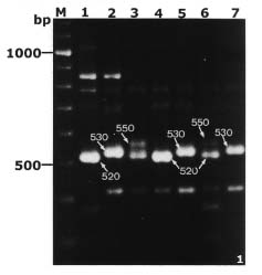
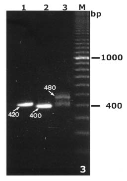
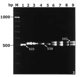
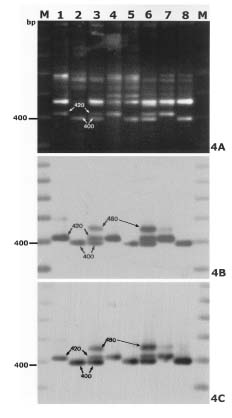
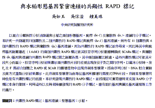
Abstract. By bulked segregant analysis, we screened for markers linked to two morphological genes, brittle culm (bc-1) and lazy (la). We identified four dominant and two co-dominant RAPD (randomly amplified polymorphic DNA) markers for bc-1 and eleven dominant and six co-dominant RAPD markers for la. One of the co-dominant RAPD markers, located 1.4cM from bc-1, was converted into co-dominant SCAR (sequence characterized amplified region). The six co-dominant markers were linked to la at various distances; two of which spanned a region with many RAPD markers. These co-dominant RAPD markers were detected by size and were a result of sequence modification between, not within, the priming sites. Non-parental bands were observed when the F1 and the heterozygous F2 RAPD and SCAR products were analyzed electrophorectically. Possible reasons for these non-parental bands were examined by sequence analysis, DNA annealing experiments, and Southern hybridization. The results indicated they were heteroduplex DNA formed by identical fragments amplified from allelic sequences. The presence of non-parental bands is further evidence of the co-dominance of the markers of interest. The co-dominant RAPD markers developed in this study overcome many of the limitations of RAPDs, and are readily verifiable and applicable. With these and additional PCR based markers, gene mapping or marker aided selection can be made more efficient.

Keywords: Co-dominant RAPD markers; Linkage; Morphological mutant; Oryza sativa; SCAR.

Introduction

Rice (Oryza sativa L.), with its small diploid genome, consists of 12 well defined genetic linkage groups (Bennett and Smith, 1976; Kinoshita, 1986; Khush and Singh, 1986; Wu et al., 1988; Ranjhan et al., 1988; Arumuganthan and Earle, 1991). A classical linkage map updated by the Rice Genetics Cooperative carries 209 genes, which include morphological, disease resistance and isozyme markers (Kinoshita, 1998). A molecular linkage map, based on restriction fragment length polymorphism (RFLP), was first reported by McCouch et al. (1988). Subsequently, several molecular genetic maps have been developed by Saito et al. (1991), Kurata et al. (1994), Causse et al. (1994), and Harushima et al. (1998). While the number of markers that can be assigned to these linkage maps is increasing (URL for http://shigen.lab.nig.ac.jp; http://www.gramene.org), the markers are not readily cross-referenced. In order to more fully exploit the information so accumulated, efforts have been made to integrate the classical linkage and molecular maps (Kishimoto et al., 1993; Abenes et al., 1994; Yu et al., 1995; Yoshimura et al., 1997). Cho et al. (1998) have integrated microsatellite, AFLP (amplified fragment length polymorphism) and SSLP (simple-sequence length polymorphism) markers into an RFLP map of rice with the aim of filling gaps, enhancing the level of saturation, and providing a reasonable start

ing point for high resolution QTL analysis, gene isolation, and molecular breeding.

RAPD (randomly amplified polymorphic DNA, Williams et al., 1990; Welsh and McClelland, 1990) markers are also promising for this purpose. RAPD requires minute quantities of DNA for detection of polymorphism based on the presence (dominant) or absence (recessive) of particular bands in electrophoresis. However, its limitation is that being dominant, RAPD markers provide less information for evaluation in linkage analysis than co-dominant RFLP markers. The recombination between dominant RAPD markers and target genes cannot be estimated effectively in a segregating population, and multi-loci inheritance may restrict the usefulness of RAPD markers. In the latter case, PCR (polymerase chain reaction) analysis based on SCAR (sequence characterized amplified region, Paran and Michelmore, 1993) would be necessary (Nair et al., 1995; Nair et al., 1996). RAPD analysis combined with BSA (bulked segregant analysis) has also been used to focus on regions of interest or areas sparsely populated with markers (Giovannoni et al., 1991; Michelmore et al., 1991; Zhang et al., 1994; Zhang et al., 1996). BSA provides information simultaneously on polymorphism of the parents, and possible linkage between a marker and a targeted gene; only two bulks and the parents are needed, reducing the cost and labor requirement several fold compared to conventional methods. In rice, RAPD markers closely linked to genes resistant to gall midge (Nair et al., 1995; Nair et al., 1996), to bacterial blight (Zhang et al., 1996), and to brown planthopper (Jena et al., 1998) have been successfully screened by these approaches.

*Corresponding author. Tel: 886-2-27899590 ext. 114; Fax: 886-2-27827954; E-mail: bomchung@ccvax.sinica.edu.tw


Botanical Bulletin of Academia Sinica, Vol. 43, 2002

Many morphological genes or mutants such as photoperiod sensitivity gene, se1 (Monna et al., 1995) and phenol staining of grain, ph and semi-dwarf habit, sd-1 (Cho et al., 1998) in rice, or sugary enhancer 1, se1 (Azanza et al., 1996) and teosinte branched locus-1, tb1 (Doebley and Stec, 1991; Doebley et al., 1995) in maize have been found to underline quantitative train loci (QTLs). To produce useful molecular markers and construct a saturated physical map near a gene of interest or in a specific region of a genome are necessary for map-base cloning.

The objective of our work is to saturate flanking regions of selected morphological gene loci with molecular markers. In this study, we elected two such genes, brittle culm (bc-1) and lazy (la), since their phenotypes could be easily recognized at the young seedling stage. The brittle culm (bc) mutant was observed with lower content of cellulose in the cell wall compared to the wild type (Nagao and Takahashi, 1963). Plants with lazy (la) gene grow horizontally (lazy growth habit) and show an inability to respond or reduced responsiveness to gravity (Abe and Suge, 1993). On the classical linkage map, bc-1 locus was assigned on chromosome 3 at 108.0 cM, and la locus was on chromosome 11 at 72.0 cM (Kinoshita, 1998). Using an RAPD combined with bulked segregant analysis (BSA) strategy, we have identified four dominant and two co-dominant RAPD markers linked to bc-1. We also found 11 RAPD markers, including six that were co-dominant, linked to la. Co-dominant RAPD markers are unusual. We propose a possible explanation for their occurrence and discuss their potential in gene mapping.

Materials and Methods

Plant Materials

Two genes, namely brittle culm (bc-1) and lazy (la), which have readily identifiable phenotypes during the growth and development of rice, were maintained in separate near-isogenic lines (NILs) of Taichung 65 (T-65, japonica type) background. The NILs were kindly provided by the late Dr. H. I. Oka of the National Institute of Genetics, Japan. T-65bc-1 and T-65la were crossed with an indica accession (no: PI161008) and their progenies were used for genetic analysis. We generated two F2 populations consisting of 150 progenies from respective crosses and 30-50 F2:3 lines, each line consisting of 20-30 plants, from dominant F2 plants. According to the phenotype segregation data in each F2:3 line, the genotype of the targeted morphological gene of its derived F2 plant could be examined. The segregating populations were established and grown at the Institute of Botany, Academia Sinica, Nankang, Taipei, Republic of China.

DNA Extraction and Quantification

Young leaves of individual plants were collected, air dried at 52°C for 48 h, ground into powder, and stored at 4°C until used for DNA extraction. DNA from each sample was extracted from 4 grams of dried leaf powders, following the protocol described by McCouch et al. (1988). DNA

concentration was quantified spectrophotometrically (GeneQuantII, Pharmacia Biotech).

RAPD-PCR Condition

The amplification conditions for RAPD analysis were as described by Williams et al. (1990), with minor modifications. The PCR reaction mixture consisted of 25ng of template DNA, 1× PCR buffer (10 mm Tris-HCl pH 8.8, 1.5 mM MgCl2, 50 mM KCl, 0.1% Triton X-100), 100 µM dNTPs, 0.2 µM of a 10-base random primers, and one unit of Taq DNA polymerase, in a total volume of 25 µl. DNA amplification was performed on a Perkin-Elmer, GeneAmp PCR System 9600. Template DNA was initially denatured at 94°C for 5 min, followed by 45 cycles of PCR amplification under the following parameters: denaturation for 5 sec at 94°C, primer annealing for 20 sec at 36°C, and primer extension for 90 sec at 72°C. A final incubation for 10 min at 72°C was performed to ensure that the primer extension reaction proceeded to completion. The PCR amplified products were separated by electrophoresis on a 1% Nusieve GTG plus 1% SeaKem LE, FMC agarose gel using 0.5X TBE buffer (44.5 mM Tris/Borate, 1 mM EDTA/disodium), and visualized by ethidium bromide staining under UV illumination. RAPD markers were named by primer origin, followed with the primer number and the size of amplified products in base pairs.

Bulked Segregant Analysis (BSA)

Equal weights of dried leaf powder from eight homozygous wild type or recessive F2 individuals were pooled to form the wild type and mutant bulks, respectively. Samples of DNA from these bulks were screened for specific sequences using a total of 820 arbitrary random primers in a primer extension reaction. The primers were obtained from Operon Technologies, Inc. Alameda, CA, USA (Kit A, C-Q, and U-Y) and the University of British Columbia, Vancouver, BC, Canada (Kit 1-4,). RAPD markers that revealed polymorphism in BSA were further analyzed in the F2 population.

SCAR Design and Analysis

Co-dominant RAPD bands, putatively linked with the targeted genes, were excised from the gel after electrophoresis; their DNA eluted and purified by a Nucleotrap Kit (Macherey-Nagel). The purified DNAs were re-amplified using the same primers under the same conditions to confirm the polymorphism. The re-amplified DNA fragments were eluted and purified by a Jetpure PCR Purified Kit (Genomed Inc.), then cloned into pCRTMII vector using a TA cloning Kit (Invitrogen) following the instructions of the suppliers. The positive (white) colonies were verified by PCR to identify the cloned fragments. Plasmids with RAPD inserts were purified by the alkaline-lysis method (Maniatis et al., 1989). Both ends of an insert were sequenced by the use of an Autoread sequencing kit and an autosequencer (Pharmacia Biotech Inc.).

We designed a pair of SCAR primers based on the sequence data obtained. Each SCAR primer contained the


Wu et al. Co-dominant RAPDs of rice morphological genes

original 5 bases of the OPA 10 primer at 5´ end and the subsequent 15 internal bases from the end. The synthesized 20 mers were: SCA10a (5´-CGCAGGTATGTGGCC GGTCA-3´) and SCA10b (5´-CGCAGCACCTAAACGCC AAC-3´). SCAR-PCR amplification of rice genomic DNA was performed in a standard PCR with an extra 10% DMSO added to the reaction mixture. Template DNA was pre-denaturated for 3 minutes at 94°C, followed by 30 cycles of polymerization reaction to amplify the DNA. DNA denaturation was rendered at 94°C, 30 seconds for primer annealing at 65°C and 90 seconds for primer extension at 72°C. A final 10 min was allowed for incubation at 72°C. The amplified DNA fragments were separated by electrophoresis with same method used for RAPD-PCR products described above.

Southern Hybridization

RAPD-PCR products generated from the segregating populations were separated on a 2% agarose gel with 0.5 X TBE buffer and blotted onto Hybond-N+ nylon membranes (Amersham Pharmacia Biotech). The interested polymorphic fragments were eluted from agarose gel and re-amplified for use as hybridization probes. Probe preparation, membrane hybridization and signal detection were performed following the instructions of ECLTM direct nucleic acid labeling and detection system (Amersham Pharmacia Biotech).

Restriction Fragment Length Polymorphism (RFLP) Mapping

Genomic DNA samples from the parents (T-65la and PI161008) and derived F1 plants were digested with restriction enzymes, which included BamHI, BglII, BstEII, DraI, EcoRI, EcoRV, HindIII, PstI, ScaI and XbaI, to screen for polymorphism. Further screening of the genomic DNA from the parents, F1, and 45 F2 progenies were made with selected restriction enzymes. Restriction products were separated on a 1% agarose gel in 0.5X TBE buffer and blotted by Southern transfer to Hybond-N+ nylon membranes for Southern hybridization. RG247, one of the rice RFLP markers near the la locus on chromosome 11 (Abenes et al., 1994) was chosen as the probe.

Linkage Analysis

The extent of linkage between the targeted morphological genes and their corresponding RAPD and RFLP markers was calculated using the MAPMAKER program (Lander et al., 1987) according to the process described by Causse et al. (1994). The distances between markers were presented in centiMorgan (cM) based on the recombination fractions and the Kosambi function (Kosambi, 1944). The distances shown on the genetic map were calculated from multi-point analysis.

Results

RAPD Markers Linked to the Morphological Marker

In this study, seven hundred of a total of 820 primers tested were shown to amplify discrete fragments from genomic DNA. The size of the amplified products ranged from 200 to 2400 base pairs (bp) with an average of 6.4 bands per primer. For the population segregating for brittle culm bc-1 gene, most of the products were identical in size (monomorphic) in both wild-type and mutant bulks. Polymorphic bands produced by five of these primers, OPA10, OPM17, UBC154, UBC269, and UBC384, were selected for further analysis (Table 1). OPA10 generated two polymorphic bands, a 520 bp fragment (OPA10520) from wild type bulk, and a 530 bp (OPA10530) fragment from the mutant bulk. Both fragments were found in the products amplified from F1 hybrid DNA. Furthermore, a non-parental band estimated to be 550 bp in size was amplified from F1 hybrid DNA (Figure 1). OPM17 amplified one 290bp fragment (OPM17290) from mutant, parent or bulk, but not wild-type parent or bulk, which was shown to link with bc-1 in the coupling phase. The remaining three primers amplified one polymorphic band each, as UBC154780, UBC269500, and UBC384600, from wild type parent or bulk (Table 1). These bands were linked with bc-1 in the repulsion phase.

For la, we selected thirteen primers and one primer combination to score. The amplified fragments generated by these primers are listed in Table 2. Nine RAPD markers, OPC11860, OPK16700, OPQ06900, OPV081000, (OPQ06+OPW02)570, UBC2461000, UBC352230 and

Figure 1. Co-dominant RAPD markers amplified from genomic DNA of different progenies and their parents of bc-1/PI161008 mapping population using OPA10 as primer. Polymorphic bands are indicated by arrows. OPA10520 and OPA10530 are co-dominant and OPA10550 present in lane 3 and 6 are non-parental band. M: molecular size marker 100bp ladder; lane 1: wild type parent; lane 2: T-65bc-1; lane3: F1; lane 4: wild type bulk; lane 5: bc-1 bulk; lane 6: heterozygous F2; lane 7: homozygous bc-1 F2.


Botanical Bulletin of Academia Sinica, Vol. 43, 2002

UBC356580, were found to be linked with la in the coupling phase, two, OPC151250 and UBC277350, in the repulsion phase. (OPQ06+OPW02)570 was amplified from DNA of mutant parent and bulk by the primer pair OPQ06 and OPW02. Each of the primers UBC154, UBC178 and UBC280 amplified two polymorphic bands, which are similar in size, as OPA10 did in bc-1/PI161008 crosses. One non-parental fragment was also found in the amplified products from F1 DNA by each of these primers; this RAPD marker was assigned putatively as co-dominant and further tested.

Conversion of RAPD Markers OPA10520/OPA10530 to SCAR Markers

Cloning and sequencing further characterized two RAPD markers, OPA10520 and OPA10530, which are linked with bc-1. Sequence data showed that these RAPD markers were bordered with the 10 bases of primer OPA10, and the length of the amplified fragment covered either 523 bp from wild type parent (PI161008) DNA or 528 bp from T-65bc-1 DNA. Both fragments shared over 95% homology with only thirteen base-substitutions and five base insertion/deletions. Based on the sequence data, we synthesized two specific 20-mer oligonucleotides as SCAR primers, SCA10a and SCA10b. The SCAR primers amplified the two

Figure 3. DNA mixing experiment of co-dominant RAPD markers UBC178400/UBC178420 shows the non-parental band UBC178480 with slower mobility (lane 3). M: molecular size marker, 100 bp ladder; lane 1: recovered UBC178400; lane 2: recovered UBC 178420; lane 3: UBC178400 and UBC178420 were mixed then heat-denatured/ re-annealled before electrophoresis.

specific bands (Figure 2). These SCAR markers were considered as a single locus and the two fragments as allelic sequences.

These SCAR primers also amplified a 550 bp non-parental band, with the DNA of heterozygous F2 individuals as the template, similar to primer OPA10. To find a possible explanation for the generation of this non-parental band, we proceeded with a mixing experiment that involved denaturing and re-annealing the SCAR-PCR products of the parents. Equal amounts of genomic DNA from the two parents were mixed and used as template to perform SCAR-PCR, and the non-parental band was found (Figure 2). The data indicated the non-parental fragment was the result of mis-annealing between two parental allelic sequences from RAPD-PCR and SCAR-PCR. Based on this experiment, primer OPA10 was determined also to be able to generate a co-dominant RAPD marker. The segregation data showed that this marker was co-segregated with bc-1 and had been converted to a co-dominant SCAR marker.

Figure 2. PCR products obtained with SCAR primers SCAR10a/b. Only specific polymorphic bands (arrows) were amplified from different progenies and their parents of bc-1/PI161008 mapping population. SCAR primers were synthesized according to the sequences of OPA10520/OPA10530. The non-parental band SCAR10550 was amplified from heterozygous individuals (lane 6 and 7) and from DNA mixing experiments (lane 8 and 9). M: molecular size marker, 100 bp ladder; lane 1: PI161008 (wild type parent); lane 2 and 3: T-65bc-1(bc-1 parent); lane 4: bc-1 bulk; lane 5: bc-1 F2 plants; lane 6: F1 plants; lane 7: heterozygous F2 plants; lane 8: both parental genomic DNAs mixed before PCR; lane 9: SCAR10523 and SCA10528 were mixed then heat-denatured/re-anneal before electrophoresis.


Wu et al. Co-dominant RAPDs of rice morphological genes

Co-Dominant RAPD Markers Linked with la Gene

Three putative co-dominant RAPD markers, UBC154660 / UBC154680, UBC178400 /UBC178420, and UBC280520 /UBC280550, linked with la were also subjected to DNA denaturation-reannealing upon mixing. One non-parental band was found, as expected, in the amplified products from each of the mixed DNA samples (Figure 3). The polymorphic fragments were individually eluted from the agarose gel and re-amplified by the same primers. The re-amplified products were eluted again from the agarose gel and used as probes to hybridize with the RAPD products generated from two parental, F1 and bulk DNA. The results showed that the probes were homologous with their derived polymorphic bands whether generated from parents, bulks DNA or the non-parental fragment from F1 plants; they shared no homology with other amplified products (Figure 4). This result confirmed that such polymorphic fragments are allelic sequences and supported the notion that they served as co-dominant RAPD markers.

Linkage Analysis of Morphological Mutants and RAPD Markers

Forty-seven brittle clum individuals randomly selected from bc-1/PI161008 F2 and forty-five lazy individuals from la/PI161008 F2 were used as the segregating populations for linkage analysis. The phenotypic and genotypic segregation data for each of the progenies are given in Tables 3 and 4, respectively. The alignment and genetic distances between bc-1 and flanking markers are shown in Figure 5. These markers spanned approximately 21.8 cM, with UBC269500, UBC154780, and OPA10520/OPA10530 clustered and located adjacent to the bc-1 locus; whereas the co-dominant markers OPA10520 /OPA10530 were 1.4 cM from the bc-1 locus.

Based on la/PI161008 segregation population, the linkage analysis allowed us to construct a map that spanned 62.1 cM, which includes la and its flanking RAPDs and one RFLP marker. The alignment and genetic distances between la and flanking markers are shown in Figure 6. RFLP marker RG247 was 16 cM from la, while RAPD markers UBC178400 /UBC178420 and OPC151250 flanked the la locus. UBC178400 /UBC178 420 was located at a distance of 5.2 cM from la locus. Ten RAPD markers, including four co-dominant markers, UBC178400/UBC178 420 and UBC154660/

Figure 4. Hybridization of co-dominant markers UBC178400 and UBC178420 to the RAPD products amplified from individuals of la/PI161008 mapping population by using UBC178 as primer. Panel A shows ethidium-stained agarose -gel electrophoretic profiles of the RAPD products. Panels B and C show the results of hybridization by using UBC178400 (B) and UBC178420 (C) as probe, respectively. UBC178400/UBC178420 are co-dominant bands and UBC178480 is non-parental band presented in heterozygous individuals. M: molecular size marker, 100 bp ladder; lane 1: wild type parent (PI161008); lane 2: mutant parent (T-65la); lane 3: F1 plants; lane 4: wild type bulk; lane 5: la mutant bulk; lane 6: heterozygous F2 plants; lane 7: homozygous wild type F2 plant; lane 8: homozygous la F2 plant.


Botanical Bulletin of Academia Sinica, Vol. 43, 2002


Wu et al. Co-dominant RAPDs of rice morphological genes

UBC154680, spanned 7.4 cM on the map. The other co-dominant marker, UBC280520/ UBC280 550, was relatively far from any of the markers studied in this linkage group. Excluding this marker, the total length of this linkage map is about 38.9 cM.

Discussion

Two segregating populations were used in this study, derived from crosses using one pollen parent and two near isogenic lines (T-65bc-1 and T-65la) as material parents. The isolines were identical in genetic background but each carried a different morphological mutant gene, bc-1 and la, respectively. The efficiency of using bulked segregant analysis to identify the RAPD markers linked with these genes was quite different. Among 820 random primers, thirteen generated polymorphic bands from wild type in la bulks, but only five in bc-1 bulks. The difference may be attributable to their respective locations on the chromosomes. Linkage analysis by telotrisomics showed that bc-1 is located on the short arm of chromosome 3 near the centromere (Singh et al., 1996). The centromere is known to suppress crossing over events in its adjacent region.

Only one RFLP marker, R19, is known thus far to be linked to bc-1, with an 8.6% recombination rate (Yoshimura et al., 1997). In this study we found five additional DNA markers that were located close to bc-1 and have developed a SCAR marker, SCA10523/SCA10528, which offers a reliable and specific assay for bc-1 in segregation analysis. In addition, UBC269500, UBC154780 and OPA10520/OPA10530 are closely linked as a cluster, 1.4 cM away from bc-1 (Figure 5). These targeted RAPDs could not be further separated because few, if any, recombinant could be identified among the segregants. The genetic distances calculated between bc-1 and the co-dominant markers, OPA10520/OPA10530, were more precise than those between bc-1 and other RAPDs. Since UBC269500 and UBC154780 were able to generate primer products only from the wild type parent, the recombination between these markers and the bc-1 locus could not be detected with efficiency in the F2 population. SCA10523/SCA10528 were identified as co-dominant SCAR markers and co-segregated with the original OPA10520 / OPA10530 RAPD markers. Both markers were mapped to the same locus, tightly linked to bc-1.

A total of six brittle culm iosgenes, bc-1~bc-6, are known in rice. One of the bc genes was recently cloned by the use of retrotransposon TOS17. A fragment that co-segregated with a brittle culm phenotype was found to have a sequence highly similar to other plant cellulose synthase (Tanaka et al., 2000). Lazy (la) gene was assigned to the long arm of chromosome 11, according to linkage analyses by telotrisomics (Singh et al., 1996) and is 13.1 cM and 28.8 cM, respectively, away from the RFLP markers XNpb202 (Kishimoto et al., 1993) and RG118 (Yu et al., 1995). It is flanked by RG1094 and RG247, spanning an interval of 9.2 cM (Abenes et al., 1994). In our study, we have identified two additional co-dominant RAPD markers,

Figure 5. Genetic map of the bc-1 region of chromosome 3 spanning 21.8 cM containing bc-1 locus and relative RAPD markers. The alignment was determined based on 47 individuals from a bc-1/PI161008 F2 mapping population. The co-dominant RAPD markers OPA10520/OPA10530 are 1.4 cM apart the from bc-1 locus. The genetic distances between loci were derived by multi-point analysis and are shown in centiMorgans (cM).

Figure 6. Genetic map of the la region on chromosome 11 containing morphological mutant la, relative RAPD markers and a RFLP marker. The genetic distances between loci were derived by multi-point analysis and shown in centiMorgans (cM). The alignment was determined based on 45 individuals from a la/PI161008 F2 mapping population and is orientated by association with RFLP marker RG247. UBC178400/UBC178420, UBC154660/UBC154680 and UBC280520/UBC280550 are co-dominant RAPD markers. The nearest co-dominant RAPD marker UBC178400/UBC178420 is 5.2cM from la locus.


Botanical Bulletin of Academia Sinica, Vol. 43, 2002

the manuscript. This work was financed by the Institute of Botany, Academia Sinica, Taipei, Taiwan ROC.

Literature Cited

Abe, K. and H. Suge. 1993. Role of gravitropic response in the dry matter production of rice (Oryza sative L.): An experiment with a line having lazy gene. J. Plant Res. 106: 337-343.

Abenes, M.L.P., R.E. Tabien, S.R. McCouch, R. Ikeda, P. Ronald, G.S. Khush, and N. Huang. 1994. Orientation and integration of the classical and molecular genetic maps of chromosome 11 in rice. Euphytica 76: 81-87.

Arumuganthan, K. and E.D. Earle. 1991. Nuclear DNA content of some important plant species. Plant Mol. Biol. Rep. 9: 208-218.

Azanza, F., Y. Tadmor, B.P. Klein, T.R. Rocheford, and J.A. Juvik. 1996. Quantitative trait loci influencing chemical and sensory characteristics of eating quality in sweet corn. Genome 39: 40-50.

Ayliffe, M.A., G.J. Lawrence, J.G. Ellis, and A.J. Pryor. 1994. Heteroduplex molecules formed between allelic sequences cause nonparental RAPD bands. Nucleic Acids Res. 22: 1632-1636.

Bennett, M.D. and J.B. Smith. 1976. Nuclear DNA amounts in angiosperms. Philos Trans R Soc. London, Ser B 274: 227-174.

Causse, M.A., T.M. Fulton, Y.G. Cho, S.N. Ahn, J. Chunwongse, K.S. Wu, J. Xiao, Z.H. Yu, P.C. Ronald, S.E. Harrington, G. Second, S.R. McCouch, and D.S. Tanksley. 1994. Saturated molecular map of the rice genome based on an interspecific backcross population. Genetics 138: 1251-1274.

Cho,Y.G., S.R. McCouch, M. Kuiper, M.P. Kang, J. Pot, J.T.M. Groenen, and M.Y. Eun. 1998. Integrated map of AFLP, SSLP and RFLP markers using a recombinant inbred population of rice (Oryza sativa L.). Theor. Appl. Genet. 97: 370-380.

Davis, T.M., H. Yu, K.M. Haigis, and P.J. McGowan. 1995. Template mixing: a method of enhancing detection and interpretation of co-dominant RAPD markers. Theor. Appl. Genet. 91: 582-588.

Doebley, J. and A. Stec. 1991. Genetic analysis of the morphological differences between maize and teosinte. Genetics 129: 285-295.

Doebley, J., A. Stec, and C. Gustus. 1995. Teosinte branched1 and the origin of maize: evidence of epistasis and the evolution of dominance. Genetics 141: 333-346.

Giovannoni, J.J., R.A. Wing, M.W. Ganal, and S.D. Tanksley. 1991. Isolation of molecular markers from specific chromosomal intervals using DNA pools from existing mapping populations. Nucleic Acids Res. 19: 6552-6558.

Harushima, Y., M. Yano, A. Shomura, M. Sato, T. Shimano, Y. Kuboki, T. Yamamoto, S.Y. Lin, B.A. Antonio, A. Parco, H. Kajiya, N. Huang, K. Yamamoto, Y. Nagamura, N. Kurata, G.S. Khush, and T. Sasaki. 1998. A High-Density rice genetic linkage map with 2275 markers using a single F2 population. Genetics 148: 479-494.

Hunt, G.J. and R.E. Jr. Page. 1992. Patterns of inheritance with RAPD molecular markers reveal novel types of polymor

UBC178400/UBC178420 , the nearest molecular markers for la, mapping at a distance of 5.2 cM (Figure 6). An adjacent region spanning 7.4 cM contained a cluster of 10 RAPD markers, four of which were co-dominant, UBC178400 /UBC178420 and UBC154660/ UBC154680. Two additional co-dominant RAPD markers, UBC280520 /UBC280550 flanked this region on the distal side, with the RFLP marker RG247 on the other. The distance between la and RG247 estimated in this study (16 cM) is greater than that observed previously (6.2 cM) by Abenes et al. (1994). The RAPD markers from the present study could facilitate molecular mapping, chromosome walking and ultimately map-based gene cloning of la or other genes around this region.

Most RAPDs are composed of co-migrating DNA fragments amplified from different sites in the genome. They may be derived from repetitive DNA sequences, rendering them unsuitable for use as locus specific probes in hybridization analysis. SCAR is an extension of RAPD and converts an RAPD into a specific single locus (Paran and Michelmore, 1993). Our study with SCAR and sequence analysis confirmed that OPA10520 /OPA10530 and SCA10523 /SCA10528 are allelic and co-dominant.

Hunt and Page (1992) and Ayliffe et al. (1994) suggested that the non-parental bands revealed in PCR resulted from the formation of heteroduplex DNA between allelic sequences. We showed also that the non-parental bands observed in F1 or F2 heterozygous individuals are formed upon denaturation and reannealing of co-dominant sequences between two allelic fragments. This was demonstrated for all of the co-dominant RAPD markers linked to la by DNA mixing experiments and Southern hybridization. These results confirmed the high degree of homology between the two parental sequences, and showed that non-parental amplicons were not the result of heteroduplex formation by non-allelic sequences. RAPD markers are generally dominant, so the formation of non-parental bands in RAPD is infrequent. The frequency of nonparental bands may reflect the frequency of minor deletions and insertions that give rise to length polymorphism in allelic RAPD products (Ayliffe et al., 1994).

The co-dominant RAPD offers a tool for better resolution in genetic mapping. Non-parental bands can provide evidence of co-dominance (Davis et al., 1995; Novy and Vorsa, 1996). Recently, a few co-dominant RAPD markers have been developed, and some of them have been studied through the SCAR analysis and DNA-mixing experiments (Hunt and Page 1992; Ayliffe et al., 1994; Davis et al., 1995; Novy et al., 1996). It is interesting to note that we have been able to generate six co-dominant RAPD markers, including two not linked to the genes targeted in this study. The high frequency of co-dominant RAPD marker identified here provides useful tools for selection in genetics and breeding and may be a reflection of the genetic similarity of the parental lines used for mapping.

Acknowledgements. We are grateful to Professor Pien-Chien Hauag (School of Hygiene and Public Health, Johns Hopkins University, USA) for critical reading and helpful comments of


Wu et al. Co-dominant RAPDs of rice morphological genes

phism in the honeybee. Theor. Appl. Genet. 85: 15-20.

Jena, K.K., I.C. Pasalu, K. Krishniah, and G.S. Khush. 1998. A RAPD marker for the gene conferring resistance to Indian biotype of BPH. Rice Genet. Newsl. 15: 133-134.

Khush, G.S. and R.J. Singh. 1986. Relationship between linkage groups and cytologically identifiable chromosome of rice. In Rice Genetics, Proc Int Rice Genet Symp, May 1985, Los Banos, Philippines, pp. 239-248.

Kinoshita, T. 1986. Standardization of gene symbols and linkage map in rice. In Rice Genetics, Proc Int Rice Genet Symp, May 1985, Los Banos, Philippines, pp. 215-228.

Kinoshita, T. 1998. Report of the committee of on gene symbolization, nomenclature and linkage groups II. Report from coordinator: linkage mapping using mutant genes in rice. Rice Genet. Newsl. 15: 13-74.

Kishimoto, N., M.R. Foolad, E. Shimosaka, S. Matsuura, and A. Saito. 1993. Alignment of molecular and classical linkage maps of rice, Oryza sativa. Plant Cell Rep. 12: 457-461.

Kosambi, D.D. 1944. The estimation of map distances from recombination values. Ann. Eugenet. 12: 172-175.

Kurata, N., Y. Nagamura, K. Yamamoto, Y. Harushima, N. Sue, J. Wu, B.A. Antonio, A. Shomura, T. Shimizu, S.Y. Lin, T. Inoue, A. Fukuda, T. Shimano, Y. Kuboki, T. Toyama, Y. Miyamoto, T. Kirihara, K. Hayasaka, A. Miyao, L. Monna, H.S. Zhong, Y. Tamura, Z.X. Wang, T. Momma, Y. Umehara, M. Yano, T. Sasaki, and Y. Monobe. 1994. A 300 kilobase interval genetic map of rice including 883 expressed sequences. Nature Genet. 8: 365-372.

Lander, E.S., P. Green, J. Abrahamson, A. Barlow, M.J. Daly, S.E. Lincoln, and L. Newburg. 1987. MAPMAKER: an interactive computer package for construction primary linakge maps of experimental and natural populatons. Genomics 1:174-181.

Maniatis, T., E.F. Fritsch, and L. Sambrook. 1989. Molecular cloning: a laboratory manual. Cold Spring Harbor Laboratory Press, Cold Spring Harbor, N.Y.

McCouch, S., G. Kochert, Z. Yu , Z. Wang, G. Khush, W. Coffman, and S. Tanksley. 1988. Molecular mapping of rice chromosomes. Theor. Appl. Genet. 76: 815-829.

Michelmore, R.W., I. Paran, and R.V. Kesseli. 1991. Identification of markers linked to disease-resistance genes by bulked segregant analysis: A rapid method to detect markers in specific genomic regions by using segregating populations. Proc. Natl. Acad. Sci. USA 88: 9828-9832.

Monna L., A. Miyao, H. S. Zhong, T. Sasaki, and Y. Minobe. 1995. Screening of RAPD markers linked to the photoperiod-sensitivity gene in rice chromosome 6 using bulked segregant analysis. DNA Res. 2: 101-106. (http://www.dna-res.kazusa.or.jp/2/3/01/HTMLA/)

Nagao, S. and M. Takahashi. 1963. Trial construction of twelve linkage group in Japanese rice. (Genetical studies on rice plant, XXVII) J. Fac. Agric. Hokkaido Univ. 53: 72-130.

Nair, S., J.S. Bentur, U. Prasada Rao, and M. Mohan. 1995. DNA markers tightly linked to a gall midge resistance gene (Gm2) are potentially useful for marker-aided selection in rice breeding. Theor. Appl. Genet. 91: 68-73.

Nair, S., A. Kumar, M.N. Srivastava, and M. Mohan. 1996. PCR-based DNA markers linked to a gall midge resistance gene, Gm4t, has potential for marker-aided selection in rice. Theor. Appl. Genet. 92: 660-665.

Novy, R.G. and N. Vorsa. 1996. Evidence for RAPD heteroduplex formation in cranberry: implication for pedigree and genetic-relateness studies and a source of co-dominant RAPD markers. Theor. Appl. Genet. 92: 840-849.

Paran, I. and R.W. Michelmore. 1993. Development of reliable PCR-based markers linked to downy mildew resistance genes in lettuce. Theor. Appl. Genet. 85: 985-993.

Ranjhan, S., J.L. Glaszman, D.A. Ramirez, and G.S. Khush. 1988. Chromosomal location of four isozyme loci by trisomic analysis in rice (Oryza sativa L.). Theor. Appl. Genet. 75: 541-545.

Saito, A., M. Yano, N. Kishimoto, M. Nakagahra, A. Yoshimura, K. Saito, S. Kuhara, Y. Ukai, M. Kawase, T. Nagamine, S. Yoshimura, O. Ideta, R. Ohsawa, Y. Hayano, N. Iwata, and M. Sugiura. 1991. Linkage map of restriction fragment length polymorphism loci in rice. Jpn. J. Breed. 41: 665-670.

Singh, K., S.D. Multani, and G.S. Khush. 1996. Secondary trisomic and telotrisomics of rice: origin, characterization, and use in determining the orientation of chromosome map. Genetics 143: 517-529.

Tanaka, K., A. Miyao, K. Murata, K. Onosato, T. Sasaki, and H. Hirochika. 2000. Ananlysis of the rice mutant with brittle culm phenotype caused by the insertion of retrotransposon TOS17. Plant and Animal Genome VIII Conference, SanDiego, CA, Jan 9-12, 2000. (http://www.intl-pag.org/pag/8/abstracts/pag8751.htlm)

Welsh, J. and M. McClelland. 1990. Fingerprinting genomes using PCR with arbitrary primers. Nucleic Acids Res. 18: 7213-7218.

Williams, J.G.K., A.R. Kubelik, K.J. Livak, J.A. Rafalski, and V. Tingey. 1990. DNA polymorphisms amplified by arbitrary primers are useful as genetic markers. Nucleic Acids Res. 18: 6531-6535.

Wu, K.S., J.C. Glaszmann, and G.S. Khush. 1988. Chromosomal location of ten isozyme loci in rice (Oryza sativa L.) through trisomic analysis. Biochem. Genet. 26: 311-328.

Yoshimura, A., O. Ideta, and N. Iwata. 1997. Linkage map of phenotype and RFLP markers in rice. Plant Mole. Biol. 35: 49-60.

Yu, Z.H., S.R. McCouch, T. Kinoshita, S. Sato, and S.D. Tanksley. 1995. Association of morphological and RFLP markers in rice (Oryza sativa L.). Genome 38: 566-574.

Zhang, Q., B.Z. Shen, X.K. Dai, M.H. Hei, M.A. Saghai-Maroof, and Z.B. Li. 1994. Using bulked extremes and recessive class to map genes for photoperiod-sensitive genetic male sterility in rice. Proc. Natl. Acad Sci. USA 91: 8675-8679.

Zhang, G., E.R. Angeles, M.L.P. Abenes, G.S. Khush, and N. Huang. 1996. RAPD and RFLP mapping of the bacterial blight resistance gene xa-13 in rice. Theor. Appl. Genet. 93: 65-70.


Botanical Bulletin of Academia Sinica, Vol. 43, 2002