Bot. Bull. Acad. Sin. (2002) 43: 211-218

Li et al. — Mycoparasitism of Gliocladium roseum on Botrytis cinerea

Ultrastructural study of mycoparasitism of Gliocladium roseum on Botrytis cinerea

G.Q. Li1, H.C. Huang*, E.G. Kokko, and S.N. Acharya

Agriculture and Agri-Food Canada, Lethbridge Research Centre, P.O. Box 3000, Lethbridge, Alberta T1J 4B1, Canada

(Received September 26, 2001; Accepted January 17, 2002)

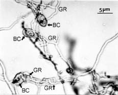
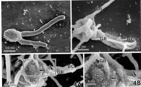
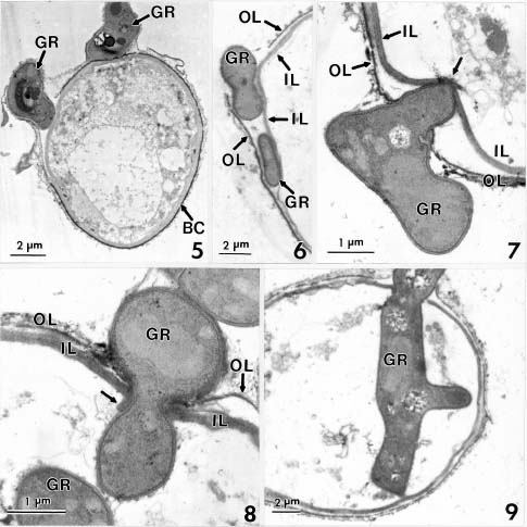
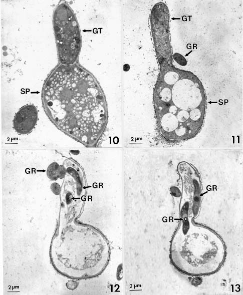
Abstract. Conidia and germ tubes of Botrytis cinerea were highly susceptible to infection by the mycoparasite Gliocladium roseum, when both organisms were grown as a mixed culture in sterile water containing 8-9×10⁴ pollen grains of alfalfa per ml solution. Scanning electron microscopic studies revealed that infection of conidia and germ tubes occurred through direct penetration by hyphal tips of G. roseum without the formation of appressoria. Transmission electron microscopic studies indicated indentation and rupture of the host cell walls at penetration sites. The parasitized conidia and germ tubes of B. cinerea showed signs of cytoplasmic disintegration and the presence of hyphae of G. roseum.

Keywords: Alfalfa pollen; Botrytis cinerea; Gliocladium roseum; Mycoparasitism.

Introduction

Botrytis cinerea Pers.: Fr. is a worldwide ubiquitous plant pathogen, which often causes severe losses to numerous crops (Jarvis, 1977). The pathogen causes shoot blight, blossom blight, and pod rot in seed alfalfa (Medicago sativa L.) (Stuteville and Erwin, 1990). Recent surveys showed that Botrytis blossom blight can be a serious problem for alfalfa seed production in western Canada, especially in the irrigated areas (Gossen et al., 1996, 1997, 1999).

Previous reports indicated that exogenous nutrients from plant tissues, such as senescent petals and pollen grains, are required for the germination of conidia, the subsequent growth and development of germ tubes and hyphae of B. cinerea, and for the infection of host plants by this fungus (Ogawa and English, 1960; Bachelder and Orton, 1962; Chou and Preece, 1968; McClellan and Hewitt, 1973; Huang et al., 1998, 1999, 2000). Moreover, Huang et al. (1999) observed that pollen grains of alfalfa were susceptible to infection by B. cinerea. Thus, pollen may play a significant role in the epidemics of Botrytis blossom blight of various crops including alfalfa (Ogawa and English, 1960; Bachelder and Orton, 1962; McClellan and Hewitt, 1973; Huang et al., 1998, 2000).

Gliocladium roseum Bainer, now reclassified as Clonostachys rosea (Link: Fr.) Schroers, Samuels, Seifert & W. Gams (Schroers et al., 1999), is a destructive

mycoparasite against several plant pathogenic fungi including Botrytis spp. (Barnett and Lilly, 1962; Pugh and Van Eden, 1969; Richard et al., 1974; Walker and Maude, 1975; Pachenari and Dix, 1980; Turhan, 1993; Yu and Sutton, 1997). It was reported as one of the promising biocontrol agents for control of B. cinerea (Sutton et al., 1997; Köhl et al., 1998). Studies by light microscopy showed that hyphae of G. roseum can parasitize B. cinerea (Yu and Sutton, 1997) and B. allii (Walker and Maude, 1975; Pachenari and Dix, 1980). Pachenari and Dix (1980) reported that cell wall degrading enzymes such as b-(1,3)-glucanase and chitinase, and other toxic substances produced by G. roseum were associated with the infection of B. allii by G. roseum. However, no information on the infection process of B. cinerea by G. roseum is available at the ultrastructural level. The objective of this study was to investigate the infection of conidia and germ tubes of B. cinerea by G. roseum using scanning and transmission electron microscopic studies.

Materials and Methods

Strains of Fungal Cultures

Botrytis cinerea strain LRC 2421 isolated from an infected plant of lentil (Lens culinaris Medik) in Alberta, Canada, and G. roseum strain GR-8 isolated from a decayed sclerotium of Sclerotinia sclerotiorum (Lib.) de Bary in Wuhan, China, were used in this study. For production of conidia of B. cinerea, an agar plug (5 mm in diam.) containing mycelial mats was removed from a 2-day-old culture grown on potato dextrose agar (PDA, Difco Laboratories, Detroit, MI, USA) and transferred onto PDA in a Petri dish (90 mm in diam.). Ten dishes were inoculated each time. After incubation at 20°C under fluores

1Present address: Department of Plant Protection, Huazhong Agricultural University, Wuhan, 430070, P.R. China.

*Corresponding author. Tel: 403-317-2226; Fax: 403-382-3156; E-mail: huangh@em.agr.ca. LRC Contribution No.: 387-01060.


Botanical Bulletin of Academia Sinica, Vol. 43, 2002

cent light for 2 to 3 wk, conidia were washed off by flooding the colony in each dish with 10 ml of sterile distilled water. The spore suspensions were filtered through a 4-layered cheesecloth and subsamples were taken to determine microscopically the spore concentration using a hemacytometer. The same procedures described above were also used for the production of conidial suspensions of G. roseum.

Preparation of Alfalfa Pollen

Alfalfa seeds, cultivar Barrier, were sown in plastic pots (15-cm diam.) filled with soil-free Cornell peat-lite mix (Boodley and Sheldrake, 1977). Plants were kept in a greenhouse and watered when needed. During the blooming period, florets were excised from racemes and tripped by pressing each keel petal against the bottom of a Petri dish (50 mm in diam.) to collect pollen grains as described by Huang et al. (1999). The pollen samples were washed in sterile distilled water containing 0.1% Tween® 20 (Fisher Chemical, Fair Lawn, NJ, USA) and filtered through a double-layered cheesecloth. The concentration of pollen was microscopically determined using a hemacytometer and a light microscope.

Light Microscopy

To study the host-mycoparasite interaction, 0.1 ml conidial suspension of B. cinerea (3.3×10⁶ conidia/ml) and 0.1 ml conidial suspension of G. roseum (6.9×10⁶ conidia/ml) were mixed with 0.8 ml of a solution containing 1.0×10⁵ alfalfa pollen grains/ml. An aliquot of 0.2 ml mixed solution was pipetted onto an autoclaved glass slide 25×7.5 mm (width×length) in size, spread gently and kept on a moist filter paper in a 90-mm Petri dish using the aseptic technique described by Huang et al. (1999). Twenty glass slides were inoculated, each serving as one replicate, in each experiment. The dishes were sealed individually with Parafilm M (American National CanÔ, Chicago, IL, USA) and incubated at 20°C. After 6, 12, 24 and 48 h, five slides were randomly taken out from the dishes at each time and the specimens on each slide were stained with lactophenol cotton blue and covered with a 22×22-mm cover slip. Each mounted specimen slide was examined for spore germination and mycoparasitism of G. roseum on B. cinerea under a compound microscope. For the controls, conidial suspensions of B. cinerea with or without alfalfa pollen and conidial suspensions of G. roseum with or without alfalfa pollen were prepared by the method described above. The experiment was repeated twice.

Scanning Electron Microscopy (SEM)

Conidial suspension of B. cinerea (2.6×10⁵ conidia/ml), conidial suspension of G. roseum (2.8×10⁵ conidia/ml), and alfalfa pollen suspension (1.0×10⁵ pollen grains /ml) were mixed throughly at a ratio of 1:1:8 (v:v:v). An aliquot of 0.1 ml mixed solution was pipetted and spread on each sterilized circular cover slip (10 mm in diameter). Ten cover slips were inoculated. The cover slips with samples were supported by glass slides (25×75 mm in size), 2 on each

slide, and kept in 90-mm Petri dishes (moist chambers) at 20°C. A solution containing a mixture of conidia of B. cinerea and pollen grains of alfalfa was used as a control. After incubation for 48 and 72 h, the cover slips were transferred to new Petri dishes (90 mm in diam.). The specimens were vapour-fixed by adding a drop of 2% aqueous solution of osmium tetroxide to the culture of each cover slip in Petri dishes (90 mm in diam.) and the dishes were sealed individually with Parafilm M. After fixation for 48 h, the cover slips were immersed in 2% glutaraldehyde fixative in 0.05 M sodium phosphate buffer, pH 7.0, at 4°C overnight (16 h), and then brought to room temperature (20±2°C). The samples were washed in a sodium phosphate buffer solution three times, 10 min each time, dehydrated in a graded series of ethanol, and dried with a critical point dryer (Polaron E3100) using liquid carbon dioxide as the transitional fluid. The materials were adhered onto aluminum specimen mounts with colloidal silver paste, air dried overnight, and sputter-coated (Denton Vacuum Desk-1) with gold (approximately 15 nm in thickness). The specimens were examined and photographed with a Hitachi S-570 SEM.

Transmission Electron Microscopy (TEM)

In order to improve germination of conidia of B. cinerea, a suspension of B. cinerea conidia (2.6×10⁶ conidia /ml) and an alfalfa pollen suspension (1.0×10 5 pollen grains/ml) were mixed at a ratio of 1:9 (v:v). An aliquot of 0.2 ml of the mixed solution was pipetted onto an alcohol-sterilized cellophane strip (6.0×1.5 cm). Each cellophane strip was placed on a sterilized glass slide, kept in a 90-mm Petri dish (moist chamber) and sealed with Parafilm M. Ten cellophane strips were inoculated. After incubation for 24 h, a 0.2 ml spore suspension of G. roseum containing 2.8×10⁶ conidia/ml was added to each strip, sealed again, and in

Figure 1. A light micrograph showing conidia and germ tubes of Botrytis cinerea (BC) colonized by hyphae of Gliocladium roseum (GR) in a mixed culture containing alfalfa pollen and incubated for 48 h. Note the thin hyphae of G. roseum in contact with the oval-shaped conidia, and the relatively coarse germ tubes of B. cinerea.


Li et al. — Mycoparasitism of Gliocladium roseum on Botrytis cinerea

Figures 2-4. Scanning electron micrographs showing a healthy conidium of Botrytis cinerea (Figure 2) and infection of conidia and germ tubes for B. cinerea by Gliocladium roseum (Figures 3-4). Figure 2. A healthy conidium of B. cinerea (SP) with two germ tubes (GT). Figures 3-4. Hyphal colonization of conidia and germ tubes of B. cinerea (BC) by G. roseum (GR). Note the hyphal penetrations of G. roseum on a conidium (Figure 4A, B; left arrow) and its germ tube (Figure 4A, B; right arrow) of B. cinerea (BC).

cubated at 20°C. Similar strips that had 0.2 ml sterile water added to each of them were used as control cultures. After incubation for another 24 and 48 h, the strips were removed from the glass slides, immersed in 2% glutaraldehyde in 0.05 M sodium cacodylate buffer, pH 7.0, at 4°C overnight (16 h), and then brought to room temperature. The specimen strips were washed in a sodium phosphate buffer solution three times, 10 min each time. The materials were post-fixed in 2% osmium tetroxide in the same buffer for 2 h, stained in 5% uranyl acetate in 50% ethanol for 1 h, dehydrated in a graded series of ethanol, infiltrated with Spurr's low-viscosity embedding medium (Spurr, 1969), and polymerized for 8 h at 70°C. Serial sections (60-90 nm in thickness) were cut with a diamond knife, using a Reichert OM-U3 ultramicrotome (Kokko et al., 1990). Sections were mounted on slotted and formvar-coated grids, stained with 5% aqueous lead citrate and 5% uranyl acetate, and examined with a Hitachi H-7100 transmission electron microscope at 75 Kv.

Results

Germination of Conidia of B. cinerea and G. roseum

In the presence of alfalfa pollen, conidia of B. cinerea and G. roseum, alone or in a mixed culture, started to ger

minate after incubation for 6 h. After 24 h, the average germination rate in the single cultures reached 81% for B. cinerea and 100% for G. roseum, and in the mixed cultures, the germination rate was 59% for B. cinerea and 73% for G. roseum. After incubation for 48 h, the germination rates increased to higher than 96% for both B. cinerea and G. roseum alone or in the mixed cultures. The two organisms were readily identified by the formation of coarse germ tubes and coarse hyphae in B. cinerea, and by the thin hyphae in G. roseum (Figure 1). In the absence of alfalfa pollen, no germination of conidia of B. cinerea was observed in the specimens incubated for 24 or 48 h, whereas 85 and 93% of conidia of G. roseum germinated after incubation for 24 and 48 h, respectively. The germ tubes of G. roseum did not form branches after incubation for 48 h in the treatment without pollen.

Parasitism of G. roseum on B. cinerea

In the cultures of B. cinerea mixed with alfalfa pollen for 48 h, germinated conidia produced elongated germ tubes (Figure 2). In the mixed cultures of B. cinerea and G. roseum containing alfalfa pollen, only the germ tubes of G. roseum developed into branched hyphae after incubation for 48 h (Figures 1, 3). Some of the hyphae of G. roseum established a firm contact with conidia or germ tubes of B. cinerea, initiating the infection process


Botanical Bulletin of Academia Sinica, Vol. 43, 2002

(Figures 1, 4A, B, 5). Infection of conidia and germ tubes of B. cinerea by G. roseum occurred by the direct hyphal tip penetration without the formation of appressoria at the penetration sites (Figures 4A, B). Signs of collapse of infected conidia and germ tubes of B. cinerea were frequently observed (Figures 3, 4A, B).

The TEM micrographs showed that the cell wall of conidia of B. cinerea consisted of an outer layer and an inner layer (Figures 6-8). Cell walls of conidia of B. cinerea infected by G. roseum showed indentations (Figures 5, 7, 8), and sometimes showed signs of separation of the inner layer from the outer layer at the penetration sites

Figures 5-9. Transmission electron micrographs showing infection of conidia of Botrytis cinerea (BC) by Gliocladium roseum (GR). Figure 5. Hyphal cells of G. roseum (GR) in firm contact with a conidium of B. cinerea (BC). Note the indentation of the inner wall at the contact site. Figure 6. Hyphae of G. roseum (GR) invading between the outer layer (OL) and the inner layer (IL) of the cell wall of a conidium of B. cinerea. Figure 7. Indentation (arrow) of the inner layer of the cell wall of a B. cinerea conidium caused by the penetrating hypha of G. roseum (GR). Figure 8. A hypha of G. roseum (GR) had penetrated the cell walls of a B. cinerea conidium. Note the inward breakage (arrow) of the inner layer (IL) of the conidium at the penetration site. Figure 9. An infected conidium of B. cinerea showing the presence of a branched hypha of G. roseum (GR) in the cell lumen. Note the cytoplasmic disintegration of the infected conidium.


Li et al. — Mycoparasitism of Gliocladium roseum on Botrytis cinerea

Figures 10-13. Transmission electron micrographs showing a healthy conidium (SP) of Botrytis cinerea germinated with a germ tube (GT) (Figure 10) and infection of germ tubes by Gliocladium roseum (GR) (Figures 11-13). Note the disintegration of cytoplasm in the infected conidia and germ tubes of B. cinerea.


Botanical Bulletin of Academia Sinica, Vol. 43, 2002

of etching of cell walls of B. cinerea by G. roseum at penetration sites was not observed in the current TEM study. Parchenari and Dix (1980) detected the activities of b-(1,3)-glucanase and chitinase in 3-week-old and 5-day-old co-inoculated cultures of B. allii and G. roseum, respectively. Bélanger et al. (1995) found that a strain of Trichoderma harzianum Rifai penetrated hyphae of B. cinerea 72 h after contact, and chitinase activity was detected after 10 days of contact using a cytochemical labelling technique. These results imply that the cell wall degrading enzymes produced by G. roseum (Pachenari and Dix, 1980) and by T. harzianum (Bélenger et al., 1995) were delayed. The lack of evidence for any enzymatic digestion of cell walls of B. cinerea by G. roseum strain GR-8 in present study might be due to the use of young cultures (24-72 h) for the SEM and TEM studies.

Huang et al. (1998) reported that plant pollen is important in the epidemiology of fungal diseases. Present study further indicates that alfalfa pollen stimulates both spore germination and hyphal development of G. roseum. These stimulatory effects suggest that pollen may be used in the formulation of G. roseum for enhancing its biocontrol efficacy against diseases caused by B. cinerea.

Acknowledgements. The senior author Dr G. Q. Li is a visiting fellow of the Natural Sciences and Engineering Research Council of Canada (NSERC). This research was funded by the Alberta Agriculture Research Institute, Farming for the Future, Project No. 19990070. The authors would like to express their sincere thanks to R. S. Erickson and B. Lee for technical support. Gliocladium roseum strain GR-8 was obtained from China with the Import Permit No. P-2000-01296 issued by Agriculture and Agri-Food Canada.

Literature Cited

Bachelder, S. and E.R. Orton. 1962. Botrytis inflorescence blight on American holly in New Jersey. Plant Dis. Reptr. 46: 320.

Barnett, H.L. and V.G. Lilly. 1962. A destructive mycoparasite, Gliocladium roseum. Mycologia 54: 72-77.

Bélanger, R.R., N. Dufour, J. Caron, and N. Benhamou. 1995. Chronological events associated with the antagonistic properties of Trichoderma harzianum against Botrytis cinerea: indirect evidence for sequential role of antibiosis and parasitism. Bio. Sci. Tech. 5: 41-53.

Boodley, J.W. and R. Sheldrake, Jr. 1977. Cornell Peat-Lie Mixes for Commercial Plant Growing. N. Y. State Coll. Agric. And Life Sci., Inform. Bull. 43, 8 pp.

Chou, M.C. and T.F. Preece. 1968. The effect of pollen grains on infections caused by Botrytis cinerea Fr. Ann. Appl. Biol. 62: 11-22.

Elad, Y., I. Chet, P. Boyle, and Y. Henis. 1983. Parasitism of Trichoderma spp. on Rhizoctonia solani and Sclerotium rolfsii-scanning electron microscopy and fluorescence microscopy. Phytopathology 73: 85-88.

Gossen, B.D. and G. Platford. 1999. Blossom blight in alfalfa seed fields in Saskatchewan and Manitoba 1998. Can. Plant Dis. Surv. 79: 94-95.

(Figures 5-9). Hyphae of G. roseum growing between the two layers of the cell wall were frequently observed (Figure 6). Penetration of conidial cell walls by G. roseum resulted in the presence of hyphae of the mycoparasite with the formation of branches in the cell lumen and the disintegration of cytoplasm (Figures 8, 9). The cytoplasm in healthy conidia and germ tubes appeared dense, and it contained numerous lipid bodies and a few vesicles (Figure 10). At the early stage of host-mycoparasite interactions, disintegration of the cytoplasm occurred in conidia of B. cinerea showing the formation of large vesicles (Figures 5, 11). The infection of germ tubes of B. cinerea by G. roseum was similar to the process of infection of conidia, except that G. roseum hyphae penetrated through a thin cell wall of germ tubes (Figure 12). At the later stage of infection, the growth and ramification of G. roseum hyphae in the cell lumen resulted in a complete disintegration of the cytoplasm of the infected germ tubes of B. cinerea (Figures 12, 13). The hyphae of G. roseum inside the cell lumen of B. cinerea appeared healthy with electronically-dense cytoplasm (Figures 5-9, 11-13).

Discussion

The ultrastructural study confirms a previous finding by light microscopy that G. roseum is a mycoparasite of B. cinerea (Yu and Sutton, 1997). The SEM study reveals that penetration of conidia and germ tubes of B. cinerea by G. roseum was achieved by direct hyphal tip penetration without the formation of appressoria. This infection process is in contrast to the light microscopic study conducted by Walker and Maude (1975) who indicated that formation of appressoria is required for infection of B. allii by G. roseum. In the study of mycoparasitism of G. cantenulatum Gilman et Abbott, on Sclerotinia sclerotiorum (Lib.) de Bary and Fusarium spp., Huang (1978) observed the formation of pseudoappressoria by the mycoparasite on the surface of the host hyphae. This TEM study demonstrates that the infection process of B. cinerea by G. roseum consists of the invasion, and the subsequent hyphal growth and branchings inside infected conidia and germ tubes of B. cinerea. This finding confirms light microscopic observations by Walker and Maude (1975) on mycoparasitism of G. roseum on B. allii and by Barnett and Lilly (1962) on mycoparasitism of G. roseum on several other fungal species. However, it is contrary to the report of Pachenari and Dix (1980) who observed that mycoparasitism of G. roseum on B. allii by was characterized by hyphal contact without any penetrations.

Evidence of indentations and breakage of cell walls of B. cinerea at each penetration sites suggest that hyphae of G. roseum produce mechanical force to overcome the cell wall barrier during the infection process. Several reports indicated that enzymatic chemical dissolution or etching of host cell walls was involved in the infection of hyphae by mycoparasites (Pachenari and Dix, 1980; Elad et al., 1983; Huang and Kokko, 1988). However, evidence


Li et al. — Mycoparasitism of Gliocladium roseum on Botrytis cinerea

Gossen, B.D., Z. Lan, L.M. Harrison, J. Holley, and S.R. Smith. 1997. Survey of blossom blight of alfalfa on the Canadian Prairies in 1996. Can. Plant Dis. Surv. 77: 91-92.

Gossen, B.D., L.M. Harrison, J. Holley, and S.R. Smith. 1996. Survey of blossom blight of alfalfa on the Canadian Prairies in 1995. Can. Plant Dis. Surv. 76: 123-125.

Huang, H.C., S.N. Acharya, and R.S. Erickson. 2000. Etiology of alfalfa blossom blight caused by Sclerotinia sclerotiorum and Botrytis cinerea. Plant Pathol. Bull. (Taiwan) 9: 11-16.

Huang, H.C., E.G. Kokko, and R.S. Erickson. 1999. Infection of alfalfa pollen by Botrytis cinerea. Bot. Bull. Acad. Sin. 40: 101-106.

Huang, H.C., E.G. Kokko, and J.W. Huang. 1998. Epidemiological significance of pollen in fungal diseases. Recent Res. Dev. Plant Pathol. 2: 91-109.

Huang, H.C. and E.G. Kokko.1988. Penetration of hyphae of Sclerotinia sclerotiorum by Coniothyrium minitans without the formation of appressoria. J. Phytopathol. 123: 133-139.

Huang, H.C. 1978. Gliocladium catenulatum: hypopaprasite of Sclerotinia sclerotiorum and Fusarium species. Can. J. Bot. 56: 2243-2246.

Jarvis, W.R. (ed). 1977. Botryotinia and Botrytis Species: Taxonomy, Physiology and Pathogenicity. Agriculture Canada, Hignell Printing Limited, 195 pp.

Köhl, J., M. Gerlagh., B.H. De Haas, and M.C. Krijger. 1998. Biological control of Botrytis cinerea in cyclamen with Ulocladium atrum and Gliocladium roseum under commercial growing conditions. Phytopathology 88: 568-575.

Kokko, E.G., B. Gowen, and C.R. Jahnke. 1990. An inexpensive water volume control device for ultramicrotomy. In Proc. XII Int. Congr. For Electr. Microscopy. San Fransisco Press, San Fransisco, USA, pp. 732-733.

McClellan, W.D. and W.B. Hewitt. 1973. Early Botrytis rot of grapes: Time of infection and latency of Botrytis cinerea Pers. in Vitis vinifera. Phytopathology 63: 1151-1157.

Ogawa, J.M. and H. English. 1960. Blossom blight and green fruit rot of almond, apricot and plum caused by Botrytis cinerea. Plant Disease Reptr. 44: 265-268.

Pachenari, A. and N.J. Dix. 1980. Production of toxins and wall degrading enzymes by Gliocladium roseum. Trans. Br. Mycol. Soc. 74: 561-566.

Pugh, G.J.F. and J.H. Van Eden. 1969. Cellulose-decomposing fungi in polder soils and their possible influence on pathogenic fungi. Neth. J. Plant Pathol. 75: 287-295.

Richard, J.L., C. Grosclaude, and N. Ale-Agha. 1974. Antangonism between Eutypa armeniacae and Gliocladium roseum. Plant Dis. Reptr. 58: 983-984.

Schroers, H. J., G.J. Samuels, K.A. Seifert, and W. Gams. 1999. Classification of the mycoparasite Gliocladium roseum in Clonostachys as G. rosea, its relationship to Bionectria ochroleuca, and notes on other Gliocladium-like fungi. Mycologia 91: 365-385.

Spurr, A.R. 1969. A low-viscosity epoxy embedding medium for electron microscopy. J. Ultrastruct. Res. 26: 31-43.

Stuteville, D.L. and D.C. Erwin (eds.). 1990. Compendium of Alfalfa Disease, 2nd edn. APS Press, ST. Paul, Minnesota, USA, 88 pp.

Sutton, J.C., D.W. Li, G. Peng, H. Yu, P.G. Zhang, and R.M. Valdebenito-Sanhueza. 1997. Gliocladium roseum: a versatile adversary of Botrytis cinerea in crops. Plant Dis. 81: 316-328.

Turhan, G. 1993. Mycoparasitism of Alternaria alternata by an additional eight fungi indicating the existence of further unknown candiates for biological control. J. Phytopathol. 138: 283-292.

Walker, J.A. and R.B. Maude. 1975. Natural occurrence and growth of Gliocladium roseum on the mycelium and sclerotia of Botrytis allii. Trans. Br. Mycol. Soc. 65: 335-338.

Yu, H. and J.C. Sutton. 1997. Morphological development and interactions of Gliocladium roseum and Botrytis cinerea in raspberry. Can. J. Plant Pathol. 19: 237-246.


Botanical Bulletin of Academia Sinica, Vol. 43, 2002