Bot. Bull. Acad. Sin. (2002) 43: 271-276

Hou et al. — SPTI degradation by aspartic type protease

An aspartic type protease degrades trypsin inhibitors, the

major root storage proteins of sweet potato (Ipomoea batatas (L.) Lam cv. Tainong 57)

Wen-Chi Hou1,*, Dong-Jiann Huang2, and Yaw-Huei Lin2,*

1Graduate Institute of Pharmacognosy Science, Taipei Medical University, Taipei 110, Taiwan

2Institute of Botany, Academia Sinica, Nankang, Taipei 115, Taiwan

(Received May 7, 2002; Accepted June 19, 2002)

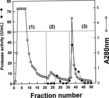
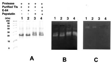
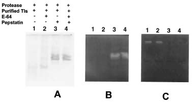
Abstract. Roots of sprouted sweet potato (Ipomoea batatas [L.] Lam) were used as materials to purify proteases which degraded trypsin inhibitors (Tis), the root storage proteins of sweet potato (SP). The commercial pepstatin-agarose (crosslinked, 6%) was chosen as an affinity column for purification. The purified protease has a molecular mass of about 64 kDa on the gelatin-SDS-PAGE gel and was inhibited by pepstatin but not by E-64 on the gelatin-SDS-PAGE gel. Therefore, it might belong to the aspartic type. Using the trypsin inhibitor activity staining method as a criterion for TI degradations, we found that this aspartic type protease could degrade purified Tis in the presence or absence of 5 mM DTT and the hydrolysis was complete in the former condition. The physiological role of aspartic type protease in the degradation of SPTis is discussed.

Keywords: Aspartic type protease; Degradation; Physiological role; Sweet potato; Trypsin inhibitor.

Abbreviations: SDS-PAGE, sodium dodecylsulfate-polyacrylamide gel electrophoresis; SP, sweet potato; TI, trypsin inhibitor.

Introduction

Proteases play important roles in post-translational modification, protein turnover, activation and inactivation of specific proteins, and nutrient supplementation (North, 1982). In plant tissues, specific proteases involved in the mobilization of reserve proteins (Chrispeels and Boulter, 1975; Wilson et al., 1986; Qi et al., 1992; Bottari et al., 1996; Senyuk et al., 1998; Davy et al., 2000), developmental processes (Lin and Tsai, 1991; Lin and Chan, 1992; Lin and Tsai, 1994; Dominguez and Cejudo, 1996; Voigt et al., 1997), and senescence (Hensel et al., 1993; Lohman et al., 1994; Smart et al., 1995; Drake et al., 1996) have been studied intensively.

Proteinaceous protease inhibitors in plants may be important in regulating and controlling endogenous proteases and in acting as protective agents against insect and/or microbial proteases (Ryan, 1973, 1989). Sohonie and Bhandarker (1954) reported for the first time the presence of trypsin inhibitors (TIs) in sweet potato (SP). Later, we indicated that TI activities in SP are positively correlated with concentrations of water-soluble protein (Lin and Chen, 1980), and that a large negative correlation exists between the natural logarithm of TI activities and cumulative rainfall, which suggests that SPTI activities may vary

in response to drought (Lin, 1989). Polyamines, including cadaverine, spermidine and spermine, were bound covalently to SPTI, which might participate in regulating the growth and developmental processes of SP (Hou and Lin, 1997a). SPTIs were also proved to have both dehydroascorbate reductase and monodehydroascorbate reductase activities and might respond to environmental stresses (Hou and Lin, 1997b). We found that TIs in SP roots accounted for about 60% of total water-soluble proteins and could be recognized as storage proteins (Lin and Chen, 1980). Maeshima et al. (1985) identified sporamin as the major storage protein in SP root, accounting for 80% of the total proteins there; however, a dramatic decrease to 2% of the original value was found during sprouting. Lin (1993) considered sporamin one form of TI in SP, a finding confirmed later by Yeh et al. (1997a). However, few reports concern the degradation of SP root storage protein during sprouting. In this work we report preliminary results showing that SP proteinaceous trypsin inhibitors were degraded by an endogenous aspartic type protease.

Materials and Methods

Plant Materials

Fresh roots of sweet potato (Ipomoea batatas L. Lam cv. Tainong 57) were purchased from a local wholesaler. After cleaning with water, the roots were either immediately cut into strips for Ti extraction according to the method of Hou and Lin (1997a) or placed in the

*Corresponding author. Prof. Yaw-Huei Lin: Fax: 886-2-2782-7954, E-mail: boyhlin@ccvax.sinica.edu.tw; or Prof. Wen-Chi Hou: Fax: 886-2-2378-0134, E-mail: wchou@tmu.edu.tw


Botanical Bulletin of Academia Sinica, Vol. 43, 2002

thermostated (30°C) growth chamber in dark and sprayed with water twice a day. After excision of the etiolated sprouts (about 5-7 cm), the roots of sprouted SP were also immediately cut into strips for protease purification.

SPTI Purification

After washing and peeling, the SP roots were cut into strips for TI extraction and purification. After extraction and centrifugation, the crude extracts were loaded directly onto a trypsin Sepharose 4B affinity column. The adsorbed TIs were eluted by pH changes with 0.2 M KCl (pH 2.0) according to the methods of Hou and Lin (1997a,b). After dialysis against deionized water, the purified TIs were concentrated with centricon 10 and then lyophilized for further use.

Isolation and Purification of an Aspartic Type Protease from Roots of Sprouted SP

The roots of sprouted SP were used as materials for isolation and purification of an aspartic type protease. After excising the sprouts, the sprouted roots were immediately cut into strips and extracted with four volumes (W/V) of 20 mM Tris-HCl buffer (pH 7.9) containing 200 mM NaCl, 10 mM EDTA and 1% ascorbate. After centrifugation twice at 14,000 g, the crude extracts were loaded directly onto a commercial pepstatin-agarose (crosslinked, 6%, PIERCE, No-20215, Illinois) affinity column (1.0 × 10 cm). After washing with 20 mM Tris-HCl buffer (pH 7.9) containing 200 mM NaCl the bound proteases were eluted batchwise firstly with the same buffer containing 450 mM NaCl for 15 fractions and then eluted batchwise with 50 mM phosphate buffer (pH 11.5) containing 500 mM NaCl for another 15 fractions. The flow rate was 32 ml/h and each fraction contained 4.8 ml. The protease activity was determined as follows: Two hundred µl of each fraction was mixed with 400 µl, 1% casein (pH 7.9) and 400 µl, 100 mM Tris buffer (pH 7.9) at 37°C. The reaction was performed for one hour, and then 400 µl of 10% trichloroacetic acid was added to stop it. The reaction mixture was then kept at 0°C for 1 h. After centrifugation at 12,000 g, the supernatants were collected, and the absorbance at 280 nm was determined. One enzyme unit was defined as the amount of enzyme that increased absorbance 0.01 at 280 nm under the reaction conditions. The active fractions were pooled and adjusted to pH 7.9 and then dialyzed against 20 mM Tris-HCl buffer (pH 7.9) for further use.

The Hydrolysis of TIs by an Aspartic Type Protease

Each 50 µl of purified protease (50 units) and SPTI (1 mg/ml) were mixed with 25 µl, 500 mM Tris-HCl buffer (pH 7.9) with or without 5 mM DTT at room temperature overnight. Either E-64 (a cysteine type protease inhibitor at a final concentration of 40 µM) or pepstatin A (an aspartic type protease inhibitor at a final concentration of 40 µM) was added to compare the extent of SPTI hydrolysis. After hydrolysis, each reaction solution was examined by SDS-PAGE.

Figure 1. The chromatogram of protease activity of sprouted sweet potato roots on a commercial pepstatin-agarose column. After washing with 20 mM Tris-HCl buffer (pH 7.9) containing 200 mM NaCl (buffer 1) the bound proteases were eluted batchwise firstly with the same buffer containing 450 mM NaCl (buffer 2) for 15 fractions and then eluted batchwise with 50 mM phosphate buffer (pH 11.5) containing 500 mM NaCl (buffer 3) for another 15 fractions. The flow rate was 32 ml/h, and each fraction contained 4.8 ml.

Protease and TI Activity Stainings on SDS-PAGE Gels

Four parts of samples were mixed with one part of sample buffer, namely 60 mM Tris-HCl buffer (pH 6.8) containing 2% SDS, 25% glycerol and 0.1% bromophenol blue without 2-mercaptoethanol for aspartic type protease and TI activity stainings at 4°C overnight. Coomassie brilliant blue R-250 was used for protein staining (Neuhoff et al., 1985). Aspartic type protease activity staining was carried out on a 12.5% SDS-PAGE gel co-polymerized with 0.1% (W/V) gelatin (Dominguez and Cejudo, 1996). After electrophoresis, gels were washed with 25% isopropanol in 10 mM Tris-HCl buffer (pH 7.9) for 10 min twice to remove SDS (Hou and Lin, 1998). For protease activity staining, the gel was shaken in 100 mM Tris-HCl buffer (pH 7.9) overnight and then stained with coomassie brilliant blue R-250. For SPTI activity staining, the gel was stained according to the method of Hou and Lin (1998).

Chemicals

All chemicals and reagents were of the highest purity available. Trypsin (TPCK-treated, 40 U/mg) was purchased from E. Merck Inc. (Darmstadt, Germany); Seeblue prestained markers for SDS-PAGE were from Novex (San Diego, CA); CNBr-activated Sepharose 4B was from Pharmacia Biotech AB (Uppsala, Sweden). Pepstatin-agarose (crosslinked, 6%, No-20215) was from Pierce Chem Co. (Rockford, USA). Other chemicals and reagents including


Hou et al. — SPTI degradation by aspartic type protease

buffer 3 fractions were pooled, adjusted to pH 7.9, and then dialyzed against 20 mM Tris-HCl buffer (pH 7.9) for further use.

Figure 2C and Figure 3C show the protease activity staining without or with 5 mM DTT, respectively, on gelatin-SDS-PAGE gels. Lane 1, the mixtures of purified protease and SPTI; lane 2, E-64 added to lane 1 mixture; lane 3, pepstatin added to lane 1 mixture; lane 4, both E-64 and pepstatin were added to lane 1 mixture. A protease activity band (lane 1) with molecular mass of about 64 kDa was found on the gelatin-SDS-PAGE gel without 5 mM DTT treatments (Figure 2C) or with 5 mM DTT treatments (Figure 3C), but the latter had a stronger protease activity band. The same protease activity band remained when inhibitors of E-64 were present (lane 2); but disappeared when inhibitor pepstatin was present (lane 3); treatment with both E-64 and pepstatin (lane 4) also inhibited protease activity. This suggested that the purified protease belongs to an aspartic type, which is inhibited by pepstatin and could be activated by 5 mM DTT.

Figure 2B and Figure 3B show the TI activity staining without or with 5 mM DTT, respectively, on SDS-PAGE gels. Lane 1, the mixtures of both purified protease and SPTI; lane 2, E-64 added to lane 1 mixture; lane 3, pepstatin added to lane 1 mixture; lane 4, both E-64 and pepstatin were added to lane 1 mixture. It was found that when the activity of aspartic type protease was inhibited by pepstatin (see lanes 3 and 4 of Figures 2C and 3C) strong TI activity appeared (see lanes 3 and 4 of Figures 2B and 3B) compared to the protein staining (see lanes 3 and 4 of Figures 2A and 3A). Meanwhile, when aspartic type protease kept full activity (lanes 1 and 2), TIs were degraded to different extents depending on whether 5 mM DTT was

protease inhibitors and synthetic substrates were from Sigma Chemical Co. (St. Louis, MO, USA).

Results and Discussion

In SP, about 60% of total water-soluble proteins were TIs which were recognized as storage proteins (Lin and Chen, 1980). Maeshima et al. (1985) pointed out that the storage proteins of SP decreased from 4.41 to 0.067 mg/g tissue after sprouting. Li and Oba (1985) also noted that the storage proteins of SP decreased from 3.22 to 0.18 mg/g tissue after one year storage at 10 to 12°C. So, it is clear that SPTIs serve as storage proteins that provide nitrogen sources during sprouting or storage. Yeh et al. (1997b) reported that SPTIs expressed in transgenic tobacco plants confer resistance against Spodoptera litura. SPTIs can also function as protective agents against insects. But so far almost no reports have dealt with the degradation of SP root storage protein during sprouting. In this work we report the preliminary results that SPTIs were degraded by an aspartic type protease. In order to start the work, we used a trypsin-Sepharose 4B affinity column (Hou and Lin, 1997a) to purify SPTIs from dormancy SP roots as substrates for purified aspartic type protease.

Figure 1 shows the chromatogram of protease purification on a commercial pepstatin-agarose column. After washing with 20 mM Tris-HCl buffer (pH 7.9) containing 200 mM NaCl (buffer 1) the bound proteases were eluted batchwise firstly with the same buffer containing 450 mM NaCl (buffer 2) for 15 fractions and then eluted batchwise with 50 mM phosphate buffer (pH 11.5) containing 500 mM NaCl (buffer 3) for another 15 fractions. We found that most of the protease activities were eluted by buffer 3. These

Figure 2. The protein staining (A), trypsin inhibitor activity staining (B) and protease activity staining (C) on 12.5% SDS-PAGE gels (A and B) or a 12.5% gelatin-SDS-PAGE gel (C) after overnight reaction at room temperature without 5 mM DTT. Lane 1, the mixtures of purified protease and SPTI; lane 2, E-64 (40 µM) added to lane 1 mixture; lane 3, pepstatin (40 µM) added to lane 1 mixture; lane 4, E-64 and pepstatin were added to lane 1 mixture.


Botanical Bulletin of Academia Sinica, Vol. 43, 2002

Figure 3. The protein staining (A), trypsin inhibitor activity staining (B) and protease activity staining (C) on 12.5% SDS-PAGE gels (A and B) or a 12.5% gelatin-SDS-PAGE gel (C) after overnight reaction at room temperature with 5 mM DTT. Lane 1, the mixtures of purified protease and SPTI; lane 2, E-64 (40 µM) added to lane 1 mixture; lane 3, pepstatin (40 µM) added to lane 1 mixture; lane 4, E-64 and pepstatin were added to lane 1 mixture.

ing the optimal pH hydrolysis, N-terminal amino acid sequences, and substrate specificity.

Acknowledgements. The authors want to acknowledge the financial support (NSC91-2313-B038-002) from the National Science Council, Republic of China.

Literature Cited

Bottari, A., A. Capocchi, D. Fontanini, and L. Galleschi. 1996. Major proteinase hydrolyzing gliadin during wheat germination. Phytochemistry 43: 39-44.

Chrispeels, M.J. and D. Boulter. 1975. Control of storage protein metabolism in the cotyledons of germinating mung beans: role of endopeptidase. Plant Physiol. 55: 1031-1037.

Davy, A., K.K. Thomsen, M.A. Julian, L.C. Alves, I.B. Svendsen, and D.J. Simpson. 2000. Purification and characterization of barley dipeptidyl peptidase IV. Plant Physiol. 122: 425-431.

Dominguez, F. and F.J. Cejudo. 1996. Characterization of the endoprotease appearing during wheat grain development. Plant Physiol. 112: 1211-1217.

Drake, R., I. John, A. Farrell, W. Copper, W. Schuch, and D. Grierson. 1996. Isolation and analysis of cDNA encoding tomato cysteine proteases expressed during leaf senescence. Plant Mol. Biol. 30: 755-767.

Hensel, L.L., V. Grbic, D.A. Baumgarten, and A.B. Bleecker. 1993. Developmental and age-related processes that inference the longevity and senescence of photosynthetic tissues in Arabidopsis. Plant Cell 5: 553-564.

Hou, W.C. and Y.H. Lin. 1997a. Polyamine-bound trypsin inhibitors in sweet potato (Ipomoea batatas [L.] Lam cv. Tainong 570 storage roots, sprouted roots and sprouts. Plant Sci. 126: 11-19.

Hou, W.C. and H.Y. Lin. 1997b. Dehydroascorbate reductase

present (Figure 3) or not (Figure 2). Without 5 mM DTT treatments (Figure 2), the TI protein bands (lanes 1 and 2 of Figure 2A) and TI activity bands (lanes 1 and 2 of Figure 2B) were changed under full aspartic protease activity in comparison with those of pepstatin treatments (lanes 3 and 4, Figure 2A and 2B). With 5 mM DTT treatment (Figure 3), it was found that the TI activity was lost completely (lanes 1 and 2 of Figure 3B), which was accompanied by the loss of TI protein bands (lanes 1 and 2, Figure 3A). Compared to pepstatin treatment (lanes 3 and 4 of Figure 3A and 3B), it was found that this aspartic type protease could degrade TIs with or without a reduced state. This is the first report of SPTI being degraded almost completely by an aspartic type protease under reducing conditions.

Soybean TI lost its inhibitory activity after DTT treatments (Trumper et al., 1994) and ovomucoid also lost its inhibitory activity when disulfide bonds were reduced (Matsuda et al., 1981). However SPTI retained its inhibitory activity under reducing conditions. Maeshima et al. (1985) indicated that the storage proteins of SP decreased from 4.41 to 0.067 mg/g tissue after sprouting. Figures 2 and 3 suggest that root storage proteins of sweet potato, TIs or sporamins, can be degraded during sprouting. Kobrehel et al. (1991, 1992) showed that TIs of both Kunitz type and Bowman-Birk type could be reduced by a NADP/thioredoxin system, which could facilitate their hydrolysis by proteases. Apart from the reported physiological functions of TIs, as storage proteins in SP roots (Maeshima et al., 1985; Yeh et al., 1997a,b; Hou and Lin, 1997a,b), this work provides the first evidence that SPTIs or sporamins can be degraded completely by an aspartic type protease purified from the roots of sprouted SP. The detailed mechanisms deserve further investigations, includ


Hou et al. — SPTI degradation by aspartic type protease

and monodehydroascorbate reductase activities of trypsin inhibitors, the major sweet potato (Ipomoea batatas [L.] Lam) root storage protein. Plant Sci. 128: 151-158.

Hou, W.C. and Y.H. Lin. 1998. Activity staining on polyacrylamide gels of trypsin inhibitors from leaves of sweet potato (Ipomoea batatas [L.] Lam) varieties. Electrophoresis 19: 212-214.

Kobrehel, K., B.C. Yee, and B.B. Buchanan. 1991. Role of the NADP/thioredoxin system in the reduction of a-amylase and trypsin inhibitor proteins. J. Biol. Chem. 266: 16135-16140.

Kobrehel, K., J.H. Wong, A. Balogh, F. Kiss, B.C. Yee, and B.B. Buchanan. 1992. Specific reduction of wheat storage proteins by thioredoxin h. Plant Physiol. 99: 919-924.

Li, H.S. and K. Oba. 1985. Major soluble proteins of sweet potato roots and changes in proteins after cutting, infection, or storage. Agri. Biol. Chem. 49: 737-744.

Lin, Y.H. and H.L. Chen. 1980. Level and heat stability of trypsin inhibitor activity among sweet potato (Ipomoea batatas Lam.) varieties. Bot. Bull. Acad. Sin. 21: 1-13.

Lin, Y.H. 1989. Relationship between trypsin-inhibitor activity and water-soluble protein and cumulative rainfall in sweet potatoes. J. Amer. Soci. Horti. Sci. 114: 814-818.

Lin, Y.H. and M. Tsai. 1991. In vitro protease activities of four parts of germinated Tainong 57 sweet potato roots. Bot. Bull. Acad. Sin. 32: 79-85.

Lin, Y.H. and H.Y. Chan. 1992. An aminopeptidase (AP1) from sprouts of sweet potato (Ipomoea batatas L. Lam cv. Tainong 64). Bot. Bull. Acad. Sin. 33: 253-261.

Lin, Y.H. 1993. Trypsin inhibitors of sweet potato: review and prospect. In Y.I. Hsing and C.H. Chou (eds.), Recent Advances in Botany, Academia Sinica Monograph series No. 13, Taipei, Taiwan, pp. 179-185.

Lin, Y.H. and M. Tsai. 1994. Purification and characterization of an enzyme hydrolyzing L-methionine-4-nitroanilide from germinated sweet potato (Ipomoea batatas L. Lam cv. Tainong 57) root. Bot. Bull. Acad. Sin. 35: 25-32.

Lohman, K.N., G. Susheng, C.J. Manorama, R.M. Amasino, 1994. Molecular analysis of natural leaf senescence in Arabidopsis thalian. Physiol. Plant. 92: 322-328.

Maeshima, M., T. Sasaki, and T. Asahi. 1985. Characterization of major proteins in sweet potato tuberous roots. Phytochemistry 24: 1899-1902.

Matsuda, T., K. Watanabe, and Y. Sato. 1981. Secondary structure of reduced ovomucoid and denaturation of reduced ovomucoid and its reduced fragment A (1-130) and B (131-186). FEBS Lett. 124: 185-188.

Neuhoff, V., R. Stamm, and H. Eibl. 1985. Clear background and highly sensitive protein staining with Coomassie blue dyes in polyacrylamide gels: a systematic analysis. Electrophoresis 6: 427-448.

North, M.J. 1982. Comparative biochemistry of the proteinases of eukaryotic microorganism. Microbiol. Rev. 46: 308-340.

Qi, X., K.A. Wilson, and A.L. Tan-Wilson. 1992. Characterization of the major protease involved in the soybean b-conglycinin storage protein mobilization. Plant Physiol. 99: 725-733.

Ryan, C.A. 1973. Proteolytic enzymes and their inhibitors in plants. Ann. Rev. Plant Physiol. 24: 173-196.

Ryan, C.A. 1989. Protease inhibitor gene families: Strategies for transformation to improve plant defenses against herbivores. BioEssays 10: 20-24.

Senyuk, V., V. Rotari, C. Becker, A. Zakharov, C. Horstmann, K. Muntz, and I. Vainttraub. 1998. Does an asparaginyl-specific cysteine endopeptidase trigger phaseolin degradation in cotyledons of kidney bean seedlings? Eur. J. Biochem. 258: 546-558.

Smart, C.M., S.E. Hosken, H. Thomas, J.A. Greaves, B.G. Blair, and W. Schuch. 1995. The timing of maize leaf senescence and characterization of senescence-related cDNAs. Physiol. Plant. 93: 673-682.

Sohonie, K. and A.P. Bhandarker. 1954. Trypsin inhibitors in Indian foodstuffs: I. Inhibitors in vegetables. J. Sci. Ind. Res. 13B: 500-503.

Trumper, S., H. Follmann, and I. Haberlein. 1994. A novel dehydroascorbate reductase from spinach chloroplasts homologous to plant trypsin inhibitor. FEBS Lett. 352: 159-162.

Voigt, G., B. Biehl, H. Heinrichs, and J. Voigt. 1997. Aspartic proteinase levels in seeds of different angiosperms. Phytochemistry 44: 389-392.

Wilson, K.A., B.R. Rightmire, J.C. Chen, and A.L. Tan-Wilson. 1986. Differential proteolysis of glycinin and b-conglycinin polypeptides during soybean germination and seedling growth. Plant Physiol. 82: 71-76.

Yeh, K.W., J.C. Chen, M.I. Lin, Y.M. Chen, and C.Y. Lin. 1997a. Functional activity of sporamin from sweet potato (Ipomoea batatas Lam.): a tuber storage protein with trypsin inhibitory activity. Plant Mol. Biol. 33: 565-570.

Yeh, K.W., M.L. Lin, S.J. Tuan, Y.M. Chen, C.Y. Lin, and S.S. Kao. 1997b. Sweet potato (Ipomoea batatas) trypsin inhibitors expressed in transgenic tobacco plants confer resistance against Spodoptera litura. Plant Cell Rep. 16: 696-699.


Botanical Bulletin of Academia Sinica, Vol. 43, 2002