Bot. Bull. Acad. Sin. (2003) 44: 79-98

Nalawade et al. — Studies on tissue culture of Chinese medicinal plant

(Review paper)

Studies on tissue culture of Chinese medicinal plant resources in Taiwan and their sustainable utilization

Satish M. Nalawade1, Abhay P. Sagare3, Chen-Yue Lee1, Chao-Lin Kao4, and Hsin-Sheng Tsay1,2,*

1Department of Applied Chemistry, Chaoyang University of Technology, Wufeng, Taichung, Taiwan 413

2Department of Agronomy, Taiwan Agricultural Research Institute, Wufeng, Taichung, Taiwan 413

3Division of Neurovascular Biology, Center for Aging and Developmental Biology, University of Rochester Medical Center, Rochester, New York 14642, USA

4Chung Hwa College of Medical Technology, Jen-Te Hsiang, Tainan Hsien, Taiwan 717

(Received September 19, 2002; Accepted December 18, 2002)

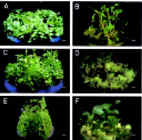
Abstract. Medicinal plants are sources of important therapeutic aid for alleviating human ailments. With increasing realization of the health hazards and toxicity associated with the indiscriminate use of synthetic drugs and antibiotics, interest in the use of plants and plant-based drugs has revived throughout the world. However, a large number of medicinal plants remain to be investigated for their possible pharmacological value. Most of the pharmaceutical industry is highly dependent on wild populations for the supply of raw materials for extraction of medicinally important compounds. Due to a lack of proper cultivation practices, destruction of plant habitats, and the illegal and indiscriminate collection of plants from these habitats, many medicinal plants are severely threatened. Advanced biotechnological methods of culturing plant cells and tissues should provide new means of conserving and rapidly propagating valuable, rare, and endangered medicinal plants. This paper describes the work carried out at the Taiwan Agricultural Research Institute and Chaoyang University of Technology on in vitro propagation of some important medicinal plants.

Keywords: Adenophora triphylla; Angelica sinensis; Anoectochilus formosanus; Bupleurum falcatum; Corydalis yanhusuo; Dendrobium linawianum; Fritillaria hupehensis; Gentiana davidii; Limonium wrightii; Medicinal plants; Pinellia ternata; Scrophularia yoshimurae; Zingiber zerumbet.

Introduction

Medicinal plants have been the subjects of man's curiosity since time immemorial (Constable, 1990). Almost every civilization has a history of medicinal plant use (Ensminger et al., 1983). Approximately 80% of the people in the world's developing countries rely on traditional medicine for their primary health care needs, and about 85% of traditional medicine involves the use of plant extracts (Vieira and Skorupa, 1993). Interest in phytomedicine has exploded in the last few years, and about 500 different plant species are used as key ingredients, and many are still being collected from the wild (Mendelsohnm and Balick, 1994). The resurgence of public interest in plant-based medicine coupled with rapid expansion of pharmaceutical industries have necessitated an increased demand for medicinal plants, leading to over-exploitation that threatens the survival of many rare species. Also, many medicinal plant species are disappearing at an alarming rate due to rapid agricultural and urban development, uncontrolled deforestation and indiscriminate collection. Combinations of in vitro propagation techniques (Fay, 1992) and cryopreservation may help in conservation of biodiversity of locally used medicinal plants. Cryopreservation is a reliable method for long-term storage of the germplasm of endangered species (Bramwell, 1990). Several medicinal plant species have been successfully cryopreserved (Bajaj, 1995; Naik, 1998). In vitro cell and tissue culture methodology is envisaged as a mean for germplasm conservation to ensure the survival of endangered plant species, rapid mass propagation for large-scale revegetation, and for genetic manipulation studies.

Plants play a dominant role in the introduction of new therapeutic agents, and also drugs from the higher plants continue to occupy an important niche in modern medicine (Dev, 1997). Many compounds used in today's medicine have a complex structure, and synthesizing these bioactive compounds chemically at a low price is not easy (Shimomura et al., 1997). With deforestation, medicinal wealth is rapidly lost, such that many valuable plants are threatened with extinction. Pharmaceutical companies depend largely upon materials procured from naturally occurring stands that are being rapidly

*Corresponding author. Dr. Hsin-Sheng Tsay, Department of Applied Chemistry, Chaoyang University of Technology, 168, Gifeng E. Road, Wufeng, Taichung, Taiwan 413. Tel: 886-4-23323000 ext. 7578; Fax: 886-4-23742371; E-mail: hstsay@mail.cyut.edu.tw


Botanical Bulletin of Academia Sinica, Vol. 44, 2003

depleted. Plant tissue culture is an alternative method of propagation (George and Sherrington, 1984) and is being used widely for the commercial propagation of a large number of plant species, including many medicinal plants (Rout et al., 2000).

The central mountain range of Taiwan is home to many highly valued medicinal herbs. These are being indiscriminately collected in large quantities from the wild to meet the ever-increasing demand for traditional crude drugs. Currently, collection of plants from within a national park is illegal in Taiwan. One of the solutions for protecting rare endemic medicinal herbs is to develop an efficient in vitro propagation method and to encourage commercial cultivation. The National Science Council (NSC) of Taiwan has been promoting research on traditional Chinese medicinal plants since 1988. The main objectives of this research programme are: (a) to collect information about important and rare traditional medicinal herbs, (b) to develop simple methods of identifying medicinal herbs, (c) to develop methods for mass propagation of medicinal herbs through tissue culture, (d) to study active principles and pharmacology for their safer use, and (e) to promote export of traditional medicinal herbs. At our research center, work has been carried out on mass propagation of some important native and traditional Chinese medicinal herbs. This review summarizes the information about tissue culture studies on Chinese medicinal plants and related species reported earlier. Also, it describes mass propagation of valuable medicinal herbs, Limonium wrightii, Adenophora triphylla, Gentiana davidii var. formosana, Anoectochilus formosanus, Scrophularia yoshimurae, Pinellia ternata, Bupleurum falcatum, Zingiber zerumbet, Dendrobium linawianum, and Fritillaria hupehensis through shoot morphogenesis, and Angelica sinensis and Corydalis yanhusuo through somatic embryogenesis done at Taiwan Agricultural Research Institute and Chaoyang University of Technology, Taiwan.

In Vitro Studies of the Traditional Chinese Medicinal Plants

Plants can be regenerated and mass propagated in vitro either by shoot morphogenesis or somatic embryogenesis. Many important Chinese traditional medicinal herbs have been successfully regenerated in vitro (Table 1). Each has a particular group of bioactive compounds. Taxol (plaxitaxol), a complex diterpene alkaloid found in the bark of Taxus tree is one of the most promising anticancer agents due to its unique mode of action on microtubular cell system (Jordan and Wilson, 1995). Latex from the opium poppy, Papaver somniferum, is a commercial source of the analgesics, morphine and codeine (Tam et al., 1980; Yoshikawa and Furuya, 1985; Siah and Doran, 1991). The root of Panax ginseng C.A. Mayer, has been widely used as a tonic and precious medicine since ancient times (Chang and Hsing, 1980b). The primary bioactive constituents of ginseng are ginsenosides, a group of triterpenoid saponins (Proctor, 1996; Sticher, 1998). Berberine is an isoquinoline

alkaloid found in roots of Coptis japonica (Nakagawa et al., 1982). Diosgenin from Dioscorea deltoidea, is a precursor for the chemical synthesis of steroidal drugs and possess tremendous importance to the pharmaceutical industry (Zenk, 1978; Yeh et al., 1994). Camptothecin, a potent antitumor alkaloid, was isolated from Camptotheca acuminata (Liu and Li, 2001). Tanshinones are a group of quinoid diterpenoids believed to be active principles of Danshen (Salvia miltiorrhiza), a well-known traditional Chinese medicine. Tanshinone I and cryptotanshinone prevent complications of myocardial ischemia; tanshinone II A has undergone successful clinical trials for the treatment of angina pectoris in China (Shimomura et al., 1991). Podophyllotoxin is an antitumor aryltetralin lignan found in Podophyllum peltatum and Podophyllum hexandrum. It also serves as a starting material for the preparation of its semi-synthetic derivatives, etoposide and teniposide, both widely used in anti-tumor therapy (Issell et al., 1984). These plants, which grow very slowly, are collected from the wild and are thus becoming increasingly rare. This limits the supply of podophyllotoxin and necessitates the search for alternative means of production. The following sections summarize the results obtained on in vitro cell and tissue culture of native and traditional Chinese medicinal herbs by our group.

Plant Regeneration Via In Vitro Induction of Shoots/Shoot Morphogenesis

In Vitro Propagation of Limonium wrightii (Hance) Ktze. (Plumbaginaceae), an Ethnomedicinal Plant, from Shoot Tip, Leaf, and Inflorescence Node Explants

Limonium wrightii (Hance) Ktze. are herbaceous perennial plants, distributed in the Japanese Bonins and Ryukyus islands and in the southern part of Taiwan, including the Taiwanese islands Lanyu and Lutao (Kan, 1978; Li, 1978). Naturally it grows among rocks along the seashore and is also cultivated on a limited scale by a few farmers on Lutao Island using seeds. However, producing a large number of elite plants within a short period using seeds is impossible because it flowers only in the autumn and seed set and germination are very poor. The dried plants, devoid of leaves, are used in traditional Chinese medicinal preparations for the treatment of asthma, tuberculosis, cold, hypertension, and backache (Kan, 1978). It is also used as a health-strengthening agent (Kan, 1978). The demand for L. wrightii is met solely by the farmers, as the plants are rarely found in the natural habitat.

We have standardized a protocol for rapid in vitro propagation of this medicinal plant using primary and lateral shoot tip, leaf bases, and inflorescence node explants (Huang et al., 2000). The explants induced adventitious shoots on Murashige and Skoog's (1962) medium (MS basal medium) supplemented with 8.87 µM N6-benzyladenine (BA) and 1.07 µM a-naphthaleneacetic acid (NAA) after two months of culture. The adventitious


Nalawade et al. — Studies on tissue culture of Chinese medicinal plant


Botanical Bulletin of Academia Sinica, Vol. 44, 2003


Nalawade et al. — Studies on tissue culture of Chinese medicinal plant

shoots were proliferated by subculturing on MS medium supplemented with BA (2.21-17.75 µM) in combination with NAA (1.07 µM). Maximum rate of shoot multiplication was observed on MS medium supplemented with 8.87 µM BA and 1.07 µM NAA (Figure 1A). The percentage of explants forming shoots and the average number of adventitious shoot buds produced per explant were stimulated by increasing the strength (1/4x, 1/2x, 1x, 2x) of the MS medium. The shoots were rooted on MS basal medium with 4.92 µM indole-3-butyric acid (IBA) and transferred to soil. The percentage survival of plantlets was 80%. The plants did not show any morphological variation. Results obtained on rapid in vitro propagation for L. wrightii would help in conserving the germplasm and commercial cultivation of this economically important species.

In Vitro Propagation of Adenophora triphylla (Thunb.) A. DC. (Campanulaceae) from Stem Internode Explants

Adenophora triphylla (Thunb.) A. DC. (Campanulaceae), commonly known as "sha shen" in Chinese, is distributed in mainland China, the Japanese Bonin and Ryukyus Islands, and Taiwan (Kao and De Vol, 1974 ). The fleshy roots of A. triphylla, which contain saponins (Read, 1982) and insulin, have antifebrile pharmacological action (Lee et al., 2000) and also have been used as an expectorant in the treatment of chronic bronchitis and whooping cough (Reid, 1986; Huang, 1993a). It stimulates the myocardial contraction and also has an antibacterial effect (Huang,


Botanical Bulletin of Academia Sinica, Vol. 44, 2003

1993a). It is also used as general tonic to restore bodily vigor (Stuart, 1979). The extracts of plant exerted tumoricidal effects on human Jurkat T cells by inducing apoptosis (Lee et al., 2000). In Taiwan, the commercial crude drug (dried roots of A. triphylla) is imported from Mainland China, as the roots collected from the plants growing naturally in the mountains of Taiwan are insufficient to meet local demand. Due to over-exploitation of the natural population for medicinal use and the lack of systematic effort for cultivation, A. triphylla is threatened with extinction in Taiwan. Hence in vitro studies were carried out for the conservation of the wild population.

In A. triphylla, we have standardized an efficient plant regeneration system from stem internode explants (Chen et al., 2001). Plants were collected from the natural habitat and replanted in pots, pruned, and grown under controlled, hygienic conditions. The stem internode explants of the new shoots, sprouted after three to four weeks of transplanting were used for the induction of adventitious shoots. Adventitious shoots were induced by culturing the explants on MS basal medium supplemented with 2.22 - 35.51 µM BA in combination with 0.54 µM NAA. The regeneration potential varied with the developmental stage of the stem explant and growth regulator combinations. The stem explants obtained from the uppermost region of the shoot, near to the shoot tip, were the most competent for the regeneration, and the competence decreased as the stem matured. Further shoot multiplication was achieved when the adventitious shoots were separated and transferred to MS medium supplemented with 17.75 µM BA for fifty days (Figure 1B). Shoots were rooted on 1/4-strength MS basal medium supplemented with 5.37 µM NAA. Plantlets produced from in vitro rooted shoots were transferred to soil and acclimatized in a growth chamber. Regenerated plants appeared morphologically normal relative to those grown naturally. This protocol could be useful in the production of a large number of uniform plants of A. triphylla for cultivation, germplasm conservation, and detailed study of the saponins for pharmacological studies.

In Vitro Propagation of Gentiana davidii var. formosana (Hayata) T.N. Ho (Gentianaceae) - an Endemic Medicinal Herb from Stem Node Explants

The genus Gentiana (Gentianaceae) comprises of about 400 species distributed throughout the world (Skrzypczak et al., 1993). Of these, eleven species and two varieties have been identified in Taiwan (Chen and Wang, 1999). Among the species in Taiwan, Gentiana davidii var. formosana (herein after referred to as G. davidii), a small perennial herb up to 12 cm tall, is mostly widespread throughout the central mountain range of the island (Chen and Wang, 1999). The whole dried herb, collected from the wild habitat, is used as a crude drug in the traditional medicine of Taiwan for the treatment of hepatic and cholesteric diseases. Considering its medicinal value, in vitro studies were carried out for the multiplication and conservation of this herb.

We have developed a highly reproducible and simple protocol for in vitro propagation of G. davidii (Chueh et al., 2000; Chueh et al., 2001). Induction of multiple shoots (6.3 shoots per explant) was achieved in the axillary buds of the stem node explants (5 mm long) on MS basal medium supplemented with 4.44 µM BA. A more than twofold increase in the number of shoots per explant (15 shoots per shoot cultured) was observed when the shoots were sub-cultured on MS medium supplemented with 1.07 µM NAA and 8.88 µM BA (Figure 1C) over the other BA and NAA combination tested. Elongated shoots from the multiple shoot cluster were rooted on MS basal medium supplemented with or without various auxins. The optimum rooting response was obtained on the growth regulator-free medium. Rooted shoots were transferred to peat moss:vermiculite mixture and acclimatized in the growth chamber under high humidity conditions. The contents of gentiopicroside and swertiamarin, the two important secoiridoid glucosides, in different plant material were determined by high performance liquid chromatography (HPLC). The analysis revealed that the content of gentiopicroside and swertiamarin in the aerial and underground parts of G. davidii var. formosana was higher than the marketed crude drug (underground parts of G. scabra) and varied with the age of the plant. Shoot cultures of G. davidii have been successfully maintained for over five years without any significant decrease in the rate of shoot multiplication.

In Vitro Propagation of Anoectochilus formosanus Hayata (Orchidaceae) Terrestrial Orchid from the Shoot Tips Explants

Anoectochilus formosanus (Orchidaceae) is an important medicinal herb and belongs to a group of terrestrial orchids commonly known as "Jewel Orchids" (Cavestro, 1994) due to their attractive foliage (Teuscher, 1978). The whole plant of A. formosanus, fresh or dried, is boiled in water and taken orally for the treatment of chest and abdominal pains (Hu, 1971), diabetes, nephritis (Chiu and Chang, 1995), fever, hypertension, impotence, liver and spleen disorders and pleurodynia (Kan, 1986). The aqueous extract of A. formosanus was found to possess anti-viral (Chan et al., 1994), anti-inflammatory, and liver-protective properties (Lin et al., 1993). Anoectochilus formosanus is a slow growing perennial herb and flowers only once in a year. To meet the ever increasing demand for the crude drug, the herbs are indiscriminately collected from the naturally grown areas, often before they have chance to bloom.

For the conservation of the germplasm and commercial cultivation of elite plants, we have optimized a method for mass propagation of A. formosanus by artificial cross-pollination and asymbiotic germination of seeds (Liu et al., 1987; Shiau et al., 2002). Fifty plants (approx. 10-15 cm in height) of A. formosanus Hayata (collected from the Shuisheta Shan mountain area of Nantou County in central Taiwan) showed uniform flowering eight months after replanting and transfer in the growth chamber. The success


Nalawade et al. — Studies on tissue culture of Chinese medicinal plant

of hand pollination and fruit set was found to be dependent on the developmental stage of male and female gametophytes. Fruit setting of hand-pollinated flowers was 86.7%. The seeds from 7-week-old capsules were germinated by culturing on half-strength MS basal medium supplemented with 0.2% activated charcoal and 8% banana homogenate for four months. Germinated seedlings were cultured in half-strength liquid MS medium containing 2 mg/l BA in 125-ml Erlenmeyer flasks for two months. Seedlings developed further into healthy plantlets and proliferated when cultured on fresh agar-gelled half-strength MS basal medium supplemented with 0.2% activated charcoal,

8% banana homogenate, and 2 mg/l BA + 0.5 mg/l NAA in 500-ml Erlenmeyer flasks for four months (Figure 1D). A high percentage of seed-derived plants (90%) survived two months after transfer to peat moss:vermiculite potting mixture and incubation in the growth chamber under high humidity conditions.

Figure 1. In vitro propagation via shoot morphogenesis (A-F). (A) Induction of multiple shoots from shoot tips of Limonium wrightii (Bar=0.74 cm); (B) Multiple shoots induced from the stem internode node explants of Adenophora triphylla (Bar=0.69 cm); (C) In vitro multiple shoots developed from the axillary buds of Gentiana davidii var. formosana (Bar=0.62 cm); (D) Proliferating shoots of Anoectochilus formosanus (Bar=0.62 cm); (E) Shoot proliferation from the node explants of Scrophularia yoshimurae (Bar=0.84 cm); (F) Adventitious shoots induced from protocorm-like bodies of Pinellia ternata cultured on liquid medium (Bar=0.71 cm).


Botanical Bulletin of Academia Sinica, Vol. 44, 2003

In Vitro Propagation of Scrophularia yoshimurae Yamazaki (Scrophulariaceae) from the Shoot Tip, Leaf Node Stem Node and Stem Internode Explants

Scrophularia yoshimurae Yamazaki (Scrophulariaceae) is a herbaceous perennial plant indigenous to Taiwan. It is used as "Xuanshen," a substitute for S. ningpoensis, in traditional Chinese medicine. The roots of S. ningpoensis have been used to treat inflammation, laryngitis, tonsillitis, abscesses of carbuncles, and constipation (Qian et al., 1991). It can lower blood pressure and blood sugar levels and also has antibacterial and antioxidant effects (Huang, 1993c; Anonymous, 1999). In Taiwan, S. yoshimurae is not cultivated on a commercial scale, and the roots collected from plants growing naturally in the mountains of Taiwan and used as crude drug are insufficient to meet local demand. Development of a rapid in vitro propagation system for S. yoshimurae would help in commercial cultivation and conserving the germplasm of this medicinally important species.

We have standardized an efficient protocol for in vitro induction of multiple shoots and complete plant regeneration of S. yoshimurae (Lin et al, 1998; Sagare et al., 2001). The explants used for the in vitro studies were obtained from the actively growing shoots of plants, growing in controlled conditions. Induction of multiple shoots was achieved by culturing various explants like shoot tip, leaf base, stem node, and stem internode on MS basal medium supplemented with 4.44 µM BA and 1.07 µM NAA. A maximum number of stem node (100%) and shoot tip (100%) explants developed multiple shoots after four weeks of culture. Multiple shoots were induced from pre-existing meristems of the shoot-tip and stem-node explants and also from the cut end of the stem-internode and leaf-base explants without an intervening callus phase. The percentage of leaf-base explants showing induction of adventitious shoots was lower than that of stem-internode explants. The induced shoots were further proliferated by subculturing them in fresh medium of similar composition (Figure 1E). Multiple shoots were transferred to MS basal medium without growth regulators and cultured for three weeks for elongation. Elongated shoots rooted on growth regulator-free MS basal medium after three weeks of culture. The plantlets were transplanted to soil and acclimatized in the growth chamber under high humidity conditions. This study may be useful in rapid micropropagation, commercial cultivation and germplasm conservation of S. yoshimurae.

In Vitro Propagation of Pinellia ternata (Araceae) from Bulbils Leaf Blade and Petiole Explants

Pinellia ternata (Araceae) is a perennial medicinal herb that grows wildly in Japan and China. The tuber of P. ternata contains homogentisic acid, its glucoside, 3,4-dihydroxybenzaldehyde, its diglucoside and ephedrine (Shoyama et al., 1983a,b) and has been used in Chinese medicine to prevent vomiting and for analgesic and sedative effects. The tubers are important component of the traditional Japanese herbal (Kampo) medicine "Sho-seiryu-

to," used for the treatment of cold syndromes (Nagai and Yamada, 1994). Pinellic acid from the tubers had shown oral adjuvant activity for nasal influenza HA vaccine (Nagai et al., 2002). The plants are not cultivated because of difficulties involving seedling- and bulbil-collection. The tubers collected from the plants growing naturally in the mountains are not sufficient. Therefore, the tissue culture studies were carried out with a view to mass propagation of this medicinal plant.

An efficient method of plant regeneration via adventitious buds or protocorm-like body formation directly from the bulbils, leaf-blade, and petiole explants without intervening callus has been developed (Tsay et al., 1989). The explants developed adventitious buds when cultured on MS basal medium supplemented with 1-15 mg/l BA and 0.0 - 0.2 mg/l NAA. The regeneration efficiency varied with the explant type. Maximum response was observed in bulbils, followed by leaf-blades and petiole explants. The protocorm-like bodies developed in BA- and NAA-containing medium multiplied at a prolific rate when cut into pieces and transferred to liquid MS basal medium supplemented with 1-15 mg/l BA and 0.2 mg/l NAA or 0.2 mg/l 2,4-D (Figure 1F). The regeneration ability of the cultures could be maintained by culturing alternately in solid and liquid medium. Plant regeneration from protocorm-like bodies was observed after continuous culture in liquid medium. Rooting was achieved in half-strength MS basal medium supplemented with 1.0 mg/l NAA. A high percentage (96%) of plants survived when transplanted to a mixture of vermiculite:loam soil:peat moss. Plants from the in vitro cultures were morphologically similar to the field-grown plants. Using this procedure it is possible to produce 1.7 × 1027 plantlets from a single bulbil in one year. This is an excellent method for mass propagation of P. ternata.

In Vitro Propagation of Zingiber zerumbet Smith (Zingerberaceae) from Shoot Tip Explant

Zingiber zerumbet (Zingerberaceae) is a perennial medicinal herb. The ginger grows well in a partly shaded, moist environment and flowers in August and September. The sweet smelling flower has an ornamental value. The herb is also known as shampoo ginger as the cone-shaped bracts contains a clear liquid which is an excellent natural hair conditioner.

Traditionally the plant was propagated using rhizome. The rhizome cannot be stored for long time as it is susceptible to fungal diseases, which affect the quality of the tubers. To achieve high productivity, homogeneity, and good quality tubers, pathogen-free planting material is crucial.

An efficient and rapid method of mass propagation has been standardized using the shoot tip explants of Z. zerumbet (Hsu et al., 1991). This study's main objective was to develop an effective in vitro method to mass propagate Z. zerumbet in the short time commercial cultivation requires. Adventitious buds developed directly on the explants on MS basal medium supplemented with 2 mg/l BA,


Nalawade et al. — Studies on tissue culture of Chinese medicinal plant

0.5 mg/l NAA, 40 mg/l adenine sulphate, 170 mg/l NaH2PO4·H2O and 3% sucrose after one month of incubation. The adventitious buds further proliferated and developed 4.78 new shoots per responding explants when cultured on MS basal medium supplemented with 4 mg/l BA for six weeks (Figure 2A). The shoot multiplication rate in liquid medium was higher than in semi-solid medium. The number of multiple shoots produced per cultured adventitious bud was higher when the growing shoot of the explants were cut and recultured, on the same medium, than the explants cultured with intact growing shoots. Using this protocol, 8 × 195 seedlings could be produced from one shoot tip explant in one year.

In Vitro Propagation of Fritillaria hupehensis Hsiao et K.C. Hsia (Liliaceae) from Bulbscales

Fritillaria hupehensis (Liliaceae), commonly known as Bei-mu in Chinese, is an important traditional medicinal plant. Bei-mu was first recorded in Shen-Nung-Pen-Ts'ao-Ching under the middle herb category and was recorded in successive Pen-ts'aos of later dynasties. It is highly effective for relieving cough, removing phlegm, and reducing fever. The plant is usually propagated by bulbscales. As the amount of bulbscales collected from the naturally grown and traditionally cultivated plants falls far short of medicinal demand, we have optimized a method of rapid mass propagation of Bei-mu using bulbscales (Chen et al., 2000; Shiau et al., 2000; Yang et al., 2001).

The bulbscales cultured on MS basal medium supplemented with 0.5 mg/l kinetin induced callus and protocorm-like bodies (PLB). PLB mass when cultured on MS basal medium supplemented with either 0-8 mg/l 2,4-D or NAA in combination with 0.5 mg/l kinetin proliferated and produced bulblets and callus. NAA was found superior to 2,4-D in both PLB mass propagation and calli formation. PLB mass was cultured for sixty days on MS medium supplemented with 0-4 mg/l NAA in combination with 0.5 mg/l BA for further growth and proliferation (Figure 2B). An increased PLB mass was observed when cultured in 125 ml Erlenmeyer flasks containing 5-15 ml of liquid MS medium supplemented with 0.5 mg/l NAA and 0.5 mg/l BA. The quantity of medium in the flasks as well as its state (solid or liquid) were found to influence the proliferation of PLB mass and bulblets. Sixty-five percent of bulblets rooted when cultured in half-strength MS basal medium supplemented with 0.5 mg/l BA and 4 mg/l NAA, in sixty days. The in vitro propagated bulblets with well-developed roots could be transplanted to the mixture of autoclaved soil, peat moss, and sand (2:1:1). Ninety percent of bulblets survived after one month of hardening in the growth chambers.

In Vitro Propagation of Bupleurum falcatum (Umbelliferae) from Terminal and Lateral Buds

The genus Bupleurum (Umbelliferae) consists of about 200 species widely cultivated in China, Japan, and Korea. Bupleuri radix, roots of two species B. chinense DC (Bei-Chaihu) and B. Scorzoneraefolium Willd. (Xiaye-Chaihu)

have been used as a Chinese medicine since ancient times. It contains three major saponins, saikosaponins a, c and d. Saikosaponins a and d posses anti-allergic activity and analgesic and anti-inflammatory action (Shibata, 1980; Yamamoto, 1980). The plant also contains many minor saponins (Ishii et al., 1980). Kusakari et al. (2000) reported the enhancement of saikosaponins by the root cultures using two-step control of suger concentration. In the studies with in vitro root cultures, Yamamoto and Kamura (1997) observed that the main roots contained more saikosaponins than the lateral roots.

We have developed a simple and effective method for plant propagation of B. falcatum using terminal and lateral buds (Hsu et al., 1993). Among the two explants tested for regeneration potential, terminal bud explants were most responsive. The induction of shoots was achieved in the one-fourth MS basal medium (containing full-strength organic constituent) supplemented with 1 mg/l BA, 0.2 mg/l NAA, and 1% sucrose. The maximum number of adventitious buds (13 adventitious buds per responding explant) developed when the terminal buds were cultured in the liquid MS basal medium supplemented with 1 mg/l BA for one month (Figure 2C). Addition of amino acids [proline (100 mg/l), tryptophan (100 mg/l), glutamine (100 mg/l) and asparagine (100 mg/l)] or coconut milk (10%) to the culture medium facilitated shoot growth and reduced browning of tissue. These results on in vitro propagation of plants could be utilized for future breeding programs of the plant and also for mass propagation.

In Vitro Propagation of Dendrobium linawianum Reichb. f. (Orchidaceae) from Lateral Buds

The genus Dendrobium is the second largest in the entire Orchidaceae and exhibits a vast diversity of vegetative and floral characteristics. It is of considerable interest due to its broad geographic distribution and the high value of hybrids as a floricultural commodity (Hawkes, 1970; Jones et al., 1998). The Chinese name of Dendrobium is Shih-hu. "Shih" means rock and "hu" means living, signifying the plant living on rocks, a reference to the saxicolous habit of the species (Hu, 1970). Dendrobium species are used in traditional Chinese medicine as a tonic to improve digestion, promote the production of body fluid, nourish "yin" and eliminate "evil-heat." The herb Dendrobium linawianum Reichb. f. is one source of the Chinese herb "Jin-Chai-Shi-Hu." Whatever herb can be collected from plants growing naturally in the mountain or propagated through traditional methods is far from able to meet medicinal needs. The establishment of a successful propagation method is a prerequisite for the use of these medicinal herbs.

We have carried out study to establish an efficient seed germination and seedling development from the seeds of D. linawianum and to evaluate the suitable culture conditions for plant growth (Chen et al., 1995). The maximum number of adventitious buds developed when the lateral buds were cultured on the MS basal medium supplemented with 3 mg/l BA, 0.2 mg/l NAA, and 3%


Botanical Bulletin of Academia Sinica, Vol. 44, 2003

activated charcoal, and 3% sucrose supported proliferation of shoots. Rooting of shoots was achieved on 1/4 MS basal medium with 5% sucrose and 9.0 g/l agar.

sucrose (Figure 2D). Incorporation of 0.3% activated charcoal in the medium was found beneficial for shoot growth. Peptone at 2 g/l in the MS basal medium supplemented with 3 mg/l BA, 0.2 mg/l NAA, 0.3%

Figure 2. In vitro propagation via shoot morphogenesis (A-D). (A) Multiple shoots induced from the shoot tip explants of Zingiber zerumbet (Bar=0.56 cm); (B) Shoot induction from the protocorm-like bodies of Fritillaria hupehensis (Bar=0.63 cm); (C) Adventitious shoots developed from the terminal bud explant of Bupleurum falcatum (Bar= 0.37 cm); (D) adventitious shoots induced from the lateral buds of Dendrobium linawianum (Bar=0.17 cm). In vitro propagation via somatic embryogenesis (E-F). (E) Development and germination of somatic embryos of Angelica sinensis on MS basal medium (Bar=0.76 cm); (F) Proliferating somatic embryos of Angelica sinensis (Bar=0.62 cm).


Nalawade et al. — Studies on tissue culture of Chinese medicinal plant

Plant Regeneration Via Somatic Embryogenesis

In Vitro Induction of Somatic Embryo and Plant Regeneration in Angelica sinensis (Oliv.) Diels (Umbelliferae)

Angelica sinensis (Umbelliferae), also known as Dang Guei, is a very valuable herb that has been used in traditional Chinese medicine prescriptions since ancient times (Chen et al., 1994). It has been used in the treatment of hypertension, rheumatism, ulcers, anemia, constipation, and to regulate menstruation and relieve pain (Zhang and Cheng, 1989). Dang Guei also contains compounds that stimulate the central nervous system, and it is also used as a mild energizer (Chen, 1973).

We have optimized an efficient system for repetitive somatic embryogenesis in Angelica sinensis using the immature embryo-derived callus (Tsay and Huang, 1998). The explants developed embryogenic calli as well as normal and abnormal plantlets, after one month on MS basal medium. Full-strength MS medium and half-strength MS basal medium were found best for the proliferation of embryogenic callus. Embryogenic competence was maintained by culturing the callus in liquid media, supplemented with 0.5 or 1.0 mg/l 2, 4-D. In this medium the embryogenic callus remained as cells and cell clumps. The cells and cell clumps grew (Figure 2F) and developed into somatic embryo upon subculture on MS basal medium devoid of 2,4-D (Figure 2E). Forty percent of the somatic embryos converted into plantlets after culturing on filter paper moistened with liquid half-strength MS basal medium containing 3% sucrose. The plants successfully survived transfer to soil. The embryogenic cell suspension has been maintained successfully, without any loss in embryogenic competence, for the past ten years in our laboratory. This regeneration system could be useful for mass production of A. sinensis plants throughout the year, without any seasonal constraints.

In Vitro Induction of Somatic Embryo and Plant Regeneration in Corydalis yanhusuo W.T. Wang (Fumariaceae) _ a Traditional Chinese Medicinal Plant

The genus Corydalis (Fumariaceae or Papaveraceae) comprises of about 320 species, widely distributed in the northern-hemisphere, of which about seventy have been used in traditional herbal remedies in China, Japan, and Korea (Kamigauchi and Iwasa, 1994). The dried and pulverized tubers of C. yanhusuo, also called Rhizoma Corydalis or yan-hu-suo, are rich sources of several pharmacologically important alkaloids (Huang, 1993b; Lee et al., 2001). These are used in traditional Chinese medicine for the treatment of gastric and duodenal ulcer, cardiac arrhythmia disease (Kamigauchi and Iwasa, 1994), rheumatism and dysmenorrhea (Tang and Eisenbrand, 1992). Corydalis yanhusuo is a slow-growing herb, susceptible to fungal diseases which cause serious crop loss

and affect the quality of tubers (Gao et al., 1991). Pathogen-free planting materials are essential to boost the productivity and obtain high tuber quality (Sagare et al., 2000).

We have standardized an efficient method for regeneration of complete plants via somatic embryogenesis in Corydalis yanhusuo (Fumariaceae) using tuber-derived callus (Sagare et al., 2000; Kuo et al., 2002). Surface sterilized mature tubers (Figure 3A) were aseptically cut into pieces (5×5×2 mm) and used as explants for the induction of primary callus. Callus was initiated by culturing explants on MS basal medium supplemented with 2.0 mg/l BA and 0.5 mg/l NAA in darkness for one month. This callus proliferated as yellow friable calli when separated from the parent tissue and transferred to fresh medium (Figure 3B). Somatic embryos were induced by subculturing the primary callus on MS medium supplemented with 0.5-4.0 mg/l BA, kinetin, or zeatin, within two weeks of culture in light (Figure 3C). The embryos progressed through the globular, late-globular, heart, early cotyledonary, and cotyledonary stages. After five weeks, somatic embryos showed development of cotyledonary leaves. For the development of roots, embryos with well-developed cotyledonary leaves were transferred in half-strength liquid MS medium supplemented with 1.0 mg/l zeatin riboside for three weeks (Figure 3D). Converted somatic embryos were cultured on half-strength MS medium supplemented with 6% sucrose and with 0.5-10.0 mg/l ABA, paclobutrazol, or ancymidol, 0.5- 5.0 mg/l GA3, and 15-100 mg/l polyethylene glycol (PEG) 4000 for further development of plantlets and in vitro tuber formation. The development of somatic embryos over the surface of tuber and/or cotyledonary leaf base region of the converted primary somatic embryo was observed in medium supplemented with 5.0 mg/l ABA (Figure 3E). A synchronized development of embryos was achieved using an appropriate concentration of ABA (Figure 3F). The conversions of somatic embryos were found to be optimum in the GA3-containing medium (Figure 3G). Converted somatic embryos developed tubers on MS basal medium devoid of phytohormones when incubated for two months (Figure 3H). More than 50 percent of cultures showed precocious in vitro flowering (Figure 3I). Before ex vitro establishment of somatic embryo-derived plants, plants with well-developed tubers were cultured on half-strength MS medium with 2% sucrose and 0.1 mg/l GA3 for three weeks. Plants with developed roots, shoots, and tuber were transplanted to a sand:peat moss mixture and kept in growth chambers for adaptation (Figure 3J).

Conclusions

The research work carried out by our group has been summarized in this review. In vitro conservation of traditional medicinal plant germplasm is important to support chemical analysis and pharmacological and genetic transformation studies. With resurgence of public interest in plant-based medicine and the rapid expansion of pharmaceutical industries, these in vitro techniques and innovative approaches will be useful. Using in vitro propagation


Botanical Bulletin of Academia Sinica, Vol. 44, 2003

Figure 3. A-J, In vitro induction of somatic embryo and plant regeneration in Corydalis yanhusuo (Fumariaceae) _ a traditional Chinese medicinal plant. (A) Mature tubers of Corydalis yanhusuo (Bar=0.78 cm); (B) Tuber-derived callus after one month culture in dark on MS basal medium supplemented with 2.0 mg/l BA and 0.5 mg/l NAA (Bar=0.33 cm); (C) Tuber-derived callus showing emergence of somatic embryos after 15 days of culture on MS medium supplemented with 1.0 mg/l zeatin (Bar=0.41 cm); (D) Converted somatic embryos with well-developed shoot and tuber (Bar=0.61 cm); (E) Development of somatic embryos directly on the surface of converted somatic embryo after two months of culture on MS medium supplemented with 2.0 mg/l ABA (Bar=0.055 cm); (F) Synchronized development of secondary somatic embryos on converted primary somatic embryos on 2.0 mg l-1 ABA (Bar=0.18 cm); (G) Converted somatic embryos after culture in MS liquid medium supplemented with 0.1 mg/l GA3 (Bar=0.36 cm); (H) Converted somatic embryos showing developed tubers on MS basal medium after four months of incubation (Bar=0.78 cm); (I) A somatic embryo-derived plantlet showing flowering and well-developed tuber after four months of culture on MS medium without phytohormones (Bar=0.61 cm); (J) Hardened somatic embryo-derived plant in pot (Bar=2.5 cm).


Nalawade et al. — Studies on tissue culture of Chinese medicinal plant

M. Clemente, and Y. Heywood (eds.), Conservation Techniques in Botanic Gardens, Koeitz Scientific Books, Koenigstein, pp. 3-15.

Cavestro, W. 1994. Cultivating Anoectochilus, Dossinia, Macodes and other jewel orchids. Am. Orchid Soc. Bull. 63: 1387-1389.

Chan, C.C., C.L. Hou, C.H. Chung, and W.T. Liu. 1994. Evaluation of an in vitro virus culture system for anti-virus study of the Chinese herb. J. Food Drug Anal. 2: 123-132.

Chang, W.C. and Y.I. Hsing. 1980a. In vitro flowering of embryoids derived from mature root callus of ginseng (Panax ginseng). Nature 284: 341-342.

Chang, W.C. and Y.I. Hsing. 1980b. Plant regeneration through somatic embryogenesis in root-derived callus of ginseng (Panax ginseng C.A. Meyer). Theor. Appl. Genet. 57: 133-135.

Chen, C.C., W.T. Chang, Y.S. Chang, and H.S. Tsay. 1994. Studies on the tissue culture of Angelica dahurica var. formosana II. Establishment of cell suspension culture and evaluation of cultural conditions. J. Chinese Medicine 5: 123-134. (in Chinese)

Chen, C.C., C. Na, C.M. Hsu, and H.S. Tsay. 1995. Studies on clonal propagation of Dendrobium linawianum. Formosan Sci. 48(1): 69-80.

Chen, C.H. and J.C. Wang. 1999. Revision of the genus Gentiana L. (Gentianaceae) in Taiwan. Bot. Bull. Acad. Sin. 40: 9-38.

Chen, J.Y.P. 1973. Chemi Chinese health foods and herb tonics. Am. J. Chin. Med. 1: 225-227.

Chen, U.C., C.D. Tai, C.C. Chen, and H.S. Tsay. 2000. Studies on the tissue culture of Fritillaria hupehensis Hsiao et K.C. Hsia. II. Influence of medium component and light treatment on in vitro rooting, and acclimatization of bulblet. J. Agric. Res. China. 49(4): 48-55.

Chen, C.C., S.J. Chen, A.P. Sagare, and H.S. Tsay. 2001. Adventitious shoot regeneration from stem internode explants of Adenophora triphylla (Thunb.) A. DC. (Campanulaceae) _ an important medicinal herb. Bot. Bull. Acad. Sin. 42: 1-7.

Chiu, N.Y. and K.H. Chang. 1995. Anoectochilus formosanus Hayata, In: The Illustrated Medicinal Plants of Taiwan, Vol. 4, SMC Publishing Inc., Taipei, Taiwan, pp. 282-283.

Choi, Y.E., S.K. Ko, K.S. Lee, and E.S. Yoon. 2002. Production of plantlets of Eleutherococcus sessiliflorus via somatic embryogenesis and successful transfer to soil. Plant Cell Tiss. Org. Cult. 69: 201-204.

Choi, Y.E. and W.Y. Soh. 1997. Enhanced somatic single embryo formation by plasmolyzing pretreatment from cultured ginseng cotyledons. Plant Sci. 130: 197-206.

Choi, Y.E., D.C. Yang, J.C. Park, W.Y. Soh, and K.T. Choi. 1998a. Regenerative ability of somatic single and multiple embryos from cotyledons of Korean ginseng on hormone-free medium. Plant Cell Rep. 17: 544-551.

Choi, Y.E., D.C. Yang, and K.T. Choi. 1998b. Induction of somatic embryos by macrosalt stress from mature zygotic embryos of Panax ginseng. Plant Cell Tiss. Org. Cult. 52: 177-181.

Choi, Y.E., J.W. Kim, and E.S. Yoon. 1999a. High frequency of plant production via somatic embryogenesis from callus or cell suspension cultures in Eleutherococcus senticosus. Ann. Bot. 83: 309-314.

techniques it is now possible to produce a large number of pathogen-free uniform clones of elite, rare, and important native medicinal plants of Taiwan for reintroduction in their natural habitat and safe exchange of germplasm across international borders.

Acknowledgements. The authors gratefully acknowledge the financial support in the form of research grants from the National Science Council (NSC-91-2317-B-324-001 and NSC-91-2313-B-324-002), Taiwan.

Literature Cited

Abou-Mandour, A.A. 1982. Studies on Ruta graveolens Spp. divaricata. (1) Communication: Initiation and cultivation of callus cultures and induction of shoot. Planta Med. 46: 105-109.

Anonymous. 1999. Scrophularia ningpoensis Hemsl. In "Chung-Hua-Ben-Tsao" vol: 7, Sanghai Science Technology Press, pp. 392-396.

Anzidei, M., A. Bennici, S. Schiff, C. Tani, and B. Mori. 2000. Organogenesis and somatic embryogenesis in Foeniculum vulgare: histological observations of developing embryogenic callus. Plant Cell Tiss. Org. Cult. 61: 69-79.

Arora, R. and S.S. Bhojwani. 1989. In vitro propagation and low temperature stroage of Saussurea lappa C.B. Clarke - An endangered, medicinal plant. Plant Cell Rep. 8: 44-47.

Arya, S., J.R. Liu, and T. Eriksson. 1991. Plant regeneration from protoplasts of Panax ginseng (C. A. Meyer) through somatic embryogenesis. Plant Cell Rep. 10: 277-281.

Arya, S., I.D. Arya, and T. Eriksson. 1993. Rapid multiplication of adventitious somatic embryos of Panax ginseng. Plant Cell Tiss. Org. Cult. 34: 157-162.

Asaka, I., I. Ii, M. Hirotani, Y. Asada, T. Yoshikawa, and T. Furuya. 1994. Mass production of ginseng (Panax ginseng) embryoids on media containing high concentrations of sugar. Planta Med. 60: 146-148.

Augustine, A. and L. D'Souza. 1997. Regeneration of an anticarcinogenic, Curculigo orchioides (Gaertn.). In Vitro Cell. Dev. Biol.-Plant 33: 111-113.

Bajaj, Y.P.S. 1995. Cryopreservation of germplasm of medicinal and aromatic plants. In Y.P.S. Bajaj (ed.), Biotechnology in Agriculture and Forestry, Cryopreservation of Plant Germplasm I, vol. 32. Springer-Verlag, Berlin, pp. 419-434.

Bang, J.W., D.S. In, S.H. Chung, and J.R. Liu. 1999. Plant regeneration from embryogenic cells-derived protoplasts of Bupleurum falcatum. Plant Cell Tiss. Org. Cult. 55: 151-154.

Bhuyan, A.K., S. Pattnaik, and P.K. Chand. 1997. Micropropagation of Curry leaf tree [Murraya koenigii (L.) Spreng.] by axillary proliferation using intact seedlings. Plant Cell Rep. 16: 779-782.

Borthakur, M., J. Hazarika, and R.S. Singh. 1999a. A protocol for micropropagation of Alpinia galanga. Plant Cell Tiss. Org. Cult. 55: 231-233.

Borthakur, M., R.S. Singh, and P. Bora. 1999b. In vitro regeneration of Houttuynia cordata: A medicinal herb. Planta Med. 65: 677.

Bramwell, D. 1990. The role of in vitro cultivation in the conservation of endangered species. In J.E. Hernández Bermejo,


Botanical Bulletin of Academia Sinica, Vol. 44, 2003

Choi, Y.E., D.C. Yang, and E.S. Yoon. 1999b. Rapid propagation of Elutherococcus senticosus via direct embryogenesis from explants of seedlings. Plant Cell Tiss. Org. Cult. 58: 93-97.

Choi, Y.E., D.C. Yang, E.S. Yoon, and K.T. Choi. 1999c. Plant regeneration via direct embryo axis-like shoot and root formation from excised cotyledon explants of ginseng seedlings. In Vitro Cell. Dev. Biol.Plant 35: 210-213.

Choi, Y.E., D.C. Yang, E.S. Yoon, and K.T. Choi. 1999d. High-efficiency plant production via direct somatic single embryogenesis from preplasmolysed cotyledons of Panax ginseng and possible dormancy of somatic embryos. Plant Cell Rep. 18: 493-499.

Chuang, M.J. and W.C. Chang. 1987a. Somatic embryogenesis and plant regeneration in callus culture derived from immature seeds and mature zygotic embryous of Dysosma pleiantha (Hance) woodson. Plant Cell Rep. 6: 484-485.

Chuang, M.J. and W.C. Chang. 1987b. Embryoid formation and plant regeneration in callus cultures derived from vegetative tissues of Dysosma pleiantha (Hance) woodson. J. Plant Physiol. 128: 279-283.

Chueh, F.S., C.C. Chen, and H.S. Tsay. 2000. Studies on factors affecting the establishment of Gentiana davidii var. formosana (Hayata) T. N. Ho cell suspension cultures. J. Food Drug Anal. 5: 297-303.

Chueh, F.S., C.C. Chen, A.P. Sagare, and H.S. Tsay. 2001. Quantitative determination of secoiridoid glucosides in in vitro propagated plants of Gentiana davidii var. formosana by high performance liquid chromatography. Planta Med. 67: 70-73.

Constable, F. 1990. Medicinal plant biotechnology. Planta Med. 56: 421-425.

Day, K.B., J. Draper, and H. Smith. 1986. Plant regeneration and thebaine content of plant derived from callus culture of Papaver bracteatum. Plant Cell Rep. 5: 471-474.

Dev, S. 1997. Ethnotherapeutics and mordern drug development: The potential of Auerveda. Curr. Sci. 73(11): 909-928.

Du, X.M., T. Yoshizawa, and Y. Shoyama. 1998. Butanoic acid glucoside composition of whole body and in vitro plantlets of Anoectochilus formosanus. Phytochemistry 49: 1925-1928.

Economou, A.S. and M.J. Spanoudaki. 1985. In vitro propagation of gardenia. HortSci. 20: 213.

Ensminger, A.H., M E. Ensminger, J E. Konlande, and J.R.K. Robson. 1983. Food & Nutrition Encyclopedia. Vol. 2, Pegus Press, Clovis, California, U.S.A., pp. 1427-1441.

Erdei, I., Z. Kiss, and P. Maliga. 1981. Rapid clonal multiplication of Digitalis lanata in tissue culture. Plant Cell Rep. 1: 34-35.

Fay, M.F. 1992. Conservation of rare and endangered plants using in vitro methods. In Vitro Cell. Dev. Biol.-Plant. 28: 1-4.

Feijoo, M.C. and I. Iglesias. 1998. Multiplication of an endangered plant: Gentiana lutea L. subsp. Aurantiaca Lainz, using in vitro culture. Plant Tissue Cult. Biotechnol. 4: 87-94.

Forsyth, C. and J. van Staden. 1982. An improved method of in vitro propagation of Dioscorea bulbifera. Plant Cell Rep. 1: 275-281.

Fujioka, N., K. Nakao, Y. Fujita, H. Miyagawa, H. Kohda, K. Yamasaki, and S. Takami. 1983. The production of plants regenerated from shoot tips of Astragalus membranaceus

Bunge. Shoyakugaku Zasshi 37: 405-411.

Fujioka, N., Y. Kurisu, H. Miyagawa, and H. Kohda. 1986. Studies on the tissue culture of Panax japonicus (1) Multiplication by somatic embryogenesis of flower bud and rhizome. Shoyakugaku Zasshi 40: 152-158.

Galewsky, S. and C.L. Nessler. 1986. Synthesis of morphinane alkaloids during opium poppy somatic embryogenesis. Plant Sci. 45: 215-222.

Gao, Q., G. Li, Z. Wan, K. Yang, X. Cheng, J. Tang, H. Zhou, and X. Ju. 1991. Preliminary infection source of downy mildew of Corydalis yanhusuo W.T. Wang [Chi]. Chung Kuo Chung Yao Tsa Chih-China J. Chinese Materia Medica 16: 211-213.

Gao, S.L., D.N. Zhu, Z.H. Cai, Y. Jiang, and D.R. Xu. 1999. Organ culture of a precious Chinese medicinal plant _ Fritillaria unibracteata. Plant Cell Tiss. Org. Cult. 59: 197-201.

George, E.F. and P.D. Sherrington. 1984. In: Plant propagation by tissue culture, Exegetics Ltd., Eversley, England, pp. 39-71.

George, P.S., G.A. Ravishankar, and L.V. Venkatraman. 1993. Clonal multiplication of Gardenia jasminoides Ellis through axillary bud culture. Plant Cell Rep. 13: 59-62.

Hatano, K., Y. Shoyama, and I. Nishioka. 1986. Multiplication of Pinellia ternata by callus culture of leaf segment. Shoyakugaku Zasshi 40: 188-192.

Hatano, K., Y. Shoyama, and I. Nishioka. 1987. Somatic embryogenesis and plant regeneration from the anther of Aconitum carmichaeli Debx. Plant Cell Rep. 6: 446-448.

Hatano, K., K. Kamura, Y. Shoyama, and I. Nishioka. 1988. Clonal multiplication of Aconitum carmichaeli by tip tissue culture and alkaloid contents of clonally propagated plant. Planta Med. 54: 152-155.

Hatano, K., Y. Shoyama, and I. Nishioka. 1990. Clonal propagation of Atractylodes japonica and A. ovata by tip tissue culture and the atractylon content of clonally propagated plants. Planta Med. 56: 131-132.

Hawkes, A.D. 1970. In: Encyclopaedia of Cultivated Orchids, an Illustrated, Descriptive Manual of the Members of the Orchidaceae Currently in Cultivation. Faber and Faber, Great Britain, pp. 153.

He, Y., C. Zhu, M. He, and S. Hao. 1996. Protoplast culture and plant regeneration of Pinellia ternata. Plant Cell Rep. 16: 92-96.

Hiraoka, N., T. Kodama, and Y. Tomita. 1983. In vitro propagation of Bupleurum falcatum. Shoyakugaku Zasshi 37: 62-67.

Hiraoka, N., N. Yamada, T. Kodama, and Y. Tomita. 1984. In vitro propagation of Atractylodes lancea. Plant Cell Rep. 3: 85-87.

Hiraoka, N., T. Kodama, M. Oyanagi, S. Nakano, Y. Tomita, N. Yamada, O. Iida, and M. Satake. 1986. Characteristics of Bupleurum falcatum plants propagated through somatic embryogenesis of callus cultures. Plant Cell Rep. 5: 319-321.

Hiraoka, N., Y. Kato, Y. Kawaguchi, and J. I. Chang. 2002. Micropropagation of Corydalis ambigua through embryogenesis of tuber sections and chemical evaluation of the ramets. Plant Cell Tiss. Org. Cult. 67: 243-249.

Hiraoka, N., N. Yamada, M. Oyanagi, and Y. Tomita. 1987. Veg


Nalawade et al. — Studies on tissue culture of Chinese medicinal plant

etative propagation of Saposhnikovia divaricata through embryogenesis of leaf callus. Shoyakugaku Zasshi 41: 43-47.

Hiraoka, N. and M. Oyanagi. 1988. In vitro propagation of Glehnia littoralis from shoot-tips. Plant Cell Rep. 7: 39-42.

Hosokawa, K., M. Nakano, Y. Oikawa, and S. Yamamura. 1996. Adventitious shoot regeneration from leaf, stem, and root explants of commercial cultivars of Gentiana. Plant Cell Rep. 15: 578-581.

Hosokawa, K., Y. Oikawa, and S. Yamamura. 1998. Mass propagation of ornamental gentian in liquid medium. Plant Cell Rep. 17: 747-751.

Hosoki, T. and Y. Sagawa. 1977. Clonal propagation of ginger (Zingiber officinale Roscoe) through tissue culture. Hortscience 12: 451-452.

Hsu, J.Y., S.Y. Liu, and H.S. Tsay. 1993. Studies on tissue culture of Bupleurum falcatum L. cv. Taichung No. 1. J. Agric. Res. China 42(3): 245-252.

Hsu, J.Y., C.C. Yeh, and H.S. Tsay. 1991. In vitro propagation of Zingiber zerumbet Smith. J. Agric. Res. China 40(2): 171-177.

Hu, S.Y. 1970. Dendrobium in Chinese medicine. Econ. Bot. 24: 165-170.

Hu, S.Y. 1971. The Orchidaceae of China. Quart. J. Taiwan Mus. 24: 67-103.

Hu, Q., S.B. Andersen, and L.N. Hansen. 1999. Plant regeneration from mesophyll protoplasts in Isatis indigotica. Plant Cell Tiss. Org. Cult. 55: 155-157.

Huang, D.D., R.C.S. Law, and O.T. Mak. 1991. Effects of tissue-cultured Anoectochilus formosanus Hay. extracts on the arachidonate metabolism. Bot. Bull. Acad. Sin. 32: 113-119.

Huang, K.C. 1993a. Sha Seng or Southern Sha Seng _ the root of Adenophora tetraphylla (Thunb.) Fisch. or A. stricta Miq. In: The Pharmacology of Chinese Herbs, CRC Press, Inc., Boca Raton, Florida, pp. 224-225.

Huang, K.C. 1993b. Yan Hu Suo _ the dried tuber of Corydalis turtschaninovii Bess f. yanhusu (Papaveraceae). In: The Pharmacology of Chinese Herbs, CRC Press, Boca Raton, Florida, pp. 141-142.

Huang, K.C. 1993c. Xuan Seng _ the dried root of Scrophularia ningpoensis Hemsl. and S. buergeriana Miq. In: The Pharmacology of Chinese Herbs, CRC Press, Boca Raton, Florida, pp. 281-282.

Huang, H.L., C.C. Chen, U.C. Chen, and H.S. Tsay. 1997. Studies on tissue culture of Angelica sinensis (Oliv.) Diels. (II) Influence of culture methods on somatic embryos formation from culture suspension cells. Chinese Agron. J. 7: 87-93.

Huang, C.L., M.T. Hsieh, W.C. Hsieh, A.P. Sagare, and H.S. Tsay. 2000. In vitro propagation of Limonium wrightii (Hance) Ktze. (Plumbaginaceae), an ethnomedicinal plant, from shoot-tip, leaf- and inflorescence-node explants. In Vitro Cell. Dev. Biol.- Plant 36: 220-224.

Ikuta, A., K. Syono, and T. Furuya. 1974. Alkaloids of callus tissues and redifferentiated plantlets in the Papaveraceae. Phytochemistry 13: 2175-2179.

Ishii, H., M. Nakamura, S. Soe, K. Tori, T. Tozoy, and Y. Yoshimura. 1980. Isolation, charecterization, and nuclear maganetic spectra of new saponins from the roots of Bupleurum falcatum L. Chem. Pharm. Bull. 28: 2367-2373.

Issell, B.F., A.R. Rudolph, and A.C. Louie. 1984. Etoposide (VP-16-213): an overview. In B.F. Issell, F.M. Muggia, and S.K. Carter (eds.), Etoposide (VP-16-213) - Current Status and New Developments. Academic Press Inc, Orlando, pp.1-13.

Jhang, J.J., E. J. Staba, and J.Y. Kim. 1974. American and Korean ginseng tissue cultures: Growth, chemical analysis, and plantlet production. In Vitro Cell. Dev. Biol.-Plant 9: 253-259.

Jia, J.F. and I. Potrykus. 1981. Mesophyll protoplasts from Solanum melongena var. depressum Bailey regenerate to fertile plants. Plant Cell Rep. 1: 71-72.

Jones W.E., A.R. Kuehnle, and K. Arumuganathan. 1998. Nuclear DNA content of 26 orchids (Orchidaceae) genera with emphasis on Dendrobium. Ann. Bot. 82: 189-194.

Jomori, H., Y. Takahata, and N. Kaizuma. 1995. Plant regeneration from leaf-derived calli of gentians and their protoplast culture. Acta Hort. 392: 81-86.

Jordon, M.A. and L. Wilson. 1995. Microtuble polymerization dynamics, mitotic, and cell death by paclitaxel at low concentration, American Chemical Society Symposium Series, Vol. 583, Chapter X, pp. 138-153.

Kamigauchi, M. and K. Iwasa. 1994. VI Corydalis spp.: In vitro culture and the biotransformation of protoberberines. In Y.P.S. Bajaj (ed.), Biotechnology in Agriculture and Forestry, vol. 26. Springer-Verlag, Berlin Heidelberg, pp. 93-105.

Kan, W.S. 1978. Manual of vegetable drugs in Taiwan, Part 2. Taipei, Taiwan: Medicine Publishing Inc. pp. 43-44.

Kan, W.S. 1986. Anoectochilus formosanus Hay. In: Pharmaceutical Botany, National Research Institute of Chinese Medicine, Taipei, Taiwan, pp. 647. (in Chinese)

Kao, M.T. and C.E. DeVol. 1974. The Campanulaceae of Taiwan. Taiwania 19: 123-146.

Kataeva, N.V. and E.A. Popowich. 1993. Maturation and rejuvenation of Coriandrum sativum shoot clones during micropropagation. Plant Cell Tiss. Org. Cult. 34: 141-148.

Kishira, H., M. Takada, and Y. Shoyama. 1992. Micropropagation of Panax ginseng C. A. Meyer by somatic embryos. Acta Hort. 319: 197-202.

Kitamura, Y., M. Dono, H. Miura, and M. Sugii. 1988. Production of swertiamarin in cultured tissues of Swertia pseudochinensis. Chem. Pharm. Bull. 36: 1575-1576.

Kitamura, Y., H. Miura, and M. Sugii. 1989. Plant regeneration from callus cultures of Swertia pseudochinensis. Shoyakugaku Zasshi 43: 256-258.

Kuo, C.L., A.P. Sagare, S.F. Lo, C.C. Chen, and H.S. Tsay. 2002. Abscisic acid (ABA) promotes development of somatic embryos on converted somatic embryos of Corydalis yanhusuo (Fumariaceae). J. Plant Physiol. 159(4): 423-427.

Kusakari, K., M. Yokoyama, and S. Inomata. 2000. Enhanced production of saikosaponins by root culture of Bupleurum falcatum L. using two-step control of sugar concentration. Plant Cell Rep. 19: 1115-1120.

Lal, N. and P.S. Ahuja. 1989. Propagation of Indian rhubarb (Rheum emodi Wall.) using shoot-tip and leaf explant culture. Plant Cell Rep. 8: 493-496.

Laurain-Matter, D., F. Gillet-Manceau, L. Bachon, S. Nabha, M-A. Fliniaux, and A. Jacquin-Dubreuil. 1999. Somatic embryogenesis and rhizogenesis of tissue cultures of two genotypes of Papaver somniferum: Relationships to alka


Botanical Bulletin of Academia Sinica, Vol. 44, 2003

loid production. Planta Med. 65: 167-170.

Lee, K.S., J.C. Lee, and W.Y. Soh. 2002. High frequency plant regeneration from Aralia cordata somatic embryos. Plant Cell Tiss. Org. Cult. 68: 241-246.

Lee, Y.L., A.P. Sagare, C.Y. Lee, H.T. Feng, Y.C. Ko, J.F. Shaw, and H.S. Tsay. 2001. Formation of protoberberine-type alkaloids by the tubers of somatic embryo-derived plants of Corydalis yanhusuo. Planta Med. 67: 839-842.

Lee, I.S., E.J. Yang, H.S. Kim, S.K. Chung, F. Furukawa, and A. Nishikawa. 2000. Suppressive effect of Adenophora triphylla extracts on in vitro tumor cell growth and in vivo gastric epithelial proliferation. Anticancer Res. 20: 3227-3232.

Li, H.L. 1978. Plumbaginaceae. In H.L. Li, T.S. Liu, T.C. Huang, T. Koyama, and C.E. Devol (eds.), Flora of Taiwan, vol. 4. Epoch Publishing Co. Ltd., Taipei, Taiwan, pp. 90-93.

Li, H., S.J. Murch, and P.K. Saxena. 2000. Thidiazuron-induced de novo shoot organogenesis on seedlings, etiolated hypocotyls and stem segments of huang-qin. Plant Cell Tiss. Org. Cult. 62: 169-173.

Lin, J.M., C.C. Lin., H.F. Chiu., J.J. Yang, and S.G. Lee. 1993. Evaluation of the anti-inflammatory and liver-protective effects of Anoectochilus formosanus, Ganoderma lucidum and Gynostemma pentaphyllum in rats. Amer. J. Chin. Med. 21: 59-69.

Lin, H.M., S.M. Chen, C.C. Chen, and H.S. Tsay. 1998. Mass propagation of Scrophularia yoshimurae Yamazaki bu tissue culture. J. Agri. Associ. China. 183: 1-12.

Liu, S.Y., H.S. Tsay, H.C. Huang, M.F. Hu, and C.C. Yeh. 1987. Comparison on growth characteristics and nutrient composition between plants of Anoectochilus species from mass vegetative propagation by tissue culture techniques. J. Agric. Res. China 36: 357-366.

Liu, Z. and Z. Li. 2001. Micropropagation of Camptotheca acuminata dacaisne from axillary buds, shoot tips and seed embryos in a tissue culture system. In Vitro Cell. Dev. Biol. - Plant 37: 84-88.

Matsumoto, M., M. Nagano, Y. Shoyama, and I. Nishioka. 1986. New vegetative propagation method of Rehmannia glutinosa. Shoyakugaku Zasshi 40: 193-197.

McHughen, A. and M. Swartz. 1984. A tissue cultured derived salt-tolerent line of flax (Linum usitatissimum). J. Plant Physiol. 117: 109-117.

Mendelsohn, R. and M. Balick. 1994. The value of undiscovered pharmaceuticals in tropical forests. Econ. Bot. 49(2): 223-228.

Meng, Y.L., Y.P. Gao, and J.F. Jia. 1996. Plant regeneration from protoplasts isolated from callus of Gentiana crassicaulis. Plant Cell Rep. 16: 88-91.

Miura, Y. and M. Tabata. 1986. Direct somatic embryogenesis from protoplasts of Foeniculum vulgare. Plant Cell Rep. 5: 310-313.

Miura, Y., H. Fukui, and M. Tabata. 1987. Clonal propagation of chemically uniform fennel plants through somatic embryoids. Planta Med. 53: 92-94.

Miura, Y., H. Fukui, and M. Tabata. 1988. Reduced inhomogeneity of Angelica acutiloba plants propagated clonally through somatic embryoids. Planta Med. 54: 79-81.

Moraes-Cerdeira, R.M., C.L. Burandt Jr, J.K. Bastos, N.P. Dhammika Nanayakkara, and J.M. McChesney. 1998. In vitro propagation of Podophyllum peltatum. Planta Med.

64: 42-45.

Murashige, T. and F. Skoog. 1962. A revised medium for rapid growth and bio-assays with tobacco tissue cultures. Physiol. Plant 15: 473-497.

Nadgauda, R.S., A.F. Mascarenhas, R.R. Hendre, and V. Jagannathan. 1978. Rapid multiplication of turmeric (Curcuma longa Linn.) plants by tissue culture. Indian J. Exp. Biol. 16: 120-122.

Nagai, T., H. Kiyohara, K. Munakata, T. Shirahata, T. Sunazuka, Y. Harigaya, and H. Yamada. 2002. Pinellic acid from the tuber of Pinellia ternata Breitenbach as an effective oral adjuvant for nasal influenza vaccine. Int. J. Immunopharmacol. 2: 1183-1193.

Nagai, T. and H. Yamada. 1994. In vivo anti-influenza virus activity of Kampo (Japnese herbal) medicine "Sho-seiryu-to" and its mode of action. Int. J. Immunopharmacol. 16: 605-613.

Naik, G.R. 1998. Micropropagation studies in medicinal and aromatic plants. In I.A. Khan and A. Khanun (eds.), Role of Biotechnology in Medicinal and Aromatic Plants, Hyderabad, pp. 50-56.

Nakagawa, K., Y. Miura, H. Fukui, and M. Tabata. 1982. Clonal propagation of medicinal plants through the induction of somatic embryogenesis from the cultured cells. In A. Fujiwara (ed.), Plant Tissue Culture, 1982, Maruzen, Tokyo, pp. 701-702.

Nakano, M., K. Hosokawa, T. Oomiya, and S. Yamamura. 1995. Plant regeneration from protoplasts of Gentiana by embedding protoplasts in gellan gum. Plant Cell Tiss. Org. Cult. 41: 221-227.

Nessler, C.L. 1982. Somatic embryogenesis in the opium poppy, Papaver somniferum. Physiol. Plant. 55: 453-458.

Nishioka, I. 1988. Clonal multiplication of medicinal plant by tissue culture. Shoyakugaku Zasshi 41: 1-11.

Ovecka, M., M. Bobak, and J. Samaj. 2000. A comparative structural analysis of direct and indirect shoot regeneration of Papaver somniferum L. in vitro. J. Plant Physiol. 157: 281-289.

Paek, K.Y. and H.N. Murthy. 2002. High frequency of bulblet regeneration from bulb scale sections of Fritillaria thunbergii. Plant Cell Tiss. Org. Cult. 68: 247-252.

Paek, K.Y., K.J. Yu, S.I. Park, N.S. Sung, and C.H. Park. 1995. Micropropagation of Rehmannia glutinosa as medicinal plant by shoot tip and root segment culture. Acta Hort. 390: 113-119.

Palacios, N., P. Christou, and M. J. Leech. 2002. Regeneration of Lonicera tatarica plants via adventitious organogenesis from cultured stem explants. Plant Cell Rep. 20: 808-813.

Proctor, J.T.A. 1996. Ginseng: Old crop, new directions. In J. Janick (ed.), Progress in New Crops. ASHS Press, Arlington, VA, pp. 565-577.

Qian, J., D. Hunkler, H. Safayhi, and H. Rimpler. 1991. New iridoid-related constituents and the anti-inflammatory activity of Scrophularia ningpoensis. Planta Med. 57(2): A56.

Raha, S. and S.C. Roy. 2001. In vitro plant regeneration in Holarrhena antidysenterica Wall through high frequency axillary shoot proliferation. In Vitro Cell Dev. Biol.-Plant 37: 232-236.

Read, B.E. 1982. Chinese Medicine Series 5. Chinese Medicinal Plants from the Pen Ts'ao Kang Mu A.D. 1596 of a Botanical, Chemical and Pharmacological Reference List.


Nalawade et al. — Studies on tissue culture of Chinese medicinal plant

Southern Materials Centre, Inc., Taipei, Republic of China, pp. 13.

Rech, E.L. and M.J.P. Pires. 1986. Tissue culture propagation of Mentha spp. by the use of axillary buds. Plant Cell Rep. 5: 17-18.

Reed, B.M. 1999. In vitro storage conditions for mint germplasm. HortSci. 34: 350-352.

Reid, D.P. 1986. Adenophora tetraphylla (Campanulaceae). In A. Amsel, M. McClellan, and D. Maitland (eds.), Chinese Herbal Medicine. Shambala Publications, Inc., Boston, Massachusetts, pp. 152.

Rout, G.R., S. Samantaray, and P. Das. 2000. In vitro manipulation and propagation of medicinal plants. Biotechnol. Advances. 18: 91-120.

Sagare, A.P., Y.L. Lee, T.C. Lin, C.C. Chen, and H.S. Tsay. 2000. Cytokinin-induced somatic embryogenesis and plant regeneration in Corydalis yanhusuo (Fumariaceae) _ a medicinal plant. Plant Sci. 160: 139-147.

Sagare, A.P., C.L. Kuo, F.S. Chueh, and H.S. Tsay. 2001. De novo regeneration of Scrophularia yoshimurae Yamazaki (Scrophulariaceae) and quantitative analysis of harpagoside, an iridoid glucoside, formed in aerial and underground parts of in vitro propagated and wild plants by HPLC. Biol. Pharm. Bull. 24: 1311-1315.

Sahoo, Y. and P. K. Chand. 1998. Micropropagation of Vitex negundo L., a woody aromatic medicinal shrub, through high frequency axillary shoot proliferation. Plant Cell Rep. 18: 301-307.

Salvi, N.D., L. George, and S. Eapen. 2000. Direct regeneration of shoots from immature inflorescence cultures of turmeric. Plant Cell Tiss. Org. Cult. 62: 235-238.

Salvi, N.D., L. George, and S. Eapen. 2002. Micropropagation and field evaluation of micropropagated plants of turmeric. Plant Cell Tiss. Org. Cult. 68: 143-151.

Sarasan, V. and G.M. Nair. 1991. Tissue culture of medicinal plants: Morphogenesis, direct regeneration and somatic embryogenesis. In J. Prakash and R.L.M.Pierik (eds.), Horticulture - new technologies and applications, Kluwer Academic Publishers, Netherlands, pp. 237-240.

Schuchmann, R. and E. Wellmann. 1983. Somatic embryognesis of tissue cultures of Papaver somniferum and Papaver orientale and its relationship to alkaloid and lipid metabolism. Plant Cell Rep. 2: 88-91.

Selvakumar, V., P. R. Anbudural, and T. Balakumar. 2001. In vitro propagation of the medicinal plant Plumbago zeylanica L. through nodal explants. In Vitro Cell. Dev. Biol.-Plant 37: 280-284.

Seong, R.S., P.H. Jo, K. H. Park, H.H. Bae, W.Y. Soh, and D.Y. Cho. 1988. Rapid propagation of Pinellia ternata (Thunb.) Breit via tissue culture. Korean J. Plant Tissue Culture 15: 75-80.

Shiau, Y.J., C.D. Tai, C.C. Chen, and H.S. Tsay. 2000. Studies on the tissue culture of Fritillaria hupehensis Hsiao et K.C. Hsia. I. The induction of bulbscale and embryogenic callus from different source explants. J. Agric. Res. China. 49(4): 29-38.

Shiau, Y.J., A.P. Sagare, U.C. Chen, S.R. Yang, and H.S. Tsay. 2002. Conservation of Anoectochilus formosanus Hayata by artificial cross-pollination and in vitro culture of seeds. Bot. Bull. Acad. Sin. 43: 123-130.

Shibata, M. 1980. Pharmacological studies on Bupleuri Radix.

Gendai Toyo Igaku. 1: 37-40.

Shimomura, K., D. Teshima, Y. Shoyama, and I. Nishioka. 1980. Studies on the breeding of Cnidium officinale MAKINO. I. The production of plants regenerated from the shoot tips (2). Shoyakugaku Zasshi 34: 306-310.

Shimomura, K., D. Teshima, Y. Shoyama, and I. Nishioka. 1981a. Studies on the breeding of Cnidium officinale MAKINO. II. The production of plants regenerated from the shoot tips (2). Shoyakugaku Zasshi 35: 33-37.

Shimomura, K., T. Kitazawa, N. Okamura, and A. Yagi. 1991. Tanshinone production in adventitious roots and regenerates of Salvia miltiorrhiza. J. Nat. Prod. 54, 1583.

Shimomura, K., Y. Shoyama, and I. Nishioka. 1981b. Studies on the breeding of Scopolia. I. The production of plants regenerated from stem segments in vitro. Shoyakugaku Zasshi 35: 115-121.

Shimomura, K., Y. Shoyama, and I. Nishioka. 1981c. Studies on the breeding of Scopolia. II. Clonal multiplication through shoot tip culture. Shoyakugaku Zasshi 35: 122-127.

Shimomura, K., K. Yoshimatsu, M. Jaziri, and K. Ishimaru. 1997. Traditional medicinal plant genetic resources and biotechnology applications. In K. Watanabe and E.R.G. Pehu (eds.), Plant Biotechnology and Plant Genetic Resources for Sustainability and Productivity, R.G. Landes Company and Academic Press Inc., Austin, Texas, pp. 209-225.

Shirgurkar, M.V., C.K. Jhon, and R.S. Nadgauda. 2001. Factors affecting in vitro microrhizome production in turmeric. Plant Cell Tiss. Org. Cult. 64: 5-11.

Shoyama, Y., K. Hatano, and I. Nishioka. 1983a. Rapid and simple multiplication of Pinellia ternata by tissue culture. Planta Med. 47: 103-105.

Shoyama, Y., K. Hatano, and I. Nishioka. 1983b. Clonal multiplication of Pinellia ternata by tissue culture. Planta Med. 49: 14-16.

Shoyama, Y., M. Nagano, and I. Nishioka. 1983c. Clonal multiplication of Rehmannia glutinosa. Planta Med. 48: 124-128.

Shoyama, Y., I. Nishioka, N. Fujioka, H. Khoda, and K. Yamasaki. 1987. Clonal multiplication of Panax japonicus by tissue culture. Shoyakugaku Zasshi 41: 333-337.

Shoyama, Y., K. Kamura, and I. Nishioka. 1988. Somatic embryogenesis and clonal multiplication of Panax ginseng. Planta Med. 54: 155-156.

Shoyama, Y., X.X. Zhu, R. Nakai, S. Shiraishi, and H. Kohda. 1997. Micropropagation of Panax notoginseng by somatic embryogenesis and RAPD analysis of regenerated plantlets. Plant Cell Rep. 16: 450-453.

Siah, C.L. and P.M. Doran. 1991. Enhanced codeine and morphine production in suspended Papaver somniferum cultures after removal of exogenous hormones. Plant Cell Rep. 10: 349-353.

Skrzypczak, L., M. Wesolowska, and E. Skrzypczak. 1993. XII Gentiana species: In vitro culture, regeneration, and production of secoiridoid glucosides. In Y.P.S. Bajaj (ed.), Biotechnology in Agriculture and Forestry, Medicinal and Aromatic Plants IV, Vol. 21. Springer Verlag, Berlin Heidelberg, pp. 172-186.

Sticher, O. 1998. Getting to the root of ginseng. Chem. Technol. 28: 26-32.

Stuart, G.A. 1979. Chinese Materia Medica; Vegetable Kingdom,


Botanical Bulletin of Academia Sinica, Vol. 44, 2003

Southern Materials Center, Inc., Taipei, Republic of China, pp. 15-18.

Su, S. 1989. Induction of callus and plant regeneration in Pinellia ternata. China J. Chinese Materia Medica 14: 655-657.

Tam, W.H.J., F. Constabel, and W.G.W. Kurz. 1980. Codeine from cell suspension cultures of Papaver somniferum. Phytochemistry 19: 486-487.

Tang, W. and G. Eisenbrand. 1992. Corydalis turtschaninovii Bess. f. yanhusuo Y.H. Chou et C.C. Hsü. In W. Tang and G. Eisenbrand (eds.), Chinese Drugs of Plant Origin, Chemistry, Pharmacology, and Use in Traditional and Modern Medicine, Berlin, Heidelberg: Springer-Verlag, pp. 377-393.

Tawfik, A.A. and G. Noga. 2002. Cumin regeneration from seedling derived embryogenic callus in response to amended kinetin. Plant Cell Tiss. Org. Cult. 69: 35-40.

Teuscher, H. 1978. Erythrodes, Goodyera, Haemaria and Macodes, with Anoectochilus. Am. Orchid Soc. Bull. 47: 121-129.

Thengane, S.R., D.K. Kulkarni, and K.V. Krishnamurthy. 1998. Micropropagation of licorice (Glycyrrhiza glabra L.) through shoot tip and nodal cultures. In Vitro Cell. Dev. Biol.-Plant 34: 331-334.

Tsay, H.S., T.G. Gau, and C.C. Chen. 1989. Rapid clonal propagation of Pinellia ternata by tissue culture. Plant Cell Rep. 8: 450-454.

Tsay, H.S. and H.L. Huang. 1998. Somatic embryo formation and germination from immature embryo-derived suspension-cultured cells of Angelica sinensis (Oliv.) Diels. Plant Cell Rep. 17: 670-674.

Tu, S., T. Tetu, R.S. Sangwan, and B.S. Sangwan-Norreel. 1996. Effect of 2-(n-morpholino) ethanesulfonic acid and myo-inositol on somatic embryogenesis and plant regeneration from zygotic embryos of Hyoscyamus niger L. Plant Sci. 114: 193-203.

Veegauwe, A., E. Van Geldre, D. Inzé, M. Van Montagu, and E. Van den Eeckhout. 1998. Factors influencing Agrobacterium tumefaciens-mediated transformation of Artemisia annua L. Plant Cell Rep. 18: 105-110.

Vincent, K.A., K.M. Mathew, and M. Hariharan. 1992. Micropropagation of Kaempferia galanga L. - a medicinal plant. Plant Cell Tiss. Org. Cult. 28: 229-230.

Vieira, R.F. and L.A. Skorupa. 1993. Brazilian medicinal plants gene bank. Acta Hort. 330: 51-58.

Viola, U. and Ch. Franz. 1989. In vitro propagation of Gentiana lutea. Planta Med. 55: 690.

Wakhlu, A.K. and P.S. Bajwa. 1986. Regeneration of uniform plants from somatic embryos of Papaver somniferum (opium poppy). Phytomorphology 36: 101-105.

Wakhlu, A.K. and P.S. Bajwa. 1987. Cytological analysis in embryogenic callus cultures and regenerated plants of Papaver somniferum L. (opium poppy). Cytologia 52: 631-638.

Walkey, D.G.A. and K.A. Matthews. 1979. Rapid clonal propagation of rhubarb (Rheum rhaponticum L.) from meristem-tips in tissue culture. Plant Sci. Lett. 14: 287-290.

Wan, M.L., H.K. Chen, Y.H. Jen, and G.Y. Chou. 1995. Studies on tissue culture and micropropagation in Pinellia ternata. China J. Chinese Materia Medica. 20: 526-529.

Wang, P.J. and C.I. Huang. 1982. Production of saikosaponins by callus and redifferentiated organs of Bupleurum falcatum

L. In A. Fujiwara (ed.), Plant Tissue Culture, Maruzen, Tokyo, pp. 71-72.

Watanabe, A., S. Araki, S. Kobari, H. Sudo, T. Tsuchida, T. Uno, N. Kosaka, K. Shimomura, M. Yamazaki, and K. Saito. 1998. In vitro propagation, restriction fragments length polymorphism, and random amplified polymorphic DNA analyses of Angelica plants. Plant Cell Rep. 18: 187-192.

Xia, G.M., Z. Li, G.Q. Guo, and H.M. Chen. 1992. Direct somatic embryogenesis and plant regeneration from protoplasts of Bupleurum scorzonerifolium Willd. Plant Cell Rep. 11: 155-158.

Xu, X.H. and M.R. Davey. 1983. Shoot regeneration from mesophyll protoplasts and leaf explants of Rehmannia glutinosa. Plant Cell Rep. 2: 55-57.

Yamada, Y., Y. Shoyama, I. Nishioka, H. Kohda, A. Namera, and T. Okamoto. 1991. Clonal micropropagation of Gentiana scabra Bunge var. buergeri Maxim. and examination of the homogeneity concerning the gentiopicroside content. Chem. Pharm. Bull. 39: 204-206.

Yamamoto, M. 1980. Biochemistry of Bupleurum falcatum L. Gendai Toyo Igaku. 1: 41-44.

Yamamoto, O. and K. Kamura. 1997. Production of saikosaponin in cultured roots of Bupleurum falcatum L. Plant Tissue Cult. Biotechnol. 3: 138-147.

Yang S.R., C.D. Tai, C.C. Chen, and Tsay. 2001. Effect of plant growth regulators and status of the medium on induction and proliferation pf protocorm-like-bodies and bulblets from bulbscale culture of Fritillaria hupehensis (Hsiao et K.C.Hsia). J. Agric. Assoc. China. 2(5): 414-422.

Yang, Y.W., Y.I. Hsing, and W.C. Chang. 1981. Clonal propagation of Stevia rebaudiana Bertoni. through axillary shoot proliferation in vitro. Bot. Bull. Acad. Sin. 22: 57-62.

Yeh, F.T., W.W. Huang, C.C. Cheng, C. Na, and H.S. Tsay. 1994. Tissue culture of Dioscorea doryophora Hance. II. Establishment of suspension culture and the measurement of diosgenin content. Chinese Agron. J. 4: 257-268.

Yonemitsu, H., M. Kusube, and M. Noguchi. 1998. Studies on propagation by tissue culture of Platycodon grandiflorum A. DC. Natural Medicines 52: 368-371.

Yoshikawa, T. and T. Furuya. 1983. Regeneration and in vitro flowering of plants derived from callus cultures of opium poppy (Papaver somniferum). Experientia 39: 1031-1033.

Yoshikawa, T. and T. Furuya. 1985. Morphinan alkaloid production by tissues differentiated from cultured cells of Papaver somniferum (1). Planta Med. 51: 110-113.

Yu, H.J., S.K. Oh, M.H. Oh, D.W. Choi, Y.M. Kwon, and S.G. Kim. 1997. Plant regeneration from callus cultures of Lithospermum erythrorhizon. Plant Cell Rep. 16: 261-266.

Zenk, M.H. 1978. The impact of plant cell culture on industry. In T.A. Thorpe (ed.), Frontiers of Plant Tissue Culture, University of Calgary; International Association for Plant Tissue Culture, pp. 1-13.

Zhang, S.Y. and K.C. Cheng. 1989. Angelica sinensis (Oliv.) Diels.: In vitro culture, regeneration, and the production of medicinal compounds. In Y.P.S. Bajaj (ed.), Biotechnology in Agriculture and Forestry, Medicinal and Aromatic Plants II, Vol. 7. Springer-Verlag, Berlin Heidelberg, pp. 1-22.


Nalawade et al. — Studies on tissue culture of Chinese medicinal plant


Botanical Bulletin of Academia Sinica, Vol. 44, 2003