Bot. Bull. Acad. Sin. (2003) 44: 129-140

Li et al. — Over-expression of AtHsfA1b enhanced chilling tolerance

Over-expression of Arabidopsis thaliana heat shock factor gene (AtHsfA1b) enhances chilling tolerance in transgenic tomato

Hsiao-Yuan Li1, Chiung-Swey Chang1, Liang-Suei Lu1, Chien-An Liu2, Ming-Tsair Chan1,*, and Yee-Yung Charng1,*

1Institutes of BioAgricultural Sciences, Academia Sinica, Nankang, Taipei, Taiwan 115, R.O.C.

2Asian Vegetable Research and Development Center, Tainan, Taiwan 741, R.O.C.

(Received September 2, 2002; Accepted November 8, 2002)

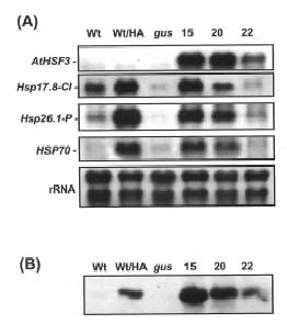
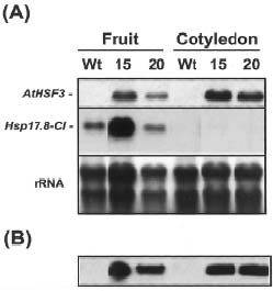
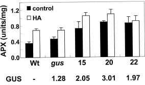
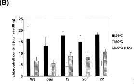
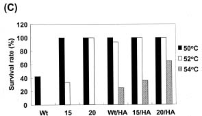
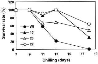
Abstract. Heat shock pre-treatment or heat acclimation can enhance the subsequent chilling tolerance of many plant species. However, the exact mechanism is currently unknown. Transgenic tomato plants harboring an Arabidopsis thaliana HsfA1b (AtHsfA1b) and b-glucuronidase (gusA) fusion gene under the control of a constitutive CaMV35S promoter were generated to evaluate whether heat shock factor (Hsf), a major transcription regulator of heat shock response, is involved in heat shock-induced chilling tolerance. The transcripts or protein product of heat-shock induced genes in the transgenic plants highly expressing AtHsfA1b-gusA accumulated to higher levels than those of the wild-type or gusA transgenic plants under non-heat shock conditions. This suggests that AtHsfA1b-GUS can cross-talk with the heat shock responsive machinery in transgenic tomato plants. In addition, the specific activity of soluble isoforms of ascorbate peroxidase of the AtHsfA1b-gusA transgenic plants was about twofold higher than that of the wild-type or gusA transgenic plants under non-heat stress conditions. Without heat acclimation, seedlings of the AtHsfA1b-gusA transgenic tomato lines showed a significantly higher level of thermal and chilling tolerance than that of the wild-type or gusA transgenic plants. Based on these results, we suggest that Hsf may play a pivotal role in heat-shock-induced chilling tolerance, and constitutive expression of the transcription regulatory gene in chilling sensitive crops may be useful in improving tolerance against chilling stress.

Keywords: Chilling stress; Heat shock factor; Transgenic tomato.

Abbreviation: HSICT, heat-shock-induced chilling tolerance; Hsf, heat shock factor; Hsp, heat shock protein; sHsp, small Hsp; GUS, b-glucuronidase;

Introduction

Chilling injury is a physiological disorder that develops in certain plants exposed to nonfreezing temperatures below about 12°C (Lyons, 1973; Saltveit and Morris, 1990). Many important crops indigenous to the tropics and subtropics—such as banana, mango, papaya, rice, and tomato—are chilling sensitive (Saltveit and Morris, 1990). Chilling injury manifests a range of visible symptoms, often used as indicators of its severity, including reduced growth vigor, abnormal ripening, stimulated respiration and ethylene production, and increased cellular membrane leakage and disease susceptibility (Lyons, 1973; Saltveit and Morris, 1990). These symptoms can be reduced by heat shock treatment before exposure to chilling temperatures (Lafuente et al., 1991; Lurie and Klein, 1991; Saltveit, 1991; McCollum et al., 1993; Collins et al., 1995; Woolf et al., 1995; Rab and Saltveit, 1996; Sato et al., 2001) in a phenomenon termed heat-shock-induced chilling tolerance (HSICT). De

novo protein synthesis seems necessary for HSICT in mung bean hypocotyls (Collins et al., 1995). The exact mechanism of HSICT is so far unknown.

The induction of genes encoding heat shock proteins (Hsps) is one of the most prominent responses at the molecular level of organisms exposed to high temperature (Kimpel and Key, 1985; Lindquist, 1986; Vierling, 1991; Waters et al., 1996). According to their approximate molecular mass in kDa, the major eukaryotic Hsps have been grouped into five conserved classes: namely Hsp100, Hsp90, Hsp70, Hsp60, and the small Hsps (~16 to 42 kDa). Plant small Hsps (sHsps) can be further divided into at least six gene families encoding several proteins localized to the cytosol and nucleus (class I, II and III) and different organelles (chloroplast, endoplasmic reticulum, and mitochondria) (Scharf et al., 2001). Accumulation of Hsps increases thermotolerance and protects cells from the detrimental effect of high temperature, probably through their functioning as molecular chaperones (Waters et al., 1996). The correlation of HSICT and the accumulation of Hsps in plant tissues have been recently characterized (Collins et al., 1995; Sabehat et al., 1996; Kadyrzhanova et al., 1998). However, the role of Hsps, if any, in HSCIT is unclear.

*Corresponding authors. Fax: 886-2-2651-5600; E-mail: yycharng@gate.sinica.edu.tw, mbmtchan@ccvax.sinica.edu.tw


Botanical Bulletin of Academia Sinica, Vol. 44, 2003

In addition to Hsps, oxidative stress-relieving enzymes, such as ascorbate peroxidase (APX) (Sato et al., 2001; Shi et al., 2001), are also induced by heat shock treatment. Heat shock-induced cytosolic APX expression is correlated with HSICT in rice seedlings (Sato et al., 2001). APX is involved in scavenging hydrogen peroxide, one of the active oxygen species that can be generated during oxidative stress imposed by chilling (Prasad et al., 1994). The expressions of apx1 gene of pea (Mittler and Zilinskas, 1992, 1994) and Arabidopsis (Storozhenko et al., 1998) and APXa gene of rice (Sato et al., 2001), which all contain heat-shock-elements (HSE) in their promoters, are induced by heat stress. It was suggested that heat shock induction of the APXa gene could be involved in reducing chilling injury in rice seedlings (Sato et al., 2001). However, more correlations like this could conceivably be revealed in future studies because heat shock stress affects the expression of a wide range of genes, as revealed by recent microarray studies (C. Guy, Univ. of Florida, Gainesville, FL, personal communication). It is plausible to suggest that HSICT may result from an effect exerted by multiple genes induced by heat shock, just as acquired thermotolerance is itself.

In general, the regulation of expression of many Hsp genes is mediated by the conserved heat shock factor (Hsf). The latent Hsf is activated upon heat treatment by induction of trimerization and high-affinity binding to the heat-shock-element (HSE), a conserved sequence in the promoter regions of many Hsp genes (Wu, 1995). The completion of the Arabidopsis thaliana genome project reveals that in the plant genome there are about 21 Hsfs, which can be classified into three classes (A, B, and C) and fourteen groups based on structural characteristics and phylogenetic comparison (Nover et al., 2001). Over-expression of class A Hsf results in constitutive synthesis of Hsps and increased basal thermotolerance in transgenic Arabidopsis (Lee et al., 1995; Prandl et al., 1998). However, overexpression of a class B Hsf did not have a similar effect (Lee et al., 1995; Prandl et al., 1998). These results indicate that certain Hsf members play a pivotal role in thermotolerance by regulating downstream Hsp genes.

In this study, we have evaluated whether Hsf are likely involved in HSICT of tomato plant. Since class A Hsf plays a major role in regulating heat shock response (Nover et al., 2001), over-expression of a transgenic Hsf may allow us to evaluate its role in HSICT, mainly by decoupling the heat shock response from the possible pleiotropic effects resulting from a high temperature treatment. From the EST libraries so far obtained, the tomato genome contains at least 19 Hsf members with representatives in each class and group defined for Arabidopsis (Nover et al., 2001), suggesting that these two species share a similar degree of complexity of Hsf network and function. We hypothesized that overexpression of an Arabidopsis class A Hsf in tomato might have an effect similar to overexpressing its tomato counterpart but without the co-suppression problem (Matzke and Matzke, 1995). A chilling-sensitive tomato line was stably trans

formed with the Arabidopsis thaliana HsfA1b (AtHsfA1b) gene (Nover et al., 2001), previously known as Hsf3 (Prandl et al., 1998), by the Agrobacterium-mediated transformation method. AtHsfA1b belongs to group A1 Hsf according to the recent classification of Nover et al. (2001). It shares about 60% amino acid sequence identity with the major tomato Hsf, LeHsfA1 (previously known as Hsf8) (Scharf et al., 1990; Nover et al., 2001). Over-expression of AtHsfA1b-gusA in transgenic tomato plants led to constitutive expression of Hsps, elevated levels of APX activity, and enhanced thermotolerance. Most interestingly, the transgenic tomato seedlings exhibited a significant increase in chilling tolerance.

Materials and Methods

Expression Plasmid Constructions

The construct for expression of AtHsfA1b-GUS fusion protein was generated according to the method described by Prandl et al. (1998) with some modification. The AtHsfA1b (also named Hsf3) open reading frame (ORF) (accession number Y14068), amplified by reverse transcriptase polymerase chain reaction (RT-PCR), was fused to gusA in pBI221 instead of pBI121.1 (Prandl et al., 1998) to yield pYC018. The ORF was sequenced to make sure no unwanted mutation generated due to PCR. The HindIII-EcoRI fragment from pYC018 containing the cauliflower mosaic virus 35S promoter, the AtHsfA1b-gusA fusion, and nos termination signal sequence was ligated into the corresponding sites of the binary vector pCAMBIA2300 (Center for the Application of Molecular Biology of International Agriculture, Australia), and the resulting plasmid was designated pYC019. The plasmid, pCAMBIA2301, with 35S-GUS-Nos construct, was employed to transform tomato as a control. The plasmids were introduced into Agrobacterium tumefaciens strain LBA4404 by the freeze-thaw method (Holsters et al., 1978).

Generation and Growth of Transgenic Tomato Plants

A local tomato inbred line, L4783, provided by the Asian Vegetable Research and Development Center, Tainan, Taiwan, was employed for genetic transformation. Tomato transformation was performed according to the method of Fillatti et al. (1987) and the medium preparation of Hamza and Chupeau (1993) with slight modification. Cotyledons from 7- to 8-day-old seedlings were cut into two pieces, placed upside down on the preculture medium (MS salts, Gamburg’s B5 vitamins, 2 mg/L BA, 0.25 mg/L IAA, and 0.3% Phytagel), and then incubated in the dark for 24 h before inoculated with A. tumefaciens cells harboring either pYC019 or pCAMBIA2301. The explants were submerged in Agrobacterium inoculum for 30 min, blotted dry, transferred to the co-cultivation medium (the pre-culture medium supplemented with 200 µM acetosyringone) and incubated in the dark for 2 days. Following co-cultivation, the explants were transferred to the AZ medium (Hamza and Chupeau, 1993), supplemented


Li et al. — Over-expression of AtHsfA1b enhanced chilling tolerance

with 100 mg/L kanamycin and 200 mg/L Timentin (Duchefa Biochemie BV, the Netherlands), for regeneration and selection. Three to four weeks later, explants with developing shoots were transferred to a shoot elongation medium, the same as the AZ medium except that zeatin and IAA were excluded. When the shoots grew to 2-3 cm tall, they were transferred to the MM medium (Hamza and Chupeau, 1993) with 50 mg/L kanamycin for rooting before being transferred to potting media. Transgenic and wild-type tomatoes were grown in a green house facility on campus during the October to May period, when average temperatures remained below 25°C during the day and 20°C during the night.

Analysis of Transgenic Tomato Plants

For comparative analysis of transgenic and wild-type plants in each experiment, the samples were harvested from the same batch of plants grown at the same time. For Southern analysis of putative transgenics, about 10 µg of purified genomic DNA was digested by restriction enzymes and resolved on a 0.6% agarose gel in 0.5× TBE buffer (Sambrook et al., 1989). Capillary blotting of the DNA from the gel to a positively charged nylon membrane (Magnacharge, MSI) was performed according to Sambrook et al. (1989). To generate a non-radioactive hybridization probe for detection of transgene, a 535 bp DNA fragment corresponding to the position 860-1394 bp of the AtHsfA1b ORF (Prandl et al., 1998) was produced by PCR and labeled with a PCR DIG-labeling kit (Roche). The hybridization and washing procedures followed the manufacturer’s protocol. For northern analysis, total RNA was isolated from plant tissues using a commercial reagent according to the manufacturer’s instructions (TRIZOL, Gibco BRL). Separation of RNA on formaldehyde-containing 1% agarose gel (20 µg of RNA per lane) and transfer to positively charged nylon membrane (Magnacharge, MSI) by capillary blotting was performed according to Sambrook et al. (1989). A 535 bp DNA fragment corresponding to the position 860-1394 bp of the AtHsfA1b ORF, tomato full length ORFs of Hsp17.8-CI (accession number: X56138), and Hsp26.1-P (U59917) cDNA were amplified by RT-PCR with gene specific primers and individually cloned into pGEM T-easy (Promega) vector for producing DIG-labeled antisense RNA probes. The probe for tomato Hsp70 was derived from a tomato EST clone (AW223426, TIGR Tomato Gene Index) that shares a 92% identity with the tobacco Hsp70 cDNA (X63106). DIG-labeled antisense RNA probes were produced by in vitro transcription using SP6 or T7 RNA polymerase (Promega) and DIG-RNA probe mix (Roche) according to the manufacturer’s instruction. Prehybridization (4 h) and hybridization (16 h) was performed at 65°C in a solution containing 50% formamide and 50 ng/mL of DIG-labelled probe. Membranes were washed twice in 2×SSC (Sambrook et al., 1989) and 0.1% SDS solution at room temperature and twice in 0.1×SSC and 0.1% SDS at 65°C for 15 min each. The amount of hybridized DIG-labelled probe on the membrane was visualized according to the manufacturer’s protocol.

Enzyme Activity Assays

Histochemical staining and fluorometric assay of GUS activity were performed according to the method of Jefferson (1987). For APX and catalase assays, the six-day-old etiolated seedlings were immediately frozen with liquid nitrogen after treatment, ground into fine powder with mortar and pestle, and homogenized in cold sodium phosphate buffer (50 mM, pH 7.0) with a homogenizer. The crude extract was then centrifuged at 4°C 12,000 g for 20 min. The supernatant was used immediately for enzyme assay. The activity of soluble isoforms of APX was determined using a spectrophotometry method previously described (Nakano and Asada, 1981). Catalase activity was analyzed according to the method of Kato and Shimizu (1987). Superoxide dismutase (SOD) isozyme activities were extracted and determined by an in gel assay method according to Chen and Pan (1996). Protein was measured by the dye-binding method (Bradford, 1976) with bovine serum albumin as standard.

Immunoblotting Analysis

SDS-PAGE was performed on 4-12% Bis-Tris precast gels (Invitrogen). After electrophoresis, proteins were electrophoretically transferred to a nitrocellulose membrane. The membrane was blocked in PBS buffer (potassium phosphate 25 mM, NaCl 150 mM, pH 7.2) with 5% skim milk and 0.2% Tween-20 and probed with 10,000-fold diluted polyclonal antibody raised against rice class I sHsp (Jinn et al., 1993), kindly provided by Prof. Chu-Yung Lin of National Taiwan University. Following secondary antibody reaction employing goat anti rabbit IgG conjugated to horseradish peroxidase, the blots were visualized using the Super Signal West Dura Extended Duration Substrate system (Pierce).

Thermotolerance Test

The thermotolerance of tomato seedlings was measured by adopting the method of Burke (1994). Tomato seeds were surface sterilized and germinated in the dark at 25°C on GM medium (MS salt, 2% sucrose, and 0.3% phytagel) in a glass jar (400 mL) capped with a glass petri dish cover. In each jar, 10 seeds were included. Six-day-old etiolated seedlings were then subjected to heat treatment at a lethal temperature, 50°C for 2 h, either with or without prior heat acclimation treatment. For heat acclimation, the samples were placed at 40°C for 2 h and recovered at 25°C for 1 h before further treatment. All treatments above were performed by placing the jars in a controlled temperature oven for an indicated length of time, and light was avoided. The air temperature inside the jar reached the desired level within 15 min as indicated by thermometer. Following heat treatment, seedlings were exposed to white light (150 µmol m-2 s-1) under a light/dark cycle (16 h/8 h) at 25°C for two days to allow chlorophyll synthesis. After photographing the seedling, the cotyledons were harvested, and the chlorophyll content was measured by the method described previously (Moran and Porath, 1980).


Botanical Bulletin of Academia Sinica, Vol. 44, 2003

Chilling-Tolerance Test

The chilling tolerance of tomato seedlings was tested by measuring radicle length or survival rate following chilling treatment. The former study was performed basically according to the method of Rab and Saltveit (1996) with some modification. T3 seeds of homozygous transgenic lines and wild-type tomato were soaked in distilled water overnight with gentle shaking at 25°C and transferred to three layers of wet paper towel between two 19 × 19.5 cm glass plates for germination. The germinated seeds with about 5 mm radicle lengths were picked out and incubated at 25 or 38°C for 15 min in a water bath. The seedlings were then positioned back into the paper towels and the glass plate sandwich for chilling treatment, set at 2.5°C for 5 days in the dark, and transferred to 25°C for 3 days, also in the dark, for continued growth of seedlings. Subsequent radical elongation was measured and recorded at the end of the 3-day regrowth period. To measure the survival rate, 3-day-old seedlings were subjected to storage in the dark at 2.5°C for 7-18 days on moistened 3MM filter paper. For heat shock acclimation, the seedlings were first incubated at 38°C for 15 min before directly subjected to chilling treatment. Following low temperature treatment, the seedlings were transferred to 25°C for 6 days under light. Seedlings showing green cotyledons and adventitious roots were considered survivors. Survival rate was calculated by the following equation: (the number of survivors/the number of tested seedlings) × 100.

Results

Identification of Transgenic Tomato Plants Harboring AtHsfA1b-gusA

To evaluate the effect of Hsf expression on chilling tolerance in tomato, we introduced the fused AtHsfA1b-gusA transgene under the control of cauliflower mosaic virus 35S promoter into tomato, using an Agrobacterium-mediated transformation. Here, the GUS fusion protein was introduced as a reporter for easy and sensitive identification of the transgene. The same AtHsfA1b-gusA chimeric gene has previously been shown to encode an AtHsfA1b-GUS fusion protein with full Hsf activity (Prandl et al., 1998). Transgenic tomato plants containing only the 35S::gusA transgene were also generated using the same method and employed as a control.

For transgenic AtHsfA1b-gusA plants, thirty-four independent R0 transformants were generated and analyzed by histochemical staining of GUS activity and Southern blot analyses to determine the presence, genomic integration, and insertion copy number of AtHsfA1b-gusA. Gene silencing was observed for some of the T0 transgenic plants with multiple insertions of the transgene (data not shown). Three independent transgenic plants, TT1-15, TT1-20 and TT1-22, each with a single insertion and different expression level of AtHsfA1b-gusA, were selected for this study. These transgenic plants were allowed to self-pollinate to generate plants homozygous for AtHsfA1b-gusA. Analyzing the segregation ratio of the T1 transgenic plants, we

Figure 1. Histochemical staining indicates the nuclear localization of AtHsfA1b-GUS in transgenic tomato cells. The red-ripe fruit pericarps of AtHsfA1b-gusA (A, B) and gusA (C, D) transgenics were free-hand sectioned, fixed in 4% paraformaldehyde, 100 mM phosphate buffer (pH 7.2), and double stained for GUS activity (A, C) and nuclei by DAPI (B, D), and observed with a Zeiss florescence microscope.

observed that all showed an approximate 3:1 ratio based on GUS activity assay. This observation was in good agreement with the result of Southern blot analysis that the foreign gene was obviously integrated into a single genetic locus (data not shown).

According to GUS histochemical staining of transgenic plants, the transgene was expressed in cotyledon, root, young leaf, stem, and fruit tissues. Histochemical staining for GUS activity showed intensified GUS staining at the site of nuclei in the AtHsfA1b-gusA transgenic lines, something not observed for the gusA transgenic plants (Figure 1). This staining pattern indicated that AtHsfA1b-gusA product was localized within the nuclei in the absence of heat shock treatment.

Over-Expression of AtHsfA1b can Derepress Heat Shock Response in Transgenic Tomato Plants

Over-expression of AtHsfA1b-gusA in Arabidopsis derepresses the heat shock response (Prandl et al., 1998). Transcript levels of three Hsp genes—Hsp17.8-CI, Hsp26.1-P, and Hsp70—were measured in immature green tomato fruits (about 30 days after anthesis) to determine whether expression of AtHsfA1b-gusA in transgenic tomato plants also conferred similar effects. A heat treatment up-regulates Hsp17.8-CI (also named tom66) and Hsp26.1-P (also named tom111), class I cytosolic and chloroplast sHsp, respectively, in mature-green tomato fruit (Sabehat et al., 1998). A tomato EST (Expressed Sequence Tags) clone (AW223426) that shares 92% identity with the tobacco Hsp70 cDNA (X63106), designated as Hsp70 here, was also induced in heat-treated fruits (Figure 2A).


Li et al. — Over-expression of AtHsfA1b enhanced chilling tolerance

ing polyclonal antibodies raised against rice class I sHsp (Jinn et al., 1993). The antibodies specifically recognized a heat inducible protein of 20 kDa as determined from its mobility on SDS-PAGE, close to the calculated size of Hsp17.8-CI (Figure 2B). This suggested that the protein recognized by the antibodies was the product encoded by Hsp17.8-CI, or at least by its homolog. The class I sHsp could be detected in all the transgenic lines, even without heat treatment, but not in the wild-type or the transgenic plants with gusA gene expression only (Figure 2B). Although the transcript levels of Hsp17.8-CI in TT1-20 and 22 were not higher than in the wild type (Figure 2A), the class I sHsp level was significantly higher than in the wild-type plant (Figure 2B). Taken together, these results indicated that high-level expression of AtHsfA1b-gusA leads to the expression of Hsps in transgenic tomato plants, even in the absence of a heat shock treatment.

The transcript levels of Hsps, however, did not increase in the cotyledons of the AtHsfA1b-gusA transgenic seedlings, as they did in immature green fruits under non-stressed conditions. Although the transcript levels of AtHsfA1b in the cotyledons of 6-day etiolated seedlings were between 1.5- to 2-fold higher than in the fruit tissue, the transcript levels of Hsp17.8-CI (Figure 3A), Hsp26.1-

Figure 2. Derepression of heat shock response in transgenic tomato under non-stressed conditions. (A) Expression of AtHsfA1b-gusA and heat shock genes in T1 transgenic tomato fruits were analyzed by northern hybridization. Twenty µg of total RNA purified from immature green tomato fruits of wild type (Wt), heat-acclimated wild type (Wt/HA), transgenic plant with gusA only (gus), and different AtHsfA1b-gusA transgenic lines (15, 20, and 22) was blotted to membrane following agarose gel electrophoresis, and the membrane was stained with methylene blue to control equal RNA loading prior to hybridization. AtHsfA1b, Hsp17.8-CI, Hsp26.1-P, and Hsp70 cDNA were employed as templates to generate RNA probes as described in Materials and Methods. (B) Immunoblot analysis of class I sHsp in the corresponding fruit tissues with an antibody raised against rice class I sHsp. About 20 µg of protein was loaded in each lane.

Northern analysis readily detected AtHsfA1b-gusA transcripts in transgenic plants (Figure 2A), the level being higher in TT1-15 and -20 than in TT1-22. Under non-heat shock condition, the transcripts of Hsp26.1-P and Hsp70 were accumulated in fruits of transgenic lines TT1-15 and 20 at a higher level than in wild-type or transgenic plants with the gusA gene only (Figure 2A). In contrast, TT1-22 showed little difference. However, the transcript levels of Hsp17.8-CI, a class I sHsp, in TT1-20 and 22 were about the same or even lower than in the wild-type plant (Figure 2A). It should be noted that at least four homologous genes share 90-99% identity with Hsp17.8-CI in the coding region according to the database of the TIGR Tomato Gene Index. Our probe derived from HSP17.8-C1 will likely cross hybridize to the transcripts of one or more of these homologous genes.

The amount of class I sHsp protein in the immature green fruits was determined by western blot analysis us

Figure 3. Differential accumulation of Hsp17.8-CI transcript (A) and class I sHsp (B) in the fruit and cotyledon of transgenic tomato. (A) The transcript levels of AtHsfA1b and Hsp17.8-CI of wild type (Wt) immature green fruit and cotyledons of 6-day old seedlings were compared with that of the AtHsfA1b-gusA transgenic lines (15 and 20). In each lane, 20 µg of total RNA was loaded and the membrane was stained previously with methylene blue to control equal loading of RNA. The probes used were the same as that described in Figure 1A. (B) Immunoblot analysis of class I sHsp in the corresponding samples with an antibody raised against rice class I sHsp. About 20 µg of protein was loaded in each lane.


Botanical Bulletin of Academia Sinica, Vol. 44, 2003

P, and Hsp70 (data not shown) were almost undetectable in the cotyledon of the transgenic plants under non-heat shock conditions. Although the transcript of Hsp17.8-CI was not detectable in the cotyledons of the transgenic AtHsfA1b-gusA plants, the class I sHsp significantly accumulated to about the same level as that of the fruit tissue (Figure 3B). Such a discrepancy between sHsp transcripts and protein levels has been previously reported between the heat-shocked callus and somatic embryo cells of carrot (Zimmerman et al., 1989). Based on these results, we suggest that effective expression of AtHsfA1b-gusA can derepress heat shock response in different parts of the transgenic plants, i.e. in fruit and in seedlings. Therefore, we limited subsequent tests to seedlings in our examination of the effect of overexpression of AtHsfA1b-gusA on heat and chilling tolerance.

Despite the constitutive heat shock response, the transgenic AtHsfA1b-gusA plants did not show significant alteration in growth or morphology and produced normal fruits and viable seeds under non-stressed growth conditions.

Higher Ascorbate Peroxidase Activity in Transgenic Tomato Plants

A heat-inducible cytosolic APX is thought to be involved with HSICT in rice seedling (Sato et al., 2001). The rice APX gene promoter contains a minimal heat shock factor-binding motif, 5´-nGAAnnTTCn-3´, the so-called heat shock element (HSE) (Sato et al., 2001). In Arabidopsis, a HSE found in the APX1 promoter was shown to be recognized by the tomato Hsf in vitro and to be responsible for the in vivo heat-shock induction of the gene (Storozhenko et al., 1998). Although it is not known whether tomato APX genes contain HSE in their promoters, we were interested in whether APX activity was affected in the transgenic to

mato plants. We observed that the activity of soluble APX was increased by about 1.4- to 1.8-fold in the gusA transgenic and wild-type tomato etiolated seedlings, respectively, by heat shock treatment (Figure 4). This is consistent with the observation in rice that soluble APX activity is up-regulated by heat stress (Sato et al., 2001). Under non-heat shock conditions, the AtHsfA1b-gusA transgenic plants exhibited up to 2-fold higher APX activity than the wild-type or gusA transgenic plants (Figure 4). Heat shock treatment can further increase APX activities in the AtHsfA1b-gusA transgenics (Figure 4). Since superoxide dismutase (SOD) and catalase are enzymes also involved in scavenging active oxygen species like APX, the activity levels of these two enzymes were also examined. No significant difference appeared in either SOD or catalase activities between the transgenic and wild-type seedlings under non-heat shock or heat shock conditions (data not shown), suggesting that these enzymes activities were not affected by constitutively expressed Hsf.

Constitutive Expression of AtHsfA1b Increased Basal Thermotolerance in Transgenic Tomato Seedlings

We have evaluated the thermotolerance of the transgenic tomatoes to determine whether AtHsfA1b could function normally in a heterogenous host. To do so, we adopted a sensitive bioassay developed by Burke (1994). This bioassay method is based on the level of inhibition of chlorophyll accumulation in etiolated seedlings following the challenges posed by lethally high temperatures. After exposure to 50°C for 2 h and then recovery at 25°C for 2 d, the cotyledons of the test etiolated seedlings of the wild-type or transgenic plants with only gusA transgene were not able to expand or turn green (Figure 5A), and more than 50% of the tested seedlings eventually failed to grow (Figure 5C). However, if the seedlings were first exposed to mild heat stress at 40°C for 2 h before being subject to lethal temperature treatment, their cotyledons obviously became greenish (Figure 5A), and all seedlings were able to continue growing, a phenomenon that has been defined as heat acclimation or acquired thermotolerance. Without heat acclimation, we found that all the T3 seedlings of the AtHsfA1b-gusA transgenic lines exhibited a better thermotolerance than the non-acclimated wild-type or gusA transgenic plants as evidenced by expanded and greenish cotyledons (Figure 5A) and continuing growth after treatment at 50°C for 2 h.

Measurement of chlorophyll content of test seedlings allows us to compare the effect quantitatively. The data are in good agreement with the observation that the AtHsfA1b-gusA transgenic lines in general accumulated significantly a higher level of chlorophyll after heat treatment without acclimation, whereas heat acclimation further increased the chlorophyll accumulation rate (Figure 5B). When tested at higher temperature, the non-acclimated AtHsfA1b-gusA transgenic lines can survive heat treatment up to 52°C, but all died at temperatures above 54°C (Figure 5C). Heat acclimation at 40°C for 2 h can further enhance

Figure 4. Elevated ascorbate peroxidase (APX) activity in AtHsfA1b-gusA transgenic tomato seedlings. Six-day old etiolated seedlings were subjected either to heat shock treatment at 40°C for 2 h (HA) or without heat treatment as controls. The APX activities in the crude extract of the tomato seedlings were then measured as described in Materials and Methods. The sample labels are the same as that in Figure 1. The bars represent means ± standard deviation (n = 6). GUS activities (nmol/min/mg) of the seedlings were indicated at the bottom of the figure.


Li et al. — Over-expression of AtHsfA1b enhanced chilling tolerance

thermotolerance of the transgenic and the wild-type plants up to 54°C (Figure 5C).

When the test was performed earlier on the T1 generation, we observed that some of the T1 seedlings of the AtHsfA1b-gusA transgenic lines showed no detectable GUS activity and displayed no enhanced thermotolerance due to segregation of the introduced transgenes in the T1 generation (data not shown), indicating that the AtHsfA1b-gusA transgene was responsible for the thermotolerance in transgenic tomato. The increased basal thermotolerance of the AtHsfA1b-gusA transgenic plants, in addition to the constitutive heat shock response, suggested that the heterologous gene functioned normally in transgenic tomato.

Constitutive Expression of AtHsfA1b Improved Chilling Tolerance in Transgenic Tomato Seedlings

Class A Hsf is the major transcription regulator of heat shock response known to date, so the effect of over-expressing AtHsfA1b-gusA on the chilling tolerance of transgenic plants was evaluated. We first measured the inhibition of tomato seedling radicle growth by chilling treatment according to the method of Rab and Saltveit (1996). After recovery of plants from storage at 2.5°C for 5 days, the radicle growth of the wild-type and transgenic gusA plants was significantly reduced compared to that of plants without chilling treatment. Heat shock at 38°C for 15 min prior to chilling treatment was able to ameliorate the growth inhibition resulting from testing chilling stress (Figure 6A). In the absence of heat shock pre-treatment, the AtHsfA1b-gusA plants exhibited a significantly better growth rate than the non-acclimated wild-type or gusA transgenic plants (Figure 6A). Following chilling treatment, the levels of class I cytosolic sHsp, Hsp17.8-CI (Figure 6B), and APX activities (Figure 6C) remained higher in the transgenic plants. Chilling treatment seemed to moderately induce the synthesis of Hsp17.8-CI (Figure 6B) in the wild-type and gusA transgenic plants.

We than examined the survival rate of seedlings following a longer term of chilling treatment of 3-day-old seedlings, from 7-18 days. After recovery at 25°C for 6 days of culture, the survivors manifested expanding greenish cotyledons and adventitious roots. The wild-type plants generally could not endure the prolonged low temperature storage, and almost all seedlings had died after 15 days of treatment. However, a mild heat shock treatment applied immediately before the chilling treatment improved survival rate by up to 30%. The AtHsfA1b-gusA transgenic plants were generally more tolerant of the chilling treatment than the wild-type plants without prior heat acclimation (Figure 7). The transgenic plants could maintain a 30-60% survival rate while the wild-type plants were all dead after 18 days of chilling treatment (Figure 7). Due to lack of seeds, we did not include the gusA transgenic plants in this experiment. Taken together, we conclude that over-expression of the AtHsfA1b gene can enhance the chilling tolerance of the transgenic plants probably due to the derepression of heat shock responses.

Figure 5. AtHsfA1b-gusA transgene enhanced thermotolerance in etiolated tomato seedlings without heat acclimation. (A) Six-day old etiolated seedlings of wild type (Wt), gusA transgenic plant (gus), and AtHsfA1b-gusA transgenic lines (15, 20, and 22) were subjected to thermotolerance test at 50°C for 2 h either without (C, upper row) or with prior heat acclimation treatment (HA, lower row) as described in Materials and Methods. The photograph was taken after the plants were exposed to light/dark cycle (16 h/8 h) at 25°C for two days following heat treatment, and three representative plants were shown here. (B) The chlorophyll contents of the seedlings were measured after 2 h treatment at 25°C, or at 50°C with or without prior heat acclimation (HA) followed by exposure to light. The bars represent means ± standard deviation (n = 10). (C) The survival rate of seedlings after exposure to indicated temperature. For each line with or without heat acclimation, 17-20 etiolated seedlings were used for heat treatment under varied temperature for 2 h and recovered at 25°C for 8 d under light/dark cycle (16 h/8 h). The survival rates were calculated by dividing the number of seedlings with emerged true leaves by the total number of seedlings tested.


Botanical Bulletin of Academia Sinica, Vol. 44, 2003

Discussion

Heat treatment has been shown to protect against chilling injury in a number of plant species, including avocado (Woolf, 1997), cucumber (Lafuente et al., 1991; McCollum et al., 1995), mango (McCollum et al., 1993), pepper (Mencarelli et al., 1993), rice (Sato et al., 2001), and tomato (Lurie and Klein, 1991; Rab and Saltveit, 1996). The HSICT is present in a wide range of plants, suggesting that it may be a general event. It has been shown that the expression of sHsps is correlated to HSICT (Sabehat et al., 1998). Recently, a rice heat-induced APX, whose promoter contains a minimal HSE, has also been shown to associate

Figure 7. Over-expression of AtHsfA1b-GUS enhanced survival rate of tomato seedlings under chilling stress. Three-day old seedlings were subjected to 2.5°C in the dark for various length of time indicated. The survival rate was determined after recovering at 25°C for 6 days. Each data point represents the result from 50-60 seedlings.

with HSICT (Sato et al., 2001). Hence, it is very likely that expression of heat shock responsive genes was responsible for the HSICT observed so far. In this report, we have evaluated and characterized the expression of a heterologous transcription factor transgene, AtHsfA1b, a class A Hsf gene, in tomato plants, and that it can derepress heat shock responses and increase chilling-tolerance of test transgenic tomato plants. To our knowledge, this is the first report to characterize the relationship between the expression of Hsf and its effect on HSICT.

In Arabidopsis, overexpression of AtHsfA1b or AtHsfA1b-gusA was shown to increase the level of transcripts of three class I sHsp (Hsp17.4, Hsp17.6, and Hsp 18.2), a class II sHsp (Hsp17.6A), and the amount of class I sHsp protein (Prandl et al., 1998). Similar results were obtained for the transgenic tomato in this study, suggesting that the heat shock response machinery is very conserved in these two species. It was suggested that the product of AtHsfA1b-gusA transgene derepresses heat shock response by functioning as a transcription factor (Prandl et al., 1998), which is in agreement with our observation that AtHsfA1b-GUS was detected in the nuclei of the transgenic tomato cells without heat shock treatment (Figure 1). Just as in Arabidopsis, high-level expression of AtHsfA1b seemed to have no negative impact on the growth or development of the transgenic plant under normal growth conditions.

A low level of Hsp17.8-CI mRNA could be detected in the wild-type immature green fruit without heat treatment (Figure 2A), but its corresponding protein was barely detectable by western blot analysis (Figure 2B). However, in two of the transgenic lines, TT1-20 and 22, the transcript levels of Hsp17.8-CI were about the same or even lower than that of the wild type, and yet the class I sHsp levels were considerably higher (Figure 2A and B). It should be noted that Hsp17.8-CI has been shown to express in ripening tomato fruit (Slater et al., 1985). We could detect

Figure 6. Over-expression of AtHsfA1b-GUS conferred chilling tolerance to tomato seedlings shown by measuring radicle length (A). The radicle regrowth assays were performed as described in Materials and Methods. Wild type that was incubated at 25°C for 3 days is indicated as Wt/C as a control. The transgenic lines exhibited radicle growth rates similar to that of the wild type without chilling stress. All samples except the Wt/C were incubated at 2.5°C for 5 days before being removed to 25°C for 3-day recovery. The Wt/HA was heat-acclimated at 38°C for 15 min before the chilling treatment. Bars represent means ± standard deviation (n = 30). (B) The level of class I sHsp, Hsp17.8-CI, of the corresponding seedlings after recovery at 25°C was measured by immunoblotting. About 20 µg of protein was loaded in each lane. (C) The level of APX activities of corresponding seedlings after recovery at 25°C was measured. Bars represent means ± standard deviation (n = 6).


Li et al. — Over-expression of AtHsfA1b enhanced chilling tolerance

and identification of the responsible APX gene in tomato is underway. Recently, an extensive study of Arabidopsis APXs expression was carried out in heat treated or AtHsfA1b transgenic plants (Panchuk et al., 2002). Unlike the case here with transgenic tomato, moderate heat shock did not induce a significant increase of total APX activity in either wild-type or transgenic plants while a novel Apx2 gene was shown to be strongly induced in heat-stressed transgenic plants (Panchuk et al., 2002).

In this report, we have demonstrated that high-level expression of AtHsfA1b-gusA can reduce the inhibition of radicle growth imposed by chilling treatment (Figure 6A) and can increase the survival rate after exposure to cold storage (Figure 7). The results suggest that Hsf is involved in HSICT by derepressing heat shock response, which may obviously trigger the expression of multiple genes to confer chilling tolerance. These genes may contribute to the enhancement of chilling tolerance with varying degrees of importance. Indeed, we did not observe a very good relationship between the level of Hsp17.8-CI and APX activity with the degrees of tolerance to chilling stress among the transgenic lines tested. This result is to be expected if HSICT is a multiple gene effect.

Since heat shock treatment could effectively alleviate chilling injury in tomato fruit (Lurie and Klein, 1991), our transgenic approach may be applicable to conferring chilling tolerance on postharvest produce without heat acclimation treatment. With a limitation of plantation scale for this study, we were not able to obtain statistically significant data on postharvest tomato fruits. The increased basal thermotolerance in the transgenic tomato shown in this study may also provide new opportunities to improve the trait against high temperature stress. Tomato is, in general, a heat-sensitive plant. A constitutive derepression of heat shock response might be beneficial to crops grown in a volitile temperature environment, though a certain time may be required for the endogenous acclimation mechanism to confer protection. Future studies are planned to address this possibility.

Acknowledgement. We thank Hui-Wen Tsai and Lin-Yun Kuang for help with production and maintenance of transgenic tomatos. We would also like to thank Dr. Chu-Yung Lin of National Taiwan University for kindly providing the antibodies against rice class I sHsp. We also are grateful to Drs. Tzyy-Jen Chiou, Kuo-Chen Yeh, Ning-Sun Yang, and David T.H. Ho for reviewing this manuscript. The Institute of Botany, Academia Sinica, provided greenhouse facilities for the growth of plant materials. This work was supported by Academia Sinica and by grants from the National Science and Technology Program for Agricultural Biotechnology of the Republic of China (88-2317-B-001-007) and from the National Science Council (NSC 89-2313-B-001-020).

Literature Cited

Apuya, N.R. and J.L. Zimmerman. 1992. Heat shock gene expression is controlled primarily at the translational level in carrot cells and somatic embryos. Plant Cell 4: 657-665.

an increasing level of class I sHsp in tomato fruits from breaker to red ripe stages by western blot analysis (Y.Y. Charng, unpublished data). However, at the immature green stage, the level of class I sHsp was either non-detectable or very low. Therefore, the level of class I sHsp detected in the transgenic fruits was not a consequence of developmental regulation.

The discrepancy between the transcript level of Hsp17.8-CI and the amount of class I sHsp in cotyledon and fruit of the transgenic plants was even more dramatic. These tissues synthesize comparable levels of class I sHsp (Figure 3B) even though they have large differences at the Hsp17.8-CI transcript levels (Figure 3A). A similar phenomenon has been reported for the callus and somatic embryo cells of carrot (Zimmerman et al., 1989), in which the undifferentiated callus cells and globular embryos synthesize a comparable amount of Hsps even though the heat shock transcript levels are quite different. It was later shown that the sHsp gene expression is controlled mainly at the translational level in these cells (Apuya and Zimmerman, 1992). It is believed that heat shock can exert translational control (Apuya and Zimmerman, 1992) probably by modifying the translational machinery (Scharf and Nover, 1982) or by other unidentified mechanisms. However, it has never been shown that over-expressing Hsf transgene alone can exert the same effect. Differential expression of Hsp17.8-CI homologues in cotyledon and fruit of the transgenic plant may be an alternative cause of the discrepancy observed. Further study is needed to verify this point.

APX is believed to be involved in the detoxification of hydrogen peroxide generated by plants due to photosynthesis or under stress conditions. To date, a number of different protein isoforms of APX have been identified, and they can be classified, according to location and structural characteristics, as cytosol soluble, cytosol membrane-bound, chloroplast soluble stromal, or thylakoid-bound APXs (Jespersen et al., 1997). It has been shown that several cytosolic APX genes contain HSE in their promoters and are heat shock inducible (Mittler and Zilinskas, 1992, 1994; Storozhenko et al., 1998; Sato et al., 2001). Recently, a cytosol menbrane-bound APX was cloned from barley and shown to be up-regulated by heat stress (Shi et al., 2001). Overexpression of this gene enhances thermotolerance in transgenic Arabidopsis (Shi et al., 2001).

According to the tomato EST records available on the TIGR Tomato Gene Index (http://www.tigr.org/tdb/lgi/), at least eight different APX cDNAs exist, putatively encoding all four major isoforms. In this study, the APX activity was found to be significantly higher in the AtHsfA1b-gusA transgenic plants (Figure 4), suggesting that the expression of at least one of the tomato APX genes was up-regulated by AtHsfA1b. The increased APX activity in the AtHsfA1b-gusA transgenics might have contributed to the enhanced thermal and chilling-tolerance shown here. At this time, we do not know which soluble isoform(s) of tomato APX contributed to the higher level of APX activity in the AtHsfA1b-gusA transgenic plants,


Botanical Bulletin of Academia Sinica, Vol. 44, 2003

Bradford, M.M. 1976. A rapid and sensitive method for the quantitation of microgram quantities of protein utilizing the principle of protein-dye binding. Anal. Biochem. 72: 248-254.

Burke, J.J. 1994. Integration of acquired thermotolerance within the developmental program of seed reserve mobilization. In J.H. Cherry (ed.), Biochemical and cellular mechanisms of stress tolerance in plants. Springer-Verlag, Berlin, Heidelberg, New York, Tokyo, pp. 191-200.

Chen, C.N. and S.M. Pan. 1996. Assay of superoxide dismutase activity by combining electrophoresis and densitometry. Bot. Bull. Acad. Sin. 37: 107-111.

Collins, G.G., X.L. Nie, and M.E. Saltveit. 1995. Heat shock proteins and chilling sensitivity of mung bean hypocotyls. J. Exp. Bot. 46: 795-802.

Fillatti, J.J., J. Kiser, B. Rose, and L. Comai. 1987. Efficient transformation of tomato and the introduction and expression of a gene for herbicide tolerance. Plant Biol. 4: 199-210.

Hamza, S. and Y. Chupeau. 1993. Re-evaluation of conditions for plant regeneration and Agrobacterium-mediated transformation from tomato (Lycopersicon esculentum). J. Exp. Bot. 44: 1837-1845.

Holsters, M., D. de Waele, A. Depicker, E. Messens, M. van Montagu, and J. Schell. 1978. Transfection and transformation of Agrobacterium tumefaciens. Mol. Gen. Genet 163: 181-187.

Jefferson, R.A. 1987. Assaying chimeric genes in plants: the GUS gene fusion system. Plant Mol. Biol. Rep. 5: 387-405.

Jespersen, H.M., I.V. Kjaersgard, L. Ostergaard, and K.G. Welinder. 1997. From sequence analysis of three novel ascorbate peroxidases from Arabidopsis thaliana to structure, function and evolution of seven types of ascorbate peroxidase. Biochem. J. 326: 305-310.

Jinn, T., S.H. Wu, C.H. Yeh, M.H. Hsieh, Y.C. Yeh, Y.M. Chen, and C.Y. Lin. 1993. Immunological kinship of class I low molecular weight heat shock proteins and thermostabilization of soluble proteins in vitro among plants. Plant Cell Physiol. 34: 1055-1062.

Kadyrzhanova, D.K., K.E. Vlachonasios, P. Ververidis, and D.R. Dilley. 1998. Molecular cloning of a novel heat induced/chilling tolerance related cDNA in tomato fruit by use of mRNA differential display. Plant Mol. Biol. 36: 885-895.

Kato, M. and S. Shimizu. 1987. Chlorophyll metabolism in higher plants. VII. Chlorophyll degradation in senescing tobacco leaves; phenolic-dependent peroxidative degradation. Can. J. Bot. 65: 729-735.

Kimpel, J.A. and J.L. Key. 1985. Heat shock in plants. Trends Biochem. Sci. 10: 353-357

Lafuente, M.T., A. Belver, M.G. Guye, and M.E. Saltveit. 1991. Effect of temperature conditioning on chilling injury of cucumber cotyledons. Possible role of abscisic acid and heat shock proteins. Plant Physiol. 95: 443-449.

Lee, J.H., A. Hubel, and F. Schoffl. 1995. Derepression of the activity of genetically engineered heat shock factor causes constitutive synthesis of heat shock proteins and increased thermotolerance in transgenic Arabidopsis. Plant J. 8: 603-612.

Lindquist, S. 1986. The heat-shock response. Annu. Rev. Biochem. 55: 1151-1191.

Lurie, S. and J.D. Klein. 1991. Acquisition of low-temperature tolerance in tomatoes by exposure to high-temperature stress. J. Am. Soc. Hortic. Sci. 116: 1007-1012.

Lyons, J.M. 1973. Chilling injury in plants. Annu. Rev. Plant Physiol. 24: 445-466.

Matzke, M.A., and A.J.M. Matzke. 1995. How and why do plants inactivate homologous (trans) genes. Plant Physiol. 107: 679-685.

McCollum, T.G., S. D’Aquino, and R.E. McDonald. 1993. Heat treatment inhibits mango chilling injury. HortSci. 28: 197-198.

McCollum, T.G., H. Doostdar, R.T. Mayer, and R.E. McDonald. 1995. Immersion of cucumber fruit in heated water alters chilling-induced physiological changes. Postharvest Biol. Technol. 6: 55-64.

Mencarelli, F., B. Ceccantoni, A. Bolini, and G. Anelli. 1993. Influence of heat treatment on the physiological response of sweet pepper kept at chilling temperature. Acta Hortic. 343: 238-243.

Mittler, R. and B.A. Zilinskas. 1992. Molecular cloning and characterization of a gene encoding pea cytosolic ascorbate peroxidase. J. Biol. Chem. 267: 21802-21807.

Mittler, R. and B.A. Zilinskas. 1994. Regulation of pea cytosolic ascorbate peroxidase and other antioxidant enzymes during the progression of drought stress and following recovery from drought. Plant J. 5: 397-405.

Moran, R. and D. Porath. 1980. Chlorophyll determination in intact tissue using N,N-dimethylformamide. Plant Physiol. 65: 478-479.

Nakano, Y. and K. Asada. 1981. Hydrogen peroxide is scavenged by ascorbate-specific peroxidase in spinach chloroplasts. Plant Cell Physiol. 22: 867-880.

Nover, L., K. Bharti, P. Doring, S.K. Mishra, A. Ganguli, and K.D. Scharf. 2001. Arabidopsis and the heat stress transcription factor world: how many heat stress transcription factors do we need? Cell Stress Chaperones 6: 177-189.

Panchuk, I.I., R.A. Volkov, and F. Schoffl. 2002. Heat stress- and heat shock transcription factor-dependent expression and activity of ascorbate peroxidase in Arabidopsis. Plant Physiol. 129: 838-853.

Prandl, R., K. Hinderhofer, G. Eggers-Schumacher, and F. Schoffl. 1998. HSF3, a new heat shock factor from Arabidopsis thaliana, derepresses the heat shock response and confers thermotolerance when overexpressed in transgenic plants. Mol. Gen. Genet. 258: 269-278.

Prasad, T.K., M.D. Anderson, B.A. Martin, and C.R. Stewart. 1994. Evidence for chilling-induced oxidative stress in maize seedlings and a regulatory role for hydrogen peroxide. Plant Cell 6: 65-74.

Rab, A. and M.E. Saltveit. 1996. Sensitivity of seedling radicles to chilling and heat-shock-induced chilling tolerance. J. Am. Soc. Hort. Sci. 121: 711-715.

Sabehat, A., S. Lurie, and D. Weiss. 1998. Expression of small heat-shock proteins at low temperatures. A possible role in protecting against chilling injuries. Plant Physiol. 117: 651-658.

Sabehat, A., D. Weiss, and S. Lurie. 1996. The correlation between heat-shock protein accumulation and persistence and chilling tolerance in tomato fruit. Plant Physiol. 110: 531-537.


Li et al. — Over-expression of AtHsfA1b enhanced chilling tolerance

Saltveit, M.E. and L.L. Morris. 1990. Overview on chilling injury of horticultural crops. In C.Y. Wang (ed.), Chilling injury of Horticultural Crops. CRC Press, Boca Raton, Fla., pp. 3-15.

Saltveit, M.E. 1991. Prior temperature exposure affects subsequent chilling sensitivity. Physiol. Plant. 82: 529-536.

Sambrook, J., E.F. Fritsch, and T. Maniatis. 1989. Molecular Cloning: A Laboratory Manual, Ed 2. Cold Spring Harbor Laboratory Press, Cold Spring Harbor, NY.

Sato, Y., T. Murakami, H. Funatsuki, S. Matsuba, H. Saruyama, and M. Tanida. 2001. Heat shock-mediated APX gene expression and protection against chilling injury in rice seedlings. J. Exp. Bot. 52: 145-151.

Scharf, K.D. and L. Nover. 1982. Heat-shock-induced alterations of ribosomal protein phosphorylation in plant cell cultures. Cell 30: 427-437.

Scharf, K.D., S. Rose, W. Zott, F. Schoffl, and L. Nover. 1990. Three tomato genes code for heat stress transcription factors with a region of remarkable homology to the DNA-binding domain of the yeast HSF. EMBO J. 9: 4495-4501.

Scharf, K.D., M. Siddique, and E. Vierling. 2001. The expanding family of Arabidopsis thaliana small heat stress proteins and a new family of proteins containing alpha-crystallin domains (Acd proteins). Cell Stress Chaperones 6: 225-237.

Shi, W.M., Y. Muramoto, A. Ueda, and T. Takabe. 2001. Cloning of peroxisomal ascorbate peroxidase gene from barley

and enhanced thermotolerance by overexpressing in Arabidopsis thaliana. Gene 273: 23-27.

Slater, A., M.J. Maunders, K. Edwards, W. Schuch, and D. Grierson. 1985. Isolation and characterisation of cDNA clones for tomato polygalacturonase and other ripening-related proteins. Plant Mol. Biol. 5: 137-147.

Storozhenko, S., P. De Pauw, M. Van Montagu, D. Inze, and S. Kushnir. 1998. The heat-shock element is a functional component of the Arabidopsis APX1 gene promoter. Plant Physiol. 118: 1005-1014.

Vierling, E. 1991. The roles of heat shock proteins in plants. Annu. Rev. Plant Physiol. Plant Mol. Biol. 42: 579-620.

Waters, E.R., G.J. Lee, and E. Vierling. 1996. Evolution, structure and function of the small heat shock proteins in plants. J. Exp. Bot. 47: 325-338.

Woolf, A.B. 1997. Reduction of chilling injuries in stored ‘Hass’ avocado fruit by 38 degrees C water treatments. HortSci. 32: 1247-1251.

Woolf, A.B., C.B. Watkins, J.H. Bowen, M. Lay Yee, J.H. Maindonald, and I.B. Ferguson. 1995. Reducing external chilling injury in stored ‘Hass’ avocados with dry heat treatments. J. Am. Soc. Hortic. Sci. 120: 1050-1056.

Wu, C. 1995. Heat shock transcription factors: structure and regulation. Annu. Rev. Cell Dev. Biol. 11: 441-469.

Zimmerman, J.L., N. Apuya, K. Darwish, and C. O’Carroll. 1989. Novel regulation of heat shock genes during carrot somatic embryo development. Plant Cell 1: 1137-1146.


Botanical Bulletin of Academia Sinica, Vol. 44, 2003