Bot. Bull. Acad. Sin. (2003) 44: 187-191

Liang et al. — Penicillinase inhibitor screenings

Screening for natural inhibitors of penicillinase by copolymerization of hydrolyzed starch or glycogen in sodium dodecylsulfate polyacrylamide gels for detecting penicillinase activity

Wen-Li Liang1, Hui-Man Huang1, Rong-Dih Lin2, and Wen-Chi Hou1,*

1Graduate Institute of Pharmacognosy, Taipei Medical University, No. 250, Wu-Hsing Street, Taipei, Taiwan 110

2Department of Internal Medicine, Taipei Municipal Ho-Ping Hospital, Taipei, Taiwan 100

(Received January 15, 2003; Accepted April 21, 2003)

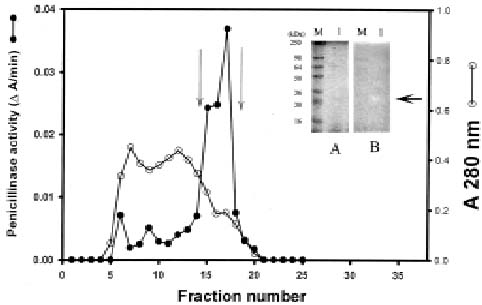
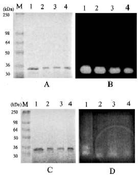
Abstract. The 0.08% hydrolyzed starch or glycogen were copolymerized in 7.5% or 10% sodium dodecylsulfate polyacrylamide gels. After electrophoresis and SDS removal, the commercial penicillinase in gels was reacted with penicillin G (100 mg in 50 mL, 0.1 M phosphate buffer, pH 7.0) for 30 min and then stained with 0.6% I2 in 6% KI solutions. The clear zone of penicillinase activity bands stood out against purple or orange-red backgrounds, respectively, for hydrolyzed starch or glycogen used. This activity staining method was used successfully to detect commercial penicillinase activities from Bacillus cereus and the cultured methicillin-resistant Staphylococcus aureus ATCC 33591 strain. This activity staining method was also applied to penicillinase natural inhibitor screenings. It was found that anthraquinone-related compounds, such as aloe-emodin, emodin and rhein, could inhibit penicillinase activity. This fast and sensitive method can be used in the process of penicillinase purification, characterization and inhibitor screening.

Keywords: Activity staining; Anthraquinone; Copolymerization; Penicillinase; SDS-PAGE.

Abbreviations: SDS-PAGE, sodium dodecylsulfate-polyacrylamide gel electrophoresis.

Introduction

The b-lactam family of antibiotics includes many of the most commonly used antibacterials in clinical medicines. The majority of clinically useful b-lactams belong to either the penicillin (penam) or the cephalosporin (cepems) group (Tyczkowska et al., 1994; Lawung et al., 2001). One of the major mechanisms of resistance to b-lactams was the expression of b-lactamases which hydrolyzed the b-lactam ring. The b-lactamases (EC 3.5.2.6), such as penicillinase and cephalosporinase, which degraded penam and cepems, respectively, have been found widely in both gram-positive and gram-negative bacteria (Livermore, 1995).

Several methods were reported for b-lactamase activity determinations, such as acidimetric methods by pH stat (Labia et al., 1973), neocuproine-copper systems by spectrophotometry (Cohenford et al., 1988), nitrocefin assays by spectrophotometry (Chen et al., 1994; Lawung et al., 2001) and fluorescent spot test (Chen et al., 1994). Despite the vital role in bacterial resistance played by b-lactam antibiotics, reports concerning the activity staining of b-lactamases in gel are few. Matthew et al. (1975) used analytical isoelectric focusing for b-lactamase separations. The isoelectric focusing gel was overlaid with Whatman No.

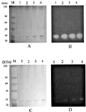
54 paper containing chromogenic substrate, cepharosporin 87/312. After reaction, the pink b-lactamases activity bands on a yellow background were found. Tai et al. (1985) used SDS-PAGE for b-lactamase separation. The SDS-PAGE gel was overlaid with Whatman 3MM filter paper. The filter paper had been prepared by dipping in starch-iodine solution and hanging it to dry overnight. It was then stored in a dark, cool, dry place. Bonnet et al. (2000) used analytical isoelectric focusing and SDS-PAGE for b-lactamase separation. The isoelectric focusing gel was revealed with iodine-agar gel by overlaying an agar containing penicillin G (0.6%), 6% potassium iodide, and 0.6% iodine. For SDS-PAGE gels, the b-lactamases activity was detected by overlaying polyacrylamide gel containing 0.5 mM nitrocefin.

Owing to the expensive, light sensitive and water-soluble properties of synthetic substrates, such as nitrocefin, it might be suitable for detecting b-lactamases in rapid screenings. The starch-iodine filter paper for b-lactamase detection was suitable, but the preparation work was tedious. In this report, we proposed a modification of a method by Tai et al. (1985) of copolymerizing hydrolyzed starch or glycogen in SDS-PAGE gels. The penicillinase activity in gels was reacted with penicillin G and revealed by I2-KI solutions. A number of traditional herb-derived medicines have been developed as anticancer drugs and free radical scavengers (Surh, 1999). Rhubarb, an important traditional Chinese medicinal herb, contained several anthraquinone-related compounds, such as aloe-emodin,

*Corresponding author. Fax: 886-2-2378-0134; E-mail: wchou@tmu.edu.tw


Botanical Bulletin of Academia Sinica, Vol. 44, 2003

emodin, and rhein. The extracts of rhubarb exhibited anti-inflammatory activities (Cuellar et al., 2001). We also used a modified penicillinase activity staining method to screen for penicillinase's natural inhibitors. We found anthraquinone-related compounds—such as aloe-emodin, emodin, and rhein—could inhibit penicillinase activity. This fast and sensitive method can be used in the process of penicillinase purification, characterization, and inhibitor screening.

Materials and Methods

Chemicals

Commercial penicillinase (type I, 5,000 units, from Bacillus cereus), penicillin G (sodium salt), aloe-emodin, emodin, rhein, I2, KI, oxacillin, hydrolyzed starch (S-5651, from potato), and glycogen (type 2, from oyster) were all purchased from Sigma Chemical Co. (St. Louis, MO, USA). Acrylamide, Bis, TEMED, APS, coomassie brilliant blue R-250, Tris, ammonium sulfate, nutrient agar (1.05450), and nutrient broth (1.05443) were obtained from E. Merck Inc. (Germany). See BlueTM pre-stained standard kit for SDS-PAGE was from Invitrogen Co. (Carlsbad, CA, USA). The kit contains myosin (250 kDa), BSA (98 kDa), glutamic dehydrogenase (64 kDa), alcohol dehydrogenase (50 kDa), carbonic anhydrase (36 kDa), myoglobin (30 kDa), and lysozyme (16 kDa). The Sephadex G-75 (F) was purchased from Pharmacia Biotech AB (Uppsala, Sweden). The strain of methicillin-resistant Staphylococcus aureus ATCC 33591 was purchased from the Food Industry Research and Development Institute (Hsinchu, Taiwan).

Activity Staining of Penicillinase on SDS-PAGE Gels

The 0.08% hydrolyzed starch or glycogen were copolymerized in 7.5% or 10% sodium dodecylsulfate polyacrylamide gels according to Weber and Osborn (1969). The vertical mini PROTEAN 3 system (Bio-Rad Inc., USA) with a 1.00 mm thickness of spacer was used. For electrophoresis, a 75 µl sample was mixed with 25 µl of 60 mM Tris buffer (pH 7.0) containing 2% SDS, 14.4 mM 2-mercaptoethanol, 25% glycerol, and 0.1% bromophenol blue and then incubated at 4°C overnight. When SDS-PAGE was finished, gels were cut into two parts. One part was fixed with 12.5% trichloroacetic acid and then stained with coomassie brilliant blue R-250 dye (Neuhoff et al., 1985). The other was immersed and shaken for 10 min twice in 25% (V/V) isopropanol in 10 mM phosphate buffer (pH 7.0) to remove SDS (Hou and Lin, 1998a, 1998b; Hou et al., 1999; Hou et al., 2001; Lin et al., 2002) and finally equilibrated in 100 mM phosphate buffer (pH 7.0) for 15 min before activity staining. The method of penicillinase activity staining was as follows: The gel was soaked in the substrate solution (100 mg penicillin G in 50 mL, 100 mM phosphate buffer, pH 7.0) with gentle shaking for 30 min. After a brief rinse, the penicillinase activity was developed with 0.6% I2 in 6% KI solutions. The clear zone of penicillinase activity bands stood out against purple or orange-red

backgrounds, respectively, for hydrolyzed starch or glycogen used. The gel was washed with 5% acetic acid for destaining.

A Methicillin-Resistant Staphylococcus aureus ATCC 33591 Cultivation

The methicillin-resistant Staphylococcus aureus ATCC 33591 strain was activated by nutrient broth, cultured on the nutrient agar following the manufacture's guidelines, and then stored at 4°C. A single colony was cultured in nutrient broth for 18 h, counted, and then diluted to 105 cfu/mL broth. One mL of this broth was mixed with 1 mL solution containing 6 µg oxacillin and 4% NaCl and then added to 100 mL nutrient broth by shaking at 125 rpm, 37°C for 18 h.

Partial Purifications of Penicillinase Isolated from Methicillin-Resistant Staphylococcus aureus ATCC 33591 Cultivation

The 18-h cultured broth was centrifuged at 5,000 rpm for 10 min. The pellets were recovered and then washed twice with 10 mM phosphate buffer (pH 7.0) before being resuspended in 100 mM phosphate buffer (pH 7.0) containing 0.1% Triton X-100 and then heated at 50°C for 1 h. After centrifugation at 12,000 g for 30 min, the supernatant was saved for ammonium sulfate fractionations. The precipitations of 30 to 95% ammonium sulfate were collected and then directly loaded onto a Sephadex G-75 column (1 × 75 cm). The column was eluted with 50 mM phosphate buffer (pH 7.0). Flow rate was 30 mL/h, and each fraction contained 2 mL. Each fraction was determined for penicillinase activity using the nitrocefin method. The active fractions were pooled and concentrated with centriprep-10 to small volumes for penicillinase activity staining.

Determinations of Penicillinase Activity Isolated from Methicillin-Resistant Staphylococcus aureus ATCC 33591 Cultivation

The penicillinase activity was determined by nitrocefin methods following Lawung et al. (2001), with some modifications. Each 100 µl of Sephadex G-75 fraction was added to 990 µl of 100 mM phosphate buffer (pH 7.0), and then 10 µl nitrocefin solution (10 mg in 2 mL N,N-dimethylformamide) was added with gentle mixing. The changes of absorbance at 486 nm during 3 min were recorded and expressed as DA/min for penicillinase activity.

Results and Discussion

A modified method of copolymerizing hydrolyzed starch or glycogen in SDS-PAGE gels was used for penicillinase activity staining and was applied to screening for penicillinase's natural inhibitors. In gauging the usefulness of this proposed method, it was considered whether the penicillinase could pass through the gel without hindrance. Figure 1 showed protein staining (A, C) and the


Liang et al. — Penicillinase inhibitor screenings

commercial penicillinase activity staining (B, D) on 7.5% SDS-PAGE gels incorporated with 0.08% hydrolyzed starch (A, B) or glycogen (C, D). Lanes 1-4 were 0.1, 0.25, 0.5 and 0.75 units, respectively. Comparing the protein staining (Figures 1A and 1C) with activity staining of penicillinase (Figures 1B and 1D), it was found that the penicillinase protein band was around 33 kDa, close to what Tai et al. (1985) reported. With increasing amounts (0.1 to 0.75 units) of penicillinase the activity bands became more intensive, meaning the incorporations of hydrolyzed starch (Figures 1A and 1B) or glycogen (Figures 1C and 1D) did not hamper the pass of penicillinase for their similar protein stainings. However, under the same conditions, the copolymerizations of hydrolyzed starch in SDS-PAGE gels had higher clear penicillinase activity than glycogen did. It might be the accessible penicillin G to penicillinase for the hydrolyzed starch used. Tai et al. (1985) used SDS-PAGE for b-lactamase separation, and then the SDS-PAGE gel was overlaid with Whatman 3MM filter paper. The filter paper had been prepared by dipping in starch-iodine solution and hanging out to dry overnight before being stored in a dark, cool, dry place. This modified method proved easier than that used by Tai et al. (1985).

This proposed activity staining method was applied to the methicillin-resistant Staphylococcus aureus ATCC 33591 strain for detections of penicillinase activity. After 18-h culture, the pellets were extracted with 100 mM phosphate buffer (pH 7.0) containing 0.1% Triton X-100 and then heated at 50°C for 1 h. The precipitations of 30 to 95% ammonium sulfate of bacteria lysates were collected and then directly loaded onto Sephadex G-75 column (1 × 75 cm) (Figure 2). Two minor and one major penicillinase

Figure 1. Protein staining (A, C) and commercial penicillinase activity staining (B, D) on 7.5% SDS-PAGE gels incorporated with 0.08% hydrolyzed starch (A, B) or glycogen (C, D). Lanes 1-4 were 0.1, 0.25, 0.5 and 0.75 units, respectively. "M" indicated the See BlueTM pre-stained markers for SDS-PAGE.

Figure 2. The chromatogram of Sephadex G-75 column (1 × 75 cm) on penicillinase activity (DA/min) of the methicillin-resistant Staphylococcus aureus ATCC 33591 strain of 30 to 95% ammonium sulfate by nitrocefin methods. The inset showed protein staining (A) and penicillinase activity staining (B) on 10% SDS-PAGE gels incorporated with 0.08% hydrolyzed starch. "M" indicated the See BlueTM pre-stained markers for SDS-PAGE. Each well contained 24 µg proteins. The arrow indicated the position of penicillinase activity band.


Botanical Bulletin of Academia Sinica, Vol. 44, 2003

activity bands were found using nitrocefin methods. These major penicillinase activity fractions were pooled, concentrated and then applied for activity staining on 10% SDS-PAGE gels incorporated with 0.08% hydrolyzed starch (Figure 2, inset). It was found that the clear zone of the penicillinase activity band (Figure 2, inset B, arrow indicated) was around 33 kDa, similar to that Tai et al. (1985) reported. One of the major mechanisms of resistance to b-lactams was the expression of b-lactamases, which hydrolyzed the b-lactam ring. It was clear that the methicillin-resistant Staphylococcus aureus ATCC 33591 strain expressed penicillinase activity against 6 µg oxacillin in the cultured medium.

Rhubarb, an important traditional Chinese medicinal herb, contained several anthraquinone-related compounds such as aloe-emodin, emodin, and rhein. The extracts of rhubarb exhibited anti-inflammatory activities (Cuellar et al., 2001). We also used the penicillinase activity staining method detailed here to screen for natural inhibitors (Figure 3). Lane 1, commercial penicillinase (0.75 units) as the controls; lanes 2 to 4 were aloe-emodin, emodin and rhein, respectively, added to commercial penicillinase for 12 h. Compared with protein staining (Figures 3A and 3C),

it was found that anthraquinone-related compounds, aloe-emodin, emodin, and rhein (1 mg/mL, lanes 2 to 4), could inhibit penicillinase activity, especially rhein (Figures 3B and 3D). Based on the results of activity staining of commercial penicillinase from Bacillus cereus (Figure 1) and from methicillin-resistant Staphylococcus aureus ATCC 33591 strain (Figure 2), the modified penicillinase activity staining method, the copolymerizing of hydrolyzed starch or glycogen in SDS-PAGE gels, presented here seems more convenient than that reported by Tai et al. (1985). We also applied this method to penicillinase inhibitor screenings. This fast and sensitive method can be used in the process of penicillinase purification, characterization, and inhibitor screening.

Acknowledgment. The authors want to thank the National Science Council, Republic of China, ROC for its financial support (NSC 91-2313-B038-002).

Literature Cited

Bonnet, R., J.L.M. Sampaio, C. Chanal, D. Sirot, C.D. Champs, J.L. Viallard, R. Labia, and J. Sirot. 2000. A novel class A extended-spectrum b-lactamase (BES-1) in Serratia marcescens isolated in Brazil. Antimicrob. Agents Chemother. 44: 3061-3068.

Chen, K.C.S., L. Chen, and J.Y. Lin. 1994. Fluorescent spot test method for specific detection of b-lactamases. Anal. Biochem. 219: 53-60.

Cohenford, M.A., J. Abraham, and A.A. Medeiros. 1988. A colorimetric procedure for measuring b-lactamase activity. Anal. Biochem. 168: 252-258.

Cuellar, M.J., R.M. Giner, M.C. Recio, S. Manez, and J.L. Rios. 2001. Topical anti-inflammatory activity of some Asian medicinal plants used in dermatological disorders. Fitoterapia 72: 221-229.

Hou, W.C. and Y.H. Lin. 1998a. Activity staining on polyacrylamide gels of trypsin inhibitors from leaves of sweet potato (Ipomoea batatas) varieties. Electrophoresis 19: 212-214.

Hou, W.C. and Y.H. Lin. 1998b. Activity staining of pectinesterase on polyacrylamide gels after acidic or sodium dodecyl sulfate electrophoresis. Electrophoresis 19: 692-694.

Hou, W.C., H.J. Chen, T.E. Chen, and Y.H. Lin. 1999. Detection of protease activities using specific aminoacyl or peptidyl p-nitroanilides after sodium dodecyl sulfate-polyacrylamide gel electrophoresis and its applications. Electrophoresis 20: 486-490.

Hou, W.C., H.J. Chen, Y.H. Lin, Y.C. Chen, L.L. Yang, and M.H. Lee. 2001. Activity staining of isocitrate lyase after electrophoresis on either native or sodium dodecyl sulfate-polyacrylamide gel. Electrophoresis 22: 2653-2655.

Labia, R., J. Andrillon, and F. LeGoffic. 1973. Computerized microacidimetric determination of beta lactamase Michaelis-Menten constants. FEBS Letts. 33: 43-44.

Lawung, R., B. Danielsson, V. Prachayasittikul, and L. Bülow. 2001. Calorimetric analysis of cepharosporins using an immobilized TEM-1 b-lactamase on Ni2+ chelating Sepharose fast flow. Anal. Biochem. 296: 57-62.

Lin, C.L., H.J. Chen, and W.C. Hou. 2002. Activity staining of

Figure 3. Protein staining (A, C) and commercial penicillinase activity staining (B, D) on 10% SDS-PAGE gels incorporated with 0.08% hydrolyzed starch (A, B) or glycogen (C, D). Lane 1, commercial penicillinase (0.75 units) as the controls; lanes 2 to 4 were aloe-emodin, emodin and rhein (1 mg/mL in dimethylformamide), respectively, added to commercial penicillinase for 12 h. "M" indicated the See BlueTM pre-stained markers for SDS-PAGE.


Liang et al. — Penicillinase inhibitor screenings

glutathione peroxidase after electrophoresis on either native or sodium dodecyl sulfate-polyacrylamide gel. Electrophoresis 23: 513-516.

Livermore, D.M. 1995. Beta-lactamases in laboratory and clinical resistance. Clin. Microbiol. Rev. 8: 557-584.

Matthew, M., A.M. Harris, M.J. Marshall, and G.W. Ross. 1975. The use of analytical isoelectric focusing for detection and identification of b-lactamase. J. Gen. Microbiol. 88: 169-178.

Neuhoff, V., R. Stamm, and H. Eibl. 1985. Clear background and highly sensitive protein staining with Coomassie blue dyes in polyacrylamide gels: a systematic analysis. Electrophoresis 6: 427- 448.

Surh, Y.J. 1999. Molecular mechanisms of chemopreventive ef

fects of selected dietry and medicinal phenolic substances. Muta. Res. 428: 305-327.

Tai, P.C., N. Zyk, and A. Citri. 1985. In situ detection of b-lactamase activity in sodium dodecylsulfate-polyacrylamide gels. Anal. Biochem. 144: 199-203.

Tyczkowska, K.L., R.D. Voyksner, R.F. Straub, and A.L. Aronson. 1994. Simultaneous multiresidue analysis of beta-lactam antibiotics in bovine milk by liquid chromatography with ultraviolet detection and confirmation by electrospray mass spectrometry. J. Assoc. Off. Anal. Chem. 77: 1122-1131.

Weber, K. and M. Osborn. 1969. The reliability of molecular weight determination by dodecyl sulphate-polyacrylamide gel electrophoresis. J. Biol. Chem. 244: 4406-4412.