Bot. Bull. Acad. Sin. (2004) 45: 61-68

Lee et al. — Comparison of Aspergillus flavus and A. oryzae by amplified fragment length polymorphism

Comparison of Aspergillus flavus and Aspergillus oryzae by amplified fragment length polymorphism

Chao-Zong Lee, Guey-Yuh Liou, and Gwo-Fang Yuan*

Bioresource Collection and Research Center, Food Industry Research and Development Institute, P.O. Box 246, Hsinchu, Taiwan 300, Republic of China

(Received April 25, 2003; Accepted October 31, 2003)

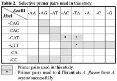
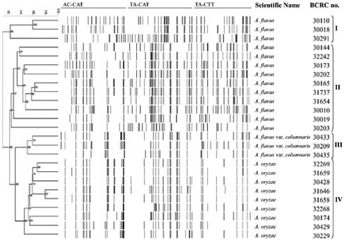
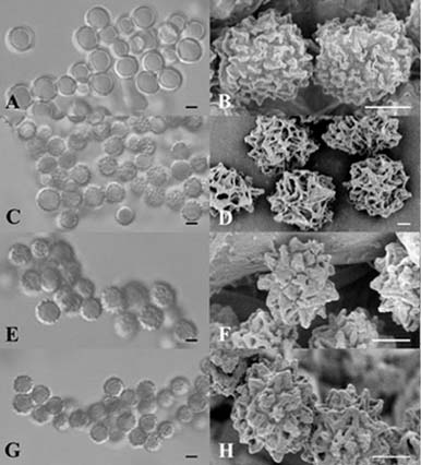
Abstract. The aflatoxin producer Aspergillus flavus and the koji mold Aspergillus oryzae are morphologically similar species that belong to the Aspergillus section Flavi. We used amplified fragment length polymorphism (AFLP) to differentiate these two species. In this study, we tested thirteen A. flavus, nine A. oryzae, and three A. flavus var. columnaris strains. DNA fragment profiles amplified with each of three selective primer pairs displayed similar patterns for the various A. flavus strains. Different patterns were observed with these primer pairs for the A. oryzae strains. We combined these data to increase the grouping of the various strains within each species and to distinguish between A. flavus and A. oryzae. By unweighted pair group method with arithmetic average (UPGMA) analysis, the AFLP data obtained from the three selective primer pairs, EcoRI-AC/MseI-CAT, EcoRI-TA/MseI-CAT, and EcoRI-TA/MseI-CTT, differentiated A. flavus from A. oryzae successfully. Three strains of A. flavus received from ATCC grouped outside of the other A. flavus strains we examined. The morphology of these isolates and our results indicated those strains were originally misidentified and they should be classified as A. parasiticus. We found that the AFLP technique is a reliable and reproducible tool with the ability to differentiate A. flavus from A. oryzae and should be generally useful in distinguishing between closely related species or strains.

Keywords: AFLP; Aspergillus flavus; Aspergillus oryzae.

Introduction

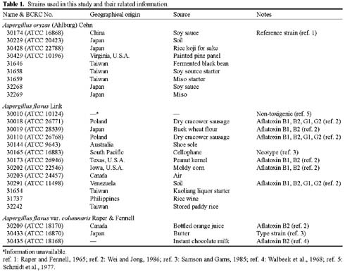
The aflatoxigenic species Aspergillus flavus Link and A. parasiticus Speare can produce the potent carcinogen aflatoxin and can pose a significant human health threat. The nonaflatoxigenic species A. oryzae (Ahlburg) Cohn and A. sojae Sakaguchi & Yamada are widely used for the production of food-grade amylase and the fermentation of sake, miso and soy sauce. All four species belong to the Aspergillus section Flavi and have many phenotypic similarities. Current methods for the identification of these species still depend primarily on cultural and morphological characteristics. However, it is often difficult to differentiate these species because the phenotypic differences are not distinct and are easily affected by the environment and are also confused by the high degree of intra- and interspecies variation.

Because of the economic value of members of the Aspergillus section Flavi and the importance of differentiating them, several molecular methods have been developed to distinguish these strains. DNA relatedness of these species in Aspergillus section Flavi was studied by Kurtzman et al. (1986) with the DNA reassociation method. The Cot value calculation results showed 100% related

ness between A. flavus and A. oryzae. Similarly, A. parasiticus and A. sojae were 91% related. The homology between these two groups was 70%. Therefore Kurtzman et al. suggested the four taxa be regarded as varieties of A. flavus. A medium containing bleomycin has been used to distinguish A. parasiticus from A. sojae (Klich and Mullaney, 1989; Yuan et al., 1995). The random amplified polymorphic DNA (RAPD) method has been used to differentiate A. parasiticus from A. sojae with three screened decamer primers (Yuan et al., 1995), but the same primers could not separate A. flavus from A. oryzae (Yuan et al., unpubl.). The sequence of a regulatory gene for the aflatoxin biosynthetic pathway was analyzed, and distinct DNA sequences within the 5' untranslated region and the zinc- finger domain were found in limited strains of A. flavus, A. oryzae, A. parasiticus, and A. sojae (Chang et al., 1995). PCR-amplified regions of ribosomal internal transcribed spacers have also been used to differentiate species of Aspergillus section Flavi with the single-strand conformation polymorphism (SSCP) method (Kumeda and Asao, 1996). The SSCP results indicated that 67 of the 68 test strains could be classified into four groups as A. flavus/A. oryzae, A. parasiticus/A. sojae, A. tamari, and A. nomius. Sixteen strains of A. flavus or A. oryzae could not be clearly separated with the sequence of ITS1-5.8S-ITS2 region of ribosomal DNA (Lin et al., 1995). Restriction site polymorphism and DNA sequences of protein-encoding genes were analyzed, and 31 A. flavus strains could be separated into two distinct groups. Five

*Corresponding author. Gwo-Fang Yuan, P.O. Box 246, Hsinchu, Taiwan 300, Republic of China. Tel: 886-3-5223191-580; Fax: 886-3-5214016; E-mail: gfy@firdi.org.tw


Botanical Bulletin of Academia Sinica, Vol. 45, 2004

of the 31 strains belonged to industrial fungus A. oryzae, and they all clustered together in one of the two groups (Geiser et al., 1998).

Previous research has indicated the difficulty of differentiating A. flavus from A. oryzae. In this study, we have used amplified fragment length polymorphism (AFLP) to differentiate strains of these two groups. The AFLP method is a relatively new molecular technique for DNA fingerprinting. The method combines the principle of restriction fragment length polymorphism (RFLP) analysis with highly specific PCR amplification (Bears et al., 1998; Vos et al., 1995; Zabeau and Vos, 1993). Total genomic DNA is digested with one or more restriction enzymes, and then double-stranded nucleotide adaptors are ligated to the DNA fragments to serve as a primer binding site for PCR amplification. Complementary primers to the adaptor and restriction site sequence with additional nucleotides at the 3'end are used to amplify a subset of the ligated fragments. In addition to fingerprinting DNA from human, animals, and plants, the AFLP technique has been used to differentiate intra- and interspecies strains of several bacteria, such as Aeromonas and Acinetobacter (Huys et al., 1996; Janssen et al., 1997), and fungi, such as Colletotrichum,

Cylindrocladium, Puccinia, and Phytophthora (Paul et al., 1996; Neill et al., 1997; Henricot and Culham, 2002; Villareal et al., 2002; Ochwo et al., 2002).

The purpose of this study was to apply the AFLP technique to strains of A. flavus and A. oryzae and evaluate the ability of this technique to differentiate between them. A total of 25 strains—including thirteen A. flavus strains, nine A. oryzae strains, and three A. flavus var. columnaris strains—were investigated in this study.

Materials and Methods

Fungal Strains and Mycelia Collection

All the fungal strains used in this study (Table 1) were originally obtained from different culture collection centers or were isolated from fermented materials and preserved in the Biosource Collection and Research Center (BCRC) of the Food Industry Research and Development Institute. BCRC 30165 (=ATCC 16883) and BCRC 30433 (=ATCC 16870) have been deposited as type strains of A. flavus and A. flavus var. columnaris, respectively. BCRC 30174 (=ATCC 16868) was deposited as a reference strain of A. oryzae.


Lee et al. — Comparison of Aspergillus flavus and A. oryzae by amplified fragment length polymorphism

Fungal cultures for genomic DNA isolation were grown as follows. Conidia were harvested from 7- day-old potato dextrose agar slant cultures growing at 28°C and inoculated into 50 ml potato dextrose broth in 250 ml flask and then incubated at 25°C for 48 to 72 h without shaking. Mycelia were collected by vacuum filtration and lyophilized.

Preparation of Fungal Genomic DNA

Frozen mycelial mats were ground with a mortar and pestle to fine powder in liquid nitrogen. Approximately 20 mg of homogenized cell powder was mixed with 600 µl of lysis buffer (100 mM Tris-HCl, pH 8.0, 100 mM NaCl, 20 mM EDTA and 2% SDS) and incubated at 60°C for 10 min. The DNA was extracted sequentially with an equal volume of phenol/chloroform (1:1), and chloroform/ isoamyl alcohol (24:1). Total nucleic acids were precipitated with 0.7 volume of isopropanol and vacuum dried. Finally, the pellets were re-dissolved in water and treated with RNase A at 37°C for 1.5 h to remove RNA, and the RNase was inactivated at 60°C for 1 h. Final DNA concentrations were determined by measuring with a Model U spectrophotometer (Hitachi, Tokyo, Japan), and the integrity of each DNA sample was examined with an agarose gel.

AFLP Analysis

AFLP assays were carried out with an AFLPTM Microbial Fingerprinting kit (Perkin Elmer Corp., Norwalk, CT, USA) according to the manufacturer's instructions. Genomic DNA (0.01 µg) was digested with EcoRI and MseI. EcoRI, and MseI adapters were ligated to the fragments to form the primary templates. Primers with complimentary sequences to the adapters and restriction site were used in the first step PCR reaction under the following cycle profile: holding at 72°C for 2 min, 20 cycles consisting of 94°C for 20 sec, 56°C for 30 sec and 72°C for 2 min. Then, the same primers, except for an additional two or three nucleotides at the 3'end as shown in Table 2, were used in the second PCR reaction with previous PCR products as a template to amplify a subset of fragments. The cycling profiles were as follows: (1) 10 cycles of 20 sec at 94 oC, 30 sec at an annealing temperature (66 to 57°C, a step-down procedure consisting of 1°C lower than that of each previous cycle), and 2 min at 72°C; (2) 20 cycles at 94°C for 20 sec, 56°C for 30 sec, and 72°C for 2 min; (3) holding for 30 min at 72°C. The PCR reactions were performed on a Perkin Elmer 9600 thermal cycler (Norwalk, CT, USA). The selective amplification products were loaded on a 6% denaturing polyacrylamide gel to analyze band patterns with an ABI 373 sequencer (Perkin Elmer Corp., Norwalk, CT, USA).

Data Analysis

The electrophoresis data of AFLP reactions were converted to MS-DOS format to make them compatible with BioNumerics Software, Version 2.5 (Applied Maths, Inc. Austin, TX, USA). The similarity between each pair of AFLP patterns was calculated by using the band-based

Dice similarity coefficient, SD. For pattern comparison, a band position tolerance value of 0.1% was optimized to compensate for misalignment of homologous bands. The strains investigated were grouped by the unweighted-pair group method with arithmetic average (UPGMA). Branch support of the tree was assessed by bootstrapping (Felsenstein, 1985). The bootstrap analysis was performed with 10,000 replicates.

Morphological Observation

The taxonomic systems of Aspergillus (Raper and Fennell, 1965; Klich and Pitt, 1988a, b) were followed for observation and identification. The isolates were cultured on CYA (Czapek Yeast Extract Agar: sucrose 3%, yeast extract 0.5%, NaNO3 0.3%, KCl 0.05%, MgSO4·7H2O 0.05%, FeSO4·7H2O 0.001% and agar 1.5%) and CzA (Czapek's Agar: sucrose 3%, NaNO3 0.3%, K2HPO4 0.1%, KCl 0.05%, MgSO4·7H2O 0.05%, FeSO4·7H2O 0.001% and agar 1.5%) at 25°C. Both macroscopical and microscopical characters were assessed when they were 3, 7, and 14 days old. The colony color was observed by naked eye with the aid of color charts in the Methuen Handbook of Colour (Kornerup and Wanscher, 1978). The material on the slide mounts was examined under light microscope (Axiophot, Zeiss, Germany). The metulae, conidial diameter and conidial rough were determined under 1,000x magnification.

For scanning electron microscopic observations, the fungi were cultured on CYA medium at 25°C for 10 days. Agar blocks containing spores were gradually dehydrated with increasing concentrations of ethyl alcohol (25 to 100%) and then placed in a solution of 100% acetone for more than 1 h (Tzean and Estey, 1978). Finally, these blocks were treated in a critical point drying apparatus (HITACHI HCP-2). Dried specimens were coated with evaporated gold (Eiko Engineering, IB-2 ion coater) and examined under scanning electron microscope (Leo 980, Leo Electron Microscopy Ltd) (Kusaka and Asano, 1987).

Results

Preliminary AFLP experiment results showed that the selective primers with only one additional nucleotide yielded band patterns too complicated to analyze. Therefore we chose selective primers with two or three additional nucleotides at the 3' end for the AFLP reaction, and the fragment length for analysis was limited to the range of 50 to 500 base pairs. Eight EcoRI and six MseI selective primers were random paired in a selective PCR reaction (Table 2).

The UPGMA analysis of the AFLP results indicated that three pairs of selective primers, EcoRI-AC/MseI-CAT, EcoRI-TA/MseI-CAT, and EcoRI-TA/MseI-CTT, could distinguish A. flavus from A. oryzae successfully. The combined data of the three primer pairs were analyzed and generated the dendrogram shown in Figure 1. None of the other primer combinations examined in this study could clearly separate A. flavus from A. oryzae with AFLP. With these multiple fungal strains, repetitive AFLP reactions


Botanical Bulletin of Academia Sinica, Vol. 45, 2004

using the three primer-sets we studied have consistently yielded similar polymorphic band numbers and patterns. Different DNA preparations yielded similar results, and although certain bands may not appear occasionally, the

structure of dendrogram clustering A. flavus and A. oryzae is maintained. The dendrogram clustered our test strains into four groups with high bootstrap values (89-95), including A. flavus in group I and II, A. flavus var. columnaris in group III, and A. oryzae in group IV.

It was found that three aflatoxin-producing A. flavus strains, BCRC 30110, 30018 and 30291, clustered together in group I and remained distinct from the other A. flavus strains in the dendrograms of AFLP data obtained with the three primer pairs. Two of them (BCRC 30018 and 30110) were originally isolated from sausage in Poland, and the other was isolated from the soil of Venezuela. We reordered the corresponding cultures from ATCC and compared their morphology with those cultures previously maintained in BCRC/FIRDI. No significant difference was found in the previous or new ordered cultures based on the light microscopic or scanning electron microscopic observation. However, the distinctly rough-walled conidia of previous and new ordered cultures (Figure 2) suggested that the three strains were originally misidentified and should be correctly identified as A. parasiticus instead.

Figure 1. Combination of amplified fragment length polymorphism analysis of thirteen A. flavus strains, nine A. oryzae strains, and three A. flavus var. columnaris strains, using EcoRI-AC/ MseI-CAT, EcoRI-TA/ MseI-CAT, and EcoRI-TA/ MseI-CTT selective primer pairs. UPGMA tree was constructed by comparing polymorphic bands generated from the three selective primer pairs. Values at branch nodes indicate bootstrap support obtained from bootstrap analysis with 10,000 replicates.


Lee et al. — Comparison of Aspergillus flavus and A. oryzae by amplified fragment length polymorphism

Figure 2. Conidia ornamentation of A. flavus strains. A, C, E, G: Light microscope micrographs, scale bar = 3 µm; B, D, F, H: Scanning electron micrographs, scale bar = 2 µm; A, B: BCRC 30165 (ATCC 16883), type strain of Aspergillus flavus; C, D: BCRC 30291 (ATCC 11498); E, F: BCRC 30110 (ATCC 26768); G, H: BCRC 30018 (ATCC 26771).

found in this study, that except with the three primer pairs described above, no primer combinations tested could clearly separate A. flavus from A. oryzae via AFLP. These results are consistent with the high relatedness of the two species.

Aspergillus oryzae is generally recognized as a safe (GRAS) microorganism. Previous research and our results indicate that the difference between A. flavus and A. oryzae is intraspecific level (Kurtzman et al., 1986; Yuan et al., 1995; Kumeda and Asao, 1996; Geiser et al., 1998). Therefore, it is worthwhile to reconsider the safety of A. oryzae. In our results, all the A. oryzae strains were tightly clustered in group IV (Figure 1), and no aflatoxin production was recorded by these strains. Thus, we suggest recognizing these strains as safe. Two A. flavus strains, BCRC 31654 and 31737, originally isolated from fermentation starter or products, were grouped with the other aflatoxigenic A.

Discussion

Many strains have been isolated and named A. flavus or A. oryzae. In order to use strains with more diversity, we selected them according to their typification, geographical origin, isolation source, and aflatoxin productivity in this study. Type strains of A. flavus and A. flavus var. columnaris were recruited. Based on the study of Kurtzman et al. (1986), we used BCRC 30174 (= ATCC 16868 = NRRL 451) as a reference strain for A. oryzae (Table 1).

The food fermentation and industrial importance of A. oryzae makes it essential to differentiate this species from the aflatoxigenic species, A. flavus. Whole genomic DNAs were compared in both the AFLP reactions and the DNA hybridization test. The 100% similarity between A. flavus and A. oryzae of Kurtzman's (1986) hybridization results indicated the close relationship of the two species. It was


Botanical Bulletin of Academia Sinica, Vol. 45, 2004

flavus strains in group II. These results indicate the importance of confirming the safety of strains used to prevent contamination during fermentation.

The variety Aspergillus flavus var. columnaris, with a columnar pattern of its conidial head, is morphologically different from A. flavus and A. oryzae (Raper and Fennell, 1965). In the dendrogram of Figure 1, strains of A. flavus var. columnaris clustered together in group III, and they displayed molecular information closer to A. oryzae than to A. flavus. These results suggest the existence of variance strains between A. flavus and A. oryzae and provide indirect support for the previous proposal that A. oryzae has been domesticated from the naturally occurring A. flavus (Kurtzman et al., 1986).

The three misidentified strains in group I have been studied for many years (Strzelecki and Badura, 1972), but the previous research did not focus on taxonomy. According to the description of Klich and Pitt (1988a, b), conidial color and microsopic characteristics strongly indicate that the three strains are A. parasiticus (Table 3). The significantly different polymorphic patterns of these three strains from the A. flavus or A. oryzae strains suggest that AFLP technique can also be applied to differentiate A. parasiticus from A. oryzae/A. flavus. On the other hand, the difference between A. parasiticus and A. sojae is more clear than between A. oryzae and A. flavus (Yuan et al., 1995). We therefore expect the AFLP method to be useful in differentiating A. parasiticus from A. sojae.

The high relatedness between A. flavus and A. oryzae suggests that the process of differentiating them requires an under-species classification accomplished by a number of different approaches (Olive and Bean, 1999). AFLP is a method for classifying strains, and we have demonstrated in this study its high differentiation power and reproducibility for differentiating A. flavus from A. oryzae. AFLP should be valuable in distinguishing other closely related species or strains.

Acknowledgments. This work was supported by grants from The Ministry of Economic Affairs, Taiwan, ROC. We thank Professor George A. Marzluf, Department Biochemistry, Ohio State University, USA for his review of the manuscript and valuable comments.

Literature Cited

Bears, M., S. De Grandis, H. Lee, and J.T. Trevors. 1998. Amplified fragment length polymorphism (AFLP): a review of the procedure and its applications. J. Ind. Microbiol. Biotech. 21: 99-114.

Chang, P., D. Bhatnagar, T.E. Cleveland, and J.W. Bennett. 1995. Sequence variability in homologs of the aflatoxin pathway gene aflR distinguishes species in Aspergillus section Flavi. Appl. Environ. Microbiol. 61: 40-43.

Felsenstein, J. 1985. Confidence limits on phylogenies: An approach using the boorstrap. Evolution 39: 783-791.

Geiser, D.M., J.I. Pitt, and J.W. Taylor. 1998. Cryptic speciation and recombination in the aflatoxin-producing fungus Aspergillus flavus. Proc. Natl. Acad. Sci. USA 95: 388-393.

Henricot, B. and A. Culham. 2002. Cylindrocladium buxicola, a new species affecting Buxus spp., and its phylogenetic status. Mycologia 94: 980-997.

Huys, G., R. Coopman, P. Janssen, and K. Kersters. 1996. High-resolution genotypic analysis of the genus Aeromonas by AFLP fingerprinting. Inter. J. Syst. Bacteriol. 46: 572-580.

Janssen, P., K. Maquelin, R. Coopman, I. Tjernberg, P. Bouvet, K. Kersters, and L. Dijkshoorn. 1997. Discrimination of Acinetobacter genomic species by AFLP fingerprinting. Int. J. Syst. Bacteriol. 47: 1179-1187.

Klich, M.A. and E.J. Mullaney. 1989. Use of a bleomycin-containing medium to distinguish Aspergillus paraciticus from A. sojae. Mycologia 81: 159-160.

Klich, M.A. and J.I. Pitt. 1988a. Differentiation of Aspergillus flavus from A. parasiticus and other closely related species. Trans. Br. Mycol. Soc. 91: 99-108.

Klich, M.A. and J.I. Pitt. 1988b. A laboratory guide to common


Lee et al. — Comparison of Aspergillus flavus and A. oryzae by amplified fragment length polymorphism

Williams & Wilkins Company, MD, U.S.A. 370-376 pp.

Samson, R.A. and W. Gams. 1985. Typification of the species of Aspergillus and associated teleomorphs. Advances in Penicillium and Aspergillus systematics, ed. by Samson, R. A. and Pitt, J. I., Plenum Publ., New York, pp. 40.

Schmidt, A.L., C.R. Curtis, and G.A. Bean. 1977. Electrophoretic comparisons of mycelial enzymes from aflatoxin-producing and non-producing strains of Aspergillus flavus and Aspergillus parasiticus. Can. J. Microbiol. 23: 60-67.

Strzelecki, E.L. and L. Badura. 1972. Occurrence of aflatoxingenic molds on "dry cravower sausage". Acta Microbiol. Pol. 4: 233-239.

Tzean, S.S. and R.H. Estey. 1978. Schizophyllum commune Fr. as a destructive mycoparasite. Can. J. Microbiol. 24: 780-784.

Villareal, L.M.M.A., C. Lannou, C. de Vallavieille-Pope, and C. Neema. 2002. Genetic variability in Puccinia striiformis f. sp. tritici populations sampled on a local scale during natural epidemics. Appl. Environ. Microbiol. 68: 6138-6145.

Vos, P., R. Hogers, M. Bleeker, M. Reijans, T. van de Lee, M. Hornes, A. Freujters, J. Pot, J. Peleman, M. Kuiper, and M. Zabeau. 1995. AFLP: a new technique for DNA fingerprinting. Nucleic Acids Res. 23: 4407-4414.

Walbeek, W.V., P.M. Scott, and F.S. Thatcher. 1968. Mycotoxins from food-borne fungi. Can. J. Microbiol. 14: 131-137.

Wei, D.L. and S.C. Jong. 1986. Production of aflatoxins by strains of the Aspergillus flavus group maintained in ATCC. Mycopathologia 93: 19-24.

Yuan, G.F., C.S. Liu, and C.C. Chen. 1995. Differentiation of Aspergillus parasiticus from Aspergillus sojae by random amplification of polymorphic DNA. Appl. Environ. Microbiol. 61: 2384-2387.

Zabeau, M. and P. Vos. 1993. Selective restriction fragment amplification: a general method for DNA fingerprinting. European Patent Application number: 9240269.7, Publication Number: 0 534 858 A1.

Aspergillus species and their teleomorphs. Commonwealth Scientific and Industrial Research Organization, Australia, 116 pp.

Kornerup, A. and J.H. Wanscher. 1978. Methuen Handbook of Colour, 3rd edn. London, U.K.

Kumeda, Y. and T. Asao. 1996. Single-strand conformation polymorphism analysis of PCR-amplified ribosomal DNA internal transcribed spacers to differentiate species of Aspergillus section Flavi. Appl. Environ. Microbiol. 62: 2947-2952.

Kurtzman, C.P., M.J. Robnett, and D.T. Wicklow. 1986. DNA relatedness among wild and domesticated species in the Aspergillus flavus group. Mycologia 78: 955-959.

Kusaka, T. and I. Asano. 1987. Morphological observation of Actinomycetes strains by scanning electron microscopy. IFO Res. Comm. 13: 35-44.

Lin, C.H., C.H. Tang, and G.F. Yuan. 1995. Differentiation of Aspergillus flavus and A. oryzae. Research Report 84-982, FIRDI.

Neill, N.R., P. van Berkum, J.-J. Lin, J. Kuo, G.N. Ude, W. Kenworthy, and J.A. Saunders. 1997. Application of amplified restriction fragment length polymorphism for genetic characterization of Collectotrichum pathogens of Alfalfa. Phytopathology 87: 745-750.

Ochwo, M.K.N., S. Kamoun, E. Adipala, P.R. Rubaihayo, K. Lamour, and M. Olanya. 2002. Genetic diversity of Phytophthora infestans (Mont.) de Bary in the eastern and western highlands of Uganda. J. Phytopathol. 150: 541-542.

Olive, D.M. and P. Bean. 1999. Principle and applications of methods for DNA-based typing of microbial organisms. J. Clinical Microbiol. 37: 1661-1669.

Paul, J., R. Coopman, G. Huys, J. Swings, M. Bleeker, P. Vos, M. Zabeau, and K. Kersters. 1996. Evaluation of the DNA fingerprinting method AFLP as a new tool in bacterial taxonomy. Microbiology 142: 1881-1893.

Raper, K.B. and D.I. Fennell. 1965. The genus Aspergillus. The


Botanical Bulletin of Academia Sinica, Vol. 45, 2004