Bot. Bull. Acad. Sin. (2004) 45: 203-212

Wang et al. — Copper-induced change in antioxidation

Copper-induced stress and antioxidative responses in roots of Brassica juncea L.

Song-Hua Wang1,2, Zhi-Min Yang1,*, Hong Yang1, Bo Lu1, Shao-Qong Li1, and Ya-Ping Lu1

1Department of Biochemistry and Molecular Biology, College of Life Science, Nanjing Agricultural University, Nanjing, 210095, P.R. China

2Department of Applied Biology, Anhui Technical Teacher's College, Fengyang, Anhui, P.R. China

(Received November 10, 2003; Accepted February 25, 2004)

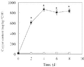
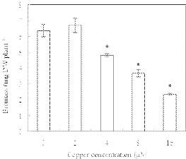
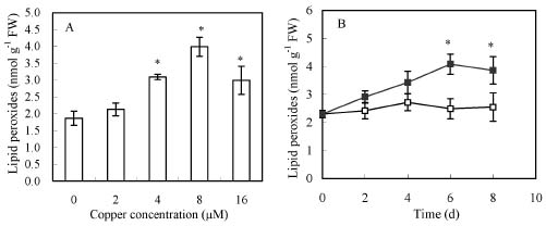
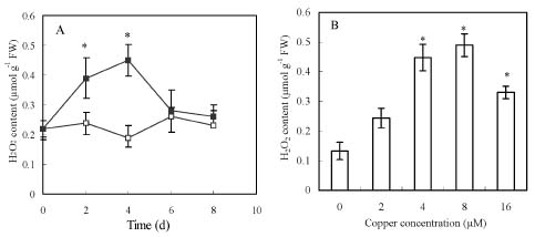
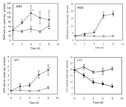
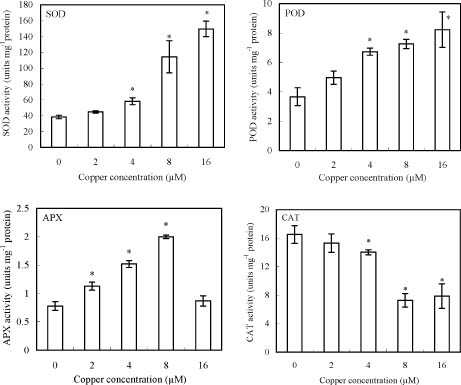
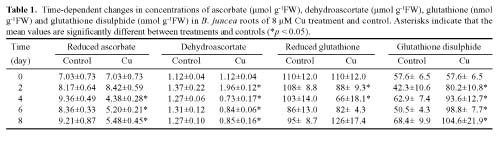
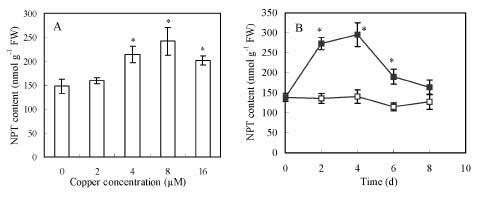
Abstract. Copper induced antioxidative reactions in the roots of Brassica juncea L. were investigated in both time-and concentration-dependent manners. The rapid uptake of Cu was observed immediately after the start of treatment. Application of Cu at 8 µM caused 50 percent reduction in biomass of Cu-treated roots as compared with control. Cu-induced root growth inhibition paralleled the level of root oxidative damage. Treatment with Cu at 8 µM induced a twofold increase in H2O2 content during the first 4 d, but it declined to the basal level thereafter. We also observed a twofold increase in superoxide dismutase activities with 8 µM Cu during the first 2 d. The stimulation lasted for 4 d and then gradually declined. Activities of both ascorbate peroxidase and guaiacol peroxidase in roots were found to be low during the first 4 d after seedling exposure to 8 µM Cu, but significantly increased after that, suggesting that increased enzyme activities would be responsible for the removal of H2O2. Catalase activities were always suppressed under Cu stress. Treatment of seedlings with 8 µM Cu induced general decreases in both reduced ascorbate and dehydroascorbate. The reduced glutathione content decreased at early stages of Cu treatment. However, it was restored to the level of controls thereafter. In contrast, the oxidized glutathione contents showed a progressive increase during the time of Cu treatment. The total non-protein thiol content was shown to increase during the first several days, but it declined at later stages.

Keywords: Antioxidative enzymes; Brassica juncea L.; Copper; Hydrogen peroxide; Non-protein thiols.

Introduction

Copper, an essential micronutrient, plays a vital role in maintaining normal metabolism in higher plants. It is involved in a wide range of biochemical and physiological processes. For example, Cu is required as a cofactor of Cu-Zn superoxide dismutase (SOD, EC 1.1.5.1.1) (Bowler et al., 1992). It also participates in electron-transfer reactions of photosynthesis in the form of plastocyanin (Raven et al., 1999). However, Cu at high levels becomes strongly phytotoxic to cells and causes inhibition of plant growth or even death (Mocquot et al., 1996; Weckx and Clijsters, 1996; Chen et al., 2000). Studies from some plant species demonstrate that excess Cu in plant growth medium induces formation of reactive oxygen species in treated-tissues (Cuyers et al., 2000; Groppa et al., 2001). Cu-induced generation of hydrogen peroxide, hydroxyl redicals, or other reactive oxygen species (ROS) has been directly correlated with the damage to proteins and lipids (De Vos et al., 1991; Murphy and Taiz, 1997). Photosynthesis is also sensitive to excessive Cu, and the pigment and protein components of photosynthetic membranes are the targets

(Pätsikkä et al., 2002). In addition, Cu toxicity is related to disturbances in the uptake of other essential elements (van Assche and Clijsters, 1990; Pätsikkä et al., 2002).

The phenomenon of Cu activating the production of ROS is described as oxidative stress (Luna et al., 1994). To repair the oxidative damage initiated by ROS, some tolerant plants have established protective mechanisms. The primary constituents of these protective mechanisms include two scavenging systems: enzymatic and non-enzymatic. Enzymatic scavengers, such as superoxide dismutase, are involved in the detoxification of O2.-; peroxidase, catalase, and enzymes of the ascorbate-glutathione cycle are related to the removal of H2O2 (De Vos et al., 1991; Murphy and Taiz, 1997); and non-enzymatic redicals-scavengers such as ascorbate and glutathione have been interpreted as the key antioxidants for the removal of H2O2 in plant cells, thus reducing the accumulation of the free radicals (van Assche and Clijsters, 1990; Foyer, 1993; Gupta et al., 1999). Evidence from several plant species reveals that Cu caused oxidative stress by mediating the activities of antioxidative enzymes (Luna et al., 1994; Gupta et al., 1999; Cuyers et al., 2000). Savouré et al. (1999) reported that Cu treatment triggered distinct oxidative defense mechanisms in Nicotiana plumbaginifolia. However, contrasting results are present. For example, in leaves of Cu-exposed Avena sativa plants, superoxide

*Corresponding author. Tel: 86-25-84395057; Fax: 86-25-84073096; E-mail: zmyang@njau.edu.cn


Botanical Bulletin of Academia Sinica, Vol. 45, 2004

dismutase activity increased while ascorbate peroxidase decreased (Luna et al., 1994). By contrast, leaves of Cu-treated Phaseolus vulgaris exhibited decreased activities of superoxide dismutase and increased ascorbate peroxidase (Pätsikkä et al., 2002). Apparently, the mechanisms by which Cu induces antioxidative responses and whether different plant species share a common defense mechanism or not, are not yet fully understood.

The inhibition of plant growth and crop production by excess heavy metals such as copper, cadmium, zinc, or nickel in contaminated-soil is a global agricultural problem. Using metal-accumulating plants to remove these excessive metals from soil and aqueous streams has been proposed as a solution (Salt et al., 1995). However, in addition to the knowledge of uptake, translocation, or compartmentation of heavy metals in plants, understanding of the tolerance mechanisms to improve plants of biotechnological interest is also important (Zenk, 1996; Salt et al., 1998). The ideal plant species to remediate heavy metal-contaminated soils is the Brassica juncea (Banuelos and Meek, 1990), a high biomass plant within the Brassicaceae family. B. juncea has been shown to accumulate Cu, Zn, Ni, Cd and Pb (Salt et al., 1996). However, to date no information has been reported on Cu-inducted oxidative stress or antioxidative responses in the species. We were therefore interested in identifying the involvement of antioxidative mechanisms responsible for the Cu-induced oxidative stress in the roots. The purpose of the study was to examine the changes in concentrations of H2O2 in the roots exposed to different levels of Cu. We also measured the several enzyme activities involved in H2O2 metabolism. In addition, we measured the antioxidative metabolites like ascorbate, glutathione, and non-protein thiols in the Cu-treated roots.

Materials and Methods

Plant Material and Treatment

Seeds of Brassica juncea L. (India mustard) were sterilized with 1% NaClO for 10 min, then washed several times with distilled water and germinated for 3d in the dark on floating plastic net. After germination, young seedlings were transferred to 1 L polyetheylene containers with 1/4-strength modified Hoagland nutrient solution with 0.7 mM Ca2+, 1.5 mM K+, 0.5 mM Mg2+, 0.25 mM NH4+, 2.9 mM NO3-, 0.25 mM H2PO4-, 0.5 mM SO42-, 4.75 µM Fe2+, 0.32 µM Cu2+, 0.2 µM Zn2+, 1.25 µM Mn2+, 11.5 µM H3BO3, 0.025 µM MoO3 (Pickering et al., 2000). Seedlings were grown at 24°C, with a light intensity of 300 µmol m-2 s-1 and a 14-h photoperiod. After growing for 10 d (four true leaves), they were treated with 0, 2, 4, 8, 16 µM Cu2+ as CuCl2 in 1/4-strength nutrient solution mentioned above for 4 days, or with 0 and 8 µM Cu2+ for 0, 2, 4, 6, 8 days. The pH for both culture and treatment solutions was adjusted to 5.5. Treatment solutions were renewed every other day. After the treatments indicated above, the roots of seedlings were harvested and immediately frozen in liquid nitrogen and stored in an -80°C freezer.

Determination of Non-Protein Thiols (NPT)

Non-protein thiols were extracted by grinding 0.5 g roots in 1 mL ice-cold 5% (w/v) sulfosalicylic acid solution. After centrifugation at 10,000 g at 4°C for 30 min, the supernatants were collected and immediately assayed. NPT was measured with Ellman's reagent (Ellman, 1959). Briefly, 300 µL of the supernatant was mixed with 1.2 mL of 0.1 M phosphate buffer solution (PBS) (pH 7.6). After a stable absorbance reading of 412 nm was obtained, 25 µM 5,5'-dithiobis (2-nitrobenzoic acid) (DTNB) solution (6 mM DTNB dissolved in 5 mM EDTA, 0.1 M PBS, pH 7.6) was added, and the increase in absorbance at 412 nm was monitored.

Determination of Lipid Peroxidation

The level of lipid peroxidation was determined by a procedure based on the method of Heath and Packer (1968). 0.5 g fresh roots were ground in 5 mL of ice-cold phosphate buffer solution (0.05 mM, pH 7.8) containing 1% PVP. The homogenate was centrifuged at 10,000 g for 30 min. 2 mL of supernatant was mixed with 2 mL of thiobarbituric acid (TBA) (0.5% TBA, 20% TCA). The mixture was heated at 100°C for 30 min, chilled on ice, and then centrifuged at 1000 g for 10 min. Absorbance of the supernatant was measured at 532 nm and adjusted for non-specific absorbance at 510 nm and 560 nm.

H2O2 Determination

The content of H2O2 was measured according to the modified method of Patterson et al. (1984). Harvested roots were ground in 6 mL ice-cold acetone. The homogenate was centrifuged at 8,000 g at 4°C for 30 min. The supernatant was collected. 0.5 mL of supernatant was mixed with 1.5 mL of mixture of CHCl3 and CCl4 (1:3, v:v). Then, 2.5 mL of distilled water was added. The mixture was centrifuged at 1,000 g for 1 min, and the water phase was collected for H2O2 determination. To set controls, 0.1 mL of catalase (0.3 unit) was added to the 1 mL of supernatant to remove the H2O2. For the treatments, catalase solution was replaced by 0.1 mL MQ (QTUM 0001 X; Millipore Co.) water. The mixtures were incubated at 37°C for 10 min. 1 mL of phosphate buffer solution (0.2 M, pH 7.8) and 1 mL of 4-(2-pyridylazo) resorcinol (200 mM) solution were added to samples. The reaction mixtures were incubated at 45°C for 20 min, and the absorbance at 508 nm was read.

Enzyme Activity Determination

Ascorbate peroxidase (APX, EC 1.11.1.11) activity was determined according to the method of Nakano and Asada (1981) with the following modification: 0.2 g fresh roots were homogenized in 50 mM ice-cold phosphate buffer (pH 7.8) containing 2 mM ascorbate and 5 mM EDTA. The homogenate was centrifuged at 10,000 g at 4°C for 30 min. The reactive solution contained 2.7 mL of 50 mM phosphate buffer (pH 7.0), 0.1 mM EDTA, 0.5 mM ascorbate, 0.1 mL of 0.1 mM H2O2 and 50 µM of enzyme extracts. The decrease in absorbance at 290 nm was read.


Wang et al. — Copper-induced change in antioxidation

Superoxide dismutase (SOD, EC 1.1.5.1.1), catalase (CAT, EC 1.11.1.6), and guaiacol peroxidase (POD, EC 1.11.1.7) activities were assayed according to the methods described by Beauchamp and Fridovich (1971), Durner and Klessig (1996), and Hammerschmidt et al. (1982), respectively. The protein in the enzyme extract was quantified by the method of Bradford (1976).

Metabolite Determination

0.5 gram of frozen root tissue was ground in 2 mL 5% (v/v) m-phosphoric acid. The homogenate was centrifuged at 10,000 g for 20 min. Total ascorbate (reduced ascorbate + dehydroascorbate) and ascorbate contents were measured according to Cakmak and Marschner (1992). The amount of dehydroascorbate (DHA) was estimated from the difference of total ascorbate and AsA. Reduced glutathione (GSH) and glutathione disulphide (GSSG) contents were determined according to the method of Anderson (1958). The amount of reduced glutathione was calculated from the difference between the total glutathione and glutathione disulphide.

Copper Measurement

Roots of intact plants were thoroughly rinsed with deionized water and blotted dry. Samples were dried at 70 °C in a forced-air oven, weighted, and digested with 1:1 nitric to perchloric acid. Cu was determined by atomic absorption spectrometry (Yang et al., 2001).

Statistical Analysis

Each result shown in a table and figure was the mean of at least three replicated treatments. The significance of differences between treatments was statistically evaluated by standard deviation and Student's t-test methods.

Results

Copper Uptake, Root Growth and Membrane Injury

Figure 1 showed the Cu content in the roots of B. juncea over the time of treatment with Cu at 8 µM. Cu accumulation occurred upon the addition of Cu to the nutrient solution. The increase in Cu uptake lasted for 4 days. After that, the uptake rate was saturated. The maximum content of Cu in the roots exposed to 8 µM Cu for 4 d was 865 mg kg-1.

The biomass of roots exposed to the various levels of Cu was measured. Treatment of seedlings with 2 µM Cu induced only a slight increase in root dry weight, and seedlings supplied with Cu at 4-16 µM had significantly lower root biomass than control (Figure 2). Treatment with Cu at 8 µM decreased the root dry weight by 42.3%. Since a moderate effect of 8 µM Cu on the root growth was observed, this concentration was used to examine the biological and physiological responses.

Exposure of seedlings to excess Cu led to lipid peroxidation in roots. The amount of lipid peroxides was

Figure 1. Time-dependent changes in the content of Cu in B. juncea roots after 8 µM Cu treatment (filled squares) and control (open squares). Vertical bars represent standard deviation of the mean (n=3). Asterisks indicate that mean values are significantly different between treatments and control (*p<0.05).

Figure 2. Effect of copper on root biomass of B. juncea. Seedlings were incubated on 1/4 strength of Hoagland solutions containing various concentrations of Cu for 4 d. Vertical bars represent standard deviation of the mean (n=30). Asterisks indicate that mean values are significantly different between treatments and control (*p < 0.05).

enhanced with the increasing Cu applied (Figure 3A). A peak was observed with Cu at 8 µM, with the lipid peroxides value being twofold higher than control. A further increase in Cu concentration to 16 µM failed to elevate the production of lipid peroxides. Addition of 8 µM Cu to the medium induced progressive increases in the lipid peroxidation level over the time (Figure 3B). However, a significant rise of lipid peroxides began to be observed on day 6, when the highest level in the presence of 8 µM Cu was found. Following that time it appeared to decline.


Botanical Bulletin of Academia Sinica, Vol. 45, 2004

Figure 3. The content of lipid peroxides in roots of B. juncea under Cu stress. Seedlings were incubated on 1/4 strength of Hoagland solutions containing various concentrations of Cu for 4 d (A) or containing 8 µM Cu (filled squares) and control (open squares) for 8 d (B). Vertical bars represent standard deviation of the mean (n=5). Asterisks indicate that mean values are significantly different between treatments and control (*p < 0.05).

H2O2

H2O2 was measured to determine whether application of excessive Cu caused oxidative stress in roots of B. juncea. In a time-course experiment, the H2O2 content increased markedly during the 8 µM Cu treatment. This stimulation reached a peak on day 4. Then, the H2O2 content fell to the level of control (Figure 4A). We also examined the dose-response changes in H2O2 levels and observed that application of Cu at 2-16 µM significantly increased the production of H2O2 (Figure 4B). However, treatment with Cu at the highest concentration (16 µM) resulted in a lower increase in the H2O2 level.

Antioxidant Enzymes

Activities of antioxidative enzymes were assayed over a period of 8 d in Cu-treated roots. During this time, control enzyme activities fluctuated slightly (Figure 5). Treatment with 8 µM Cu resulted in nearly doubling SOD

activities during the first 2 d. The stimulation lasted for 8 d. In a dose-response experiment, treatment with Cu at 4-16 µM progressively increased the SOD activities, and 16 µM of Cu was found to be most effective concentration for inducing increases in SOD activities (Figure 6).

Activities of both APX and POD in roots were low during the initial 4 d after exposure of seedlings to 8 µM Cu, but significantly increased after that (Figure 5). At the time of day 8, APX and POD activities were enhanced by 2.7- and 5.1-fold respectively. Activities of POD displayed a progressive increase in response to Cu in a dose-response experiment, and the peak activity was found at 16 µM of Cu (Figure 6). Although APX activities were greatly promoted in the presence of 8 µM Cu, a further increasing Cu concentration to 16 µM resulted in a lack of the inhibition of the enzyme activities (Figure 6). In contrast to activities of SOD, APX and POD, the activities of CAT in the root were diminished under Cu stress (Figures 5-6).

Figure 4. The content of H2O2 in roots of B. juncea under Cu stress. Seedlings were incubated on 1/4 strength of Hoagland solutions containing 8 mM Cu (filled squares) and control (open squares) for 8 d (A) and containing various concentrations of Cu for 4 d (B). Vertical bars represent standard deviation of the mean (n=4). Asterisks indicate that mean values are significantly different between treatments and control (*p < 0.05).


Wang et al. — Copper-induced change in antioxidation

Figure 5. Time-dependent changes in activities of SOD, POD, APX and CAT in B. juncea roots of 8 µM Cu treatment (filled squares) and control (open squares). Vertical bars represent standard deviation of the mean (n=3). Asterisks indicate that mean values are significantly different between treatments and control (*p < 0.05).

Ascorbate, Glutathione and Non-Protein Thiols

Treatment of seedlings of B. juncea with 8 µM Cu had no marked effect on the ascorbate content in the root during the first 2 d of the treatment (Table 1). Following that time however, ascorbate content fell significantly. On day 4, the content of ascorbate with 8 µM Cu was 46.8% that of controls. Following that day, the Cu-induced low level of ascorbate was maintained up to the end of experiment. In contrast to the reduced ascorbate content, the dehydroascorbate content showed a significant increase during the first 2 d, but it sharply declined thereafter as compared with controls (Table 1). We also measured the content of glutathione. Results showed that glutathione contents decreased after the start of Cu treatment, but the significant reduction was observed on day 4, at which time the reduced glutathione content decreased by 36% compared with control. However, the content of reduced glutathione was restored after that, and at the end of experiment it exceeded the controls. By contrast, the oxidized glutathione contents showed a progressive increase (Table 1).

The levels of total non-protein thiols (NPT) were enhanced by 44.8% and 63.6% with Cu at 4 and 8 µM, respectively (Figure 7A). Neither lower nor higher concentrations (16 µM) of Cu induced accumulation of thiol compounds. A time-dependent change in thiol contents was observed. As shown in Figure 7B, the changes in the total NPT content showed a pattern similar to that of H2O2 over the time of 8 µM Cu treatment.

Discussion

In the present study, we examined the Cu uptake and several biochemical and physiological responses representing the oxidative damage and protection in roots of B. juncea. Seedlings of the plant readily took up Cu from the growth medium (Figure 1). The adverse effect of excess Cu in the roots probably caused the decrease in biomass of Cu-treated roots. The enhanced levels of lipid peroxides in roots indicated that excess Cu accumulation triggered the production of ROS, which caused the oxidative damage to plasma membrane. An increase in ROS pro


Botanical Bulletin of Academia Sinica, Vol. 45, 2004

Figure 6. Effect of copper on activities of SOD, POD, APX and CAT in roots of B. juncea. Seedlings were incubated on 1/4 strength of Hoagland solutions containing various concentrations of Cu for 4 d. Vertical bars represent standard deviation of the mean (n=3). Asterisks indicate that mean values are significantly different between treatments and control (*p < 0.05).

It is well known that under certain conditions some transition metals like copper and iron can induce oxidative stress. However, understanding of the antioxidative mechanisms for plant resistance to Cu toxicity is poor. The current study with B. juncea indicated that treatment with Cu resulted in a general increase in H2O2 level in root tissues (Figure 4A-B). The time-dependent changes in H2O2 indicated that Cu-induced H2O2 increases occurred immediately after exposure of seedlings to Cu, suggesting that accumulation of H2O2 was an early event. Because occurrence of the highest accumulation of H2O2 preceded the highest accumulation of lipid peroxides, it was possible that excessive H2O2 would be the cause of root lipid peroxidation.

duction was also evidenced by the enhanced SOD activities (Figures 5-6). Our understanding of oxidative resistance to Cu in B. juncea was related to the experimental design. To approach a situation in which both Cu toxicity and resistance were exhibited, the seedlings were exposed to Cu at a moderate concentration (8 µM), where the root biomass decreased to 50% of the control (Figure 2). This was designed to examine a resistance mechanism responsible for the Cu-induced enhancement of antioxidation. In this case, treatment with Cu at the moderate level was shown effectively to activate antioxidative systems, which consequently alleviated the oxidative damage in roots of B. juncea.


Wang et al. — Copper-induced change in antioxidation

Figure 7. The content of non-protein thiols (NPT) in roots of B. juncea under Cu stress. Seedlings were incubated on 1/4 strength of Hoagland solutions containing various concentrations of Cu for 4 d (A) and containing 8 µM Cu (filled squares) and control (open squares) for 8 d (B). Vertical bars represent standard deviation of the mean (n=5). Asterisks indicate that mean values are significantly different between treatments and control (*p < 0.05).

could not be compared. There might be two possibilities for the decreased CAT activity. One was Cu2+ that bound or replaced some components such as Fe2+ in the enzyme. Another reason for the decrease in CAT activity might be the increase in H2O2 that in turn inactivated the enzyme (Luna et al., 1994; Mashoudi et al., 1997). Also, it was possible that catalase in B. juncea would be more sensitive to excess Cu2+ since it readily bound to thiol groups and thereby inactivated the thiol-containing enzyme.

The activities of APX were progressively activated by Cu exposure (Figure 5). This suggested that the antioxidative capacity stimulated by Cu was involved in conversion of H2O2 to water and O2. APX is a component of the ascorbate-glutathione pathway, which plays a role in scavenging H2O2 (Asada, 1992). H2O2 is a systemic signal for the induction of APX (Morita et al., 1999). In callus cultures of rice embryos, H2O2 transiently induced mRNA for cytosolic APX (Morita et al., 1999). Cd-induced H2O2 accumulation in pine seedlings was also found to significantly elevate the APX activity (Schützendübel et al., 2001). Additionally, there is a report indicating that treatment of Phaseolus vulgaris roots with 15 µM Cu stimulated the activities of APX (Gupta et al., 1999). In the present study, the low level of APX activity during the initial Cu treatment suggested that plants appeared to require several days for induction of the enzyme (Figure 5).

The peroxidase exhibited an inducible activity in the presence of 8 µM Cu. The significant increase in activities of the enzyme at latter stages might greatly contribute to the degradation of H2O2 (Figure 5). This indicated that the moderate concentration of Cu (8 µM) activated a sufficiently defensive mechanism against oxidative stress by inducing the potent antioxidant enzyme in the roots of B. juncea. The guaiacol peroxidase has been demonstrated to catalyze the oxidation of various organic compounds like phenolics to form lignin or suberin (Lagrimini, 1991; Quiroga et al., 2000). Schützendübel et al. (2001) reported

Before the accumulation of Cu in roots, the significant and rapid increase in SOD activities had occurred. By the time the roots were saturated on day 4, the activities of SOD had already peaked (Figure 5). Therefore, the Cu-induced increases in SOD activities might be mediated by the ROS. Reactive oxygen species like O2.- and H2O2 have been considered central components of signal transduction, which triggers the defense genes responsible for antioxidant enzymes, including SOD. Conversely, the increased enzyme activities contribute to the removal of O2.- (Prasad et al., 1994; Alvarez and Lamb, 1997). It was noted that although the treatment with 8 µM Cu initially resulted in doubling SOD activities, this stimulation lasted only for 4-6 d. (Figure 5). The following decrease in SOD activities suggested that the degree of oxidative stress was greatly alleviated due to the strong capability of the plant for recovery from Cu stress. In fact, the time-course experiment on root growth indicated that after an 8-d exposure of seedlings to 8 µM Cu, root growth gradually recovered, and no visible difference between the control and Cu-treated roots remained (data not shown).

The reduction of H2O2 content in roots of plants after a 4-d period of 8 µM Cu treatment suggested that some H2O2-scavening enzymes would work effectively for the removal of H2O2. For this reason, we examined the activities of CAT, one of the major antioxidant enzymes that eliminate hydrogen peroxide by converting it into oxygen and water. However, CAT activities were always suppressed under copper stress (Figures 5 and 6). This was not consistent with the results previously reported in Silene cucubalus roots, in which the capacity of CAT with toxic concentrations of Cu was increased (C.H.R.De Vos 1991. Thesis, Univ. of Amsterdam, Amsterdam, the Netherlands). Although a decrease in CAT activity was observed in oat leaf segments after exposure to the toxic level of Cu (Luna et al., 1994), the results between oat leaves and the B. juncea roots presented here apparently


Botanical Bulletin of Academia Sinica, Vol. 45, 2004

that treatment of Cd at 50 µM induced increases in POD activities in pine root tips, which was accompanied by accumulation of phenolics and lignification. The processes have been involved in a variety of physiological responses like pathogen defense (Cosgrove, 1997), salt stress (Lin and Kao, 2001), and heavy metal stress (Schützendübel et al., 2001). While a variety of reactions were catalyzed by POD for cell wall rigidification, H2O2 served as a necessary substrate for these processes (Schopfer, 1996; Lin and Kao, 2001; Schützendübel et al., 2001). Thus, the consumption of H2O2 might lead to a decrease in Cu-induced oxidative stress in plants.

The control of reactive oxygen species levels can be also obtained by non-enzymitic antioxidants composed of metabolites such as ascorbate, glutathione, or tocopherol (Schützendübel et al., 2002). Therefore, we measured the contents of ascorbate and glutathione, the two components for plant cells to dispose of H2O2 in some cellular compartments (del Río et al., 2002). Our results indicated that the reduced ascorbate content in roots was significantly reduced 4d after the start of Cu treatment (Table 1), suggesting the pronounced consumption of reduced ascorbate during scavenging of H2O2, a reaction catalyzed by APX. Dehydroascorgate, the fully oxidized product of ascorbate, was also shown to be low at latter stages of Cu treatment, although the amount of dehydroascorgate increased at the time of 2d (Table 1). The decreased level of dehydroascorgate was most likely the result of irreversible hydrolysis to 2,3-diketogulonic acid (Washko et al., 1992) under Cu stress.

The non-protein thiol (NPT) compounds are comprised of several acid-soluble sulfhydryl-components, such as cysteine, g-glutamylcysteine, glutathione (GSH), and phytochelatin (De Vos et al., 1992). GSH is a well-known antioxidant playing a prominent role in the defense against ROS. The metal-induced depletion of GSH in plants due to phytochelatin synthesis may increase the susceptivility of cells to oxidative stress (De Vos et al., 1992). Since the phytochelatin has the ability to bind the toxic metals, e.g. Cd or Cu, the induction of phytochelatin production is considered to confer on the plant a tolerance to heavy metals (Cobbett, 2000). We measured the total NPT content in the roots of B. juncea exposed to 8 µM Cu and observed a stimulation of its production during first several days of treatment (Figure 7A-B). This suggested that Cu-induced increase in the level of thiol compounds represented another defensive mechanism against oxidative stress. On the other hand, Cu has been reported to catalyze the oxidation of GSH (De Vos et al., 1992). Our results showed a transient decline of GSH (on day 4) and progressive increases in Glutathione disulphide (GSSG) contents (Table 1). This indicated that a large amount of thiol-containing compounds (including GSH) must be consumed and the consumed GSH would be coupled to the generation of GSSG. In addition, despite of the possibility that a high level of Cu in the roots affected the thiol compound synthesis, the loss of thoil content in the late days of experiment did not mean a breakdown of the defensive system against Cu toxicity. In fact, multiple defense

mechanisms exist (Clemens, 2001). For example, some Cu-treated species contain induced metallothioneins-like (MTs-like) proteins (Rauser, 1999). These ubiquitous low-molecular-weight proteins are rich with cysteine and also have the ability to bind metal ions (Rauser, 1999).

Acknowlegements. This study was supported by the National Natural Science Foundation of China (No. 30070447)

Literature Cited

Anderson, M.E. 1958. Determinatin of glutathione and glutathioine disulphide in biological samples. Methods Enzymol. 113: 548-555.

Alvarez, M.E. and C. Lamb. 1997. Oxidative burst mediated defense responses in plant disease resistance. In J.G. Scandalios (eds.), Oxidative Stress and the Molecular Biology of Antioxidant Defenses. Cold Spring Harbor Laboratory Press, New York, pp. 815-839.

Asada, K. 1992. Ascorbate peroxidase: a hydrogen peroxide-scavenging enzyme in plants. Physiol Plant. 85: 235-241.

Banuelos, G.S. and D.W. Meek. 1990. Accumulation of selenium in plants grown on selenium-treated soil. J. Environ. Qual. 19: 772-777.

Beauchamp, C. and I. Fridovich. 1971. Superoxide dismutase: improved assays and an assay applicable to acrylamide gels. Anal. Biochem. 44: 276-287.

Bowler, C., M. Van Montagu, and D. Inzé. 1992. Superoxide-dismutase and stress tolerance. Annu. Rev. Plant Physiol. Plant Mol. Biol. 43: 83-116.

Bradford, M.W. 1976. A Rapid and sensitive method for the quantitation of microgram quantities protein using the principle of protein-dye binding. Anal. Biochem. 72: 248-259.

Cakmak, I. and H. Marschner. 1992. Magnesium deficiency and high light intensity enhance activities of superoxide dismutase, ascorbate peroxidase, and glutathione reductase in bean leaves. Plant Physiol. 98: 1222-1227.

Chen, L.M., C.C. Lin, and C.H. Kao. 2000. Copper toxicity in rice seedlings: Changes in antioxidative enzyme activies, H2O2 level, and cell wall peroxidase activity in roots. Bot. Bull. Acad. Sin. 41: 99-103.

Clemens, S. 2001. Molecular mechanism of plant metal tolerance and homeostasis. Planta 212: 475-486.

Cobbett, C.S. 2000. Phytochelatins and their roots in heavy metal detoxification. Plant Physiol. 123: 825-832.

Cosgrove, D.J. 1997. Assembly and enlargement of the primary cell wall in plants. Annu. Rev. Cell Biol. 13: 171-201.

Cuyers, A., J. Vangronsveld, and H. Clijsters. 2000. Biphasic effect of copper on the ascorbate-glutathione pathway in primary leaves of Phaseolus vulgaris seedlings during the early stages of metal assimilation. Physiol. Plant. 110: 512-517.

del Río, L.A., J.F. Corpas, L.M. Sandalio, J.M. Palma, M. Gómez, and J.B. Barroso. 2002. Reactive oxygen species, antioxidant systems and nitric oxide in peroxisomes. J. Exper.Bot. 53: 1255-1272.

De Vos, C.H.R., M.J. Vonk, R.V. Vooijs, and H. Schat. 1992. Glutathione depletion due to copper-induced phytochelatin synthesis causes oxidative stress in Silene cucubalus. Plant Physiol. 98: 853-858.


Wang et al. — Copper-induced change in antioxidation

De Vos, C.H.R, H. Schat, M.A.M. De Waal, R. Vooijs, and W.H.O. Ernst. 1991. Increased resistance to copper-induced damage of the root cell plasmalemma in copper tolerant Silene cucubalus. Physiol. Plant. 82: 523-528.

Durner, J. and D.F. Klessig. 1996. Salicylic acid is a modulator of tobacco and mammalian catalases. J. Biol. Chem. 271: 28482-28502.

Ellman, G.L. 1959. Tissue sulfhydryl groups. Archives Biochem. Biophys. 82: 70-77.

Foyer, C.H. 1993. Ascorbic acid. In R.G. Alscher and J.L. Hess (eds.), Antioxidants in Higher Plants. CRC Press, Boca Raton, FL., pp. 31-58.

Groppa, M.D., M.L. Tomaro, and M.P. Benavides. 2001. Polyamines as protectors against cadmium or coper-induced oxidative damage in sunfloer leaf discs. Plant Sci. 161: 481-488.

Gupta, M., A. Cuypers, J. Vangronsveld, and H. Clijsters. 1999. Copper affects the enzymes of the ascorbate-glutathione cycle and its related metabolites in the roots of Phaseolus vulgaris. Physiol. Plant. 106: 262-267.

Hammerschmidt, R., E.M. Nuckles, and J. Kuc. 1982. Association of enhanced peroxidase activity with induced systemnic resistance of cucumber to collectotrchum lagenarium. Physiol. Mol. Plant. Pathol. 20: 73-82.

Heath, R.L. and K. Packer. 1968. Leaf senescense: correlated with increased levels of membrane permeability and lipid peroxidation, and decreased levels of superoxide dismutase and catalase. J. Exp. Bot. 32: 93-101.

Lagrimini, L.M. 1991. Wound-induced deposition of polyphenols in transgenic plants overexpressing peroxidase. Plant Physiol. 96: 577-583.

Lin, C.C. and C.H. Kao. 2001 Cell wall peroxidase activity, hydrogen peroxide level and NaCl-inhibited root growth of rice seedlings. Plant Soil. 230: 135-143.

Luna, C.M., C.A. González, and V.S. Trippi. 1994. Oxidative damage caused by an excess of copper in oat leaves. Plant Cell Physiol. 35: 11-15.

Mashoudi, S., A. Chaoui, M.H. Ghorbal, and E.E. Ferjani. 1997. Response of antioxidant enzymes to excess copper in tomato (Lycopersicon esculentum, Mill). Plant Sci. 127: 129-137.

Mocquot, B., J. Vangronsveld, H. Clijsters, and M. Mench. 1996. Copper toxicity in young maize (Zea mays L.) plants: Effects on growth, mineral and chlorophyll contents, and enzyme activities. Plant Soil. 182: 287-300.

Morita, S., H. Kaminaka, T. Masumura, and K. Yanaka. 1999. Induction of rice cytosolic ascorbate peroxidase mRNA by oxidative stress signaling. Plant Cell Physiol. 40: 417-422.

Murphy, A. and L. Taiz. 1997. Correlation between potassium efflux and copper sensitivity in ten Arabidopsis ecotypes. New Phytol. 136: 211-222.

Nakano, Y. and K. Asada. 1981. Hydrogen peroxide is scavenged by ascorbate-specific peroxidase in spinach chloroplasts. Plant Cell Physiol. 22: 867-880.

Pätsikkä E, M. Kairavuo, F. Šeršen, E.M. Aro, and E. Tyystjärvi. 2002. Excess copper predisposes photosysten II to photoinhibition in Vivo by out competing iron and causing decrease in leaf chlorophyll. Plant Physiol. 129: 1359-1367.

Patterson, B.D. 1984. Estimation of hydrogen peroxide in plant

extracts using titanium (IV). Anal. Biochem. 139: 487-492.

Pickering, I.J., R.C. Prince, M.J. George, R.D. Smith, G.N. George, and D.E. Salt. 2000. Reduction and coordination of arsenic in Indian mustard. Plant Physiol. 122: 1171-1177.

Prasad, T.K., M.D. Anderson, B.A. Martin, and C.R. Stewart. 1994. Evidence for chilling-induced oxidative stress in maize seedlings and a regulatory role for hydrogen peroxide. Plant Cell. 6: 65-74.

Quiroga, M., C. Guerrero, A. Botella, M. Barceló, I. Amaya, M.I. Medina, F.J. Alonso, S.M. Forchetti, H. Tigier, and V. Valpuesta. 2000. A tomato peroxidase involved in the synthesis of lignin and suberin. Plant Physiol. 122: 1119-1127.

Rauser, W.E. 1999. Structure and function of metal chelators produced by plants. Cell Biochem. Biophys. 31: 19-48.

Raven, J.A., M.C.W. Evans, and R.E. Korb. 1999. The role of trace metals in photosynthetic electron transport in O2-evolving organisms. Photosynthesis Res. 60: 111-150.

Salt, D.E., I.J. Pickering, R.C. Prince, D. Gleba, S. Dushenkov, R.D. Smith, and I. Raskin. 1996. Phytoremediation: a novel strategy for the removal of toxic metals from the environment using plants. Biotechnology 13: 468-474.

Salt, D.E., R.C. Prince, I.J. Pickering, and I. Raskin. 1995. Mechanisms of cadmium mobility and accumulation in Indian mustard. Plant Physiol. 109: 1427-1433.

Salt, D.E., R.D. Smith, and I. Raskin. 1998. Phytoremediation. Annu. Rev. Plant Physiol. Plant Mol. Bio. 49: 643-668.

Savouré, A., D. Thorin, M. Davey, X.J. Hua, S. Mauro, M. Van Montagu, D. Inzé, and N. Verbruggen. 1999. NaCl and CuSO4 treatments trigger distinct oxidative defense mechanisms in Nicotiana plumbaginifolia L. Plant Cell Environ. 22: 387-396.

Schopfer, P. 1996. Hydrogen peroxide-mediated cell-wall stiffening in vitro in maize coleoptiles. Planta 199: 43-49.

Schützendübel, A. and A. Polle. 2002. Plant responses to abiotic stresses: heavy metal-induced oxidative stress and protection by mycorrhization. J Exp. Bot. 53: 1351-1365.

Schützendübel, A., P. Schwanz, T. Teichmann, and K. Gross. 2001. Cadmium-induced changes in antioxidative systems, hydrogen peroxide content, and differentiation in scots pine roots. Plant Physiol. 127: 887-898.

Van Assche, F. and H. Clijsters. 1990. Effects of metals on enzyme activity in plants. Plant Cell Environ. 13: 195-206.

Washko, P.W., R.W. Welch, K.R. Dhariwal, Y. Wang, and M. Levine. 1992. Ascorbate acid and dehdroascorbic acid analysis in biological samples. Anal. Biochem. 204: 1-14.

Weckx, J.E.J. and H.M.M. Clijsters. 1996. Oxidative damage and defense mechanisms in primary leaves of Phaseolus vulgaris as result of root assimilation of toxic amounts of copper. Physiol. Plant. 96: 506-512.

Yang, H., J.W.C. Wong, Z.M. Yang, and L.X. Zhou. 2001. Ability of Agrogyron elongatum to accumulate the single metal of cadmium, copper, nickel and lead and root exudation of organic acids. J. Environ. Sci. 13: 368-375.

Yruela, I., M. Alfonso, M. Barón, and R. Picorel. 2000. Copper effect on the protien composition of photosystem II. Physiol. Plant. 110: 551-557.

Zenk, M.H. 1996. Heavy-metal detoxification in higher plants: a review. Gene 179: 21-30.


Botanical Bulletin of Academia Sinica, Vol. 45, 2004