Bot. Bull. Acad. Sin. (2004) 45: 275-283

Chen et al. — b-N-acetylhexosaminidases from mungbean seedlings

Purification and characterization of isoforms of b-N-acetylhexosaminidase from mungbean seedlings

Yi-Ching CHEN1,6, Wei-Liang LIU2,6, Hui-Ching HSU3, Yung-An LEE2, and Ching-San CHEN3,4,5,*

1Graduate Institute of Bioscience and Biotechnology, National Ocean University, Keelung 202, Taiwan

2Department of Life Science, Fu-Jen Catholic University, HsinChuang, Taipei 242, Taiwan

3Institute of Botany, Academia Sinica, Nankang, Taipei 115, Taiwan

4Department of Agricultural Chemistry, National Taiwan University, Taipei 106, Taiwan

5Institute of Microbiology and Biochemistry, National Taiwan University, Taipei 106, Taiwan

(Received June 2, 2004; Accepted July 19, 2004)

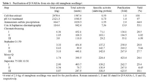
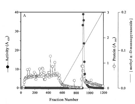
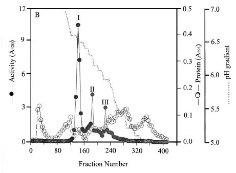
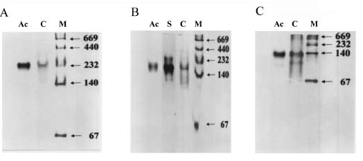
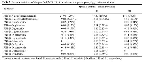
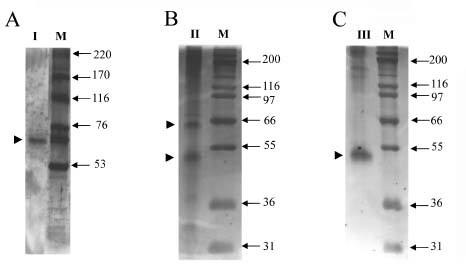
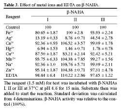
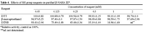
Abstract. Three isoforms of b-N-acetylhexosaminidase (b-NAHA), named b-NAHAs I, II and III, were isolated from six-day-old etiolated mungbean (Vigna radiata) seedlings. b-NAHA I was purified to apparent homogeneity by a procedure involving Con A-Sepharose chromatography, chromatofocusing, and gel filtration. b-NAHAs II and III were highly purified. b-NAHAs I, II and III had molecular masses of 135,127 and 110 kDa, respectively. b-NAHA I was dissociated into a single 67 kDa protein band. II was dissociated into two protein bands corresponding to 60 and 48 kDa, and III was dissociated into a single 48 kDa protein band in SDS-polyacrylamide gel electrophoresis. The results suggest that isoforms I and III are homodimeric enzymes, each comprising two identical subunits with molecular masses of 67 kDa and 48 kDa, respectively, while isoform II is a heterodimeric enzyme, comprising two non-identical subunits with molecular masses of 60 kDa and 48 kDa. All the enzymes were active against para- nitrophenyl-b-N-acetylglucosaminide (PNP-b-N-acetylglucosaminide) and PNP-b-N-galactosaminide. The enzymes were inhibited by 5, 5'-dithiobis (2-nitrobenzoic acid)(DTNB), Ag+, Hg2+, and N, N'-diacetylchitobiose. Km values for isoforms I, II and III were 0.67 mM, 1.04 mM and 1.76 mM, respectively, using PNP-b-N-acetylglucosaminide as a substrate. These three isoforms had acidic pI values (I, 6.3; II, 6.1; and III, 5.9). Their optimal pH in the reaction towards PNP-b-N-acetylglucosaminide was 5.4, 4.7 and 5.7, and optimal temperatures were 65°C, 65°C and 50°C for isoforms I, II and III, respectively.

Keywords: b-N-acetylhexosaminidase; Enzyme purification; Germination; Oligomeric structure; Vigna radiata.

Abbreviations: DTNB, 5,5'-dithiobis-2-nitrobenzoic acid; DTT, dithiothreitol; b-GlcNAcase, N-acetyl-b-D-glucosaminidase; b-GalNAcase, N-acetyl-b-D-galactosaminidase; b-NAHA, N-acetyl-b-D-hexosaminidase; PMSF, phenylmethylsulphonyl fluoride; PNP, p-nitrophenyl.

Introduction

b-N-Acetylglucosaminidase (b-GlcNAcase) (EC3,2,1,30) catalyzes the release of N-aetylglucosaminyl residue from the non-reducing terminus of oligosaccharides. It is also referred to as b-N-acetylhexosaminidase (b-NAHA) because it can also cleave terminal N-acetyl-b-D-galactosaminyl residues from oligosaccharides (Dey and Campillo, 1984; Conzelmann and Sandhoff, 1987). This enzyme occurs widely in animals, microorganisms, and plants and has been implicated in a number of biological processes, including the degradation of glycoproteins (Bahl and Agrawal, 1968; Li and Li, 1970; Neely and Beevers, 1980; Poulton et al., 1985), glycolipids (Fernandes et al., 1997) , and N-glycans (Choi and Gross, 1994). In

plants, b-NAHA is thought to play a vital role during seed germination by cleaving N-acetylglucosaminyl residues from storage glycoproteins which have undergone prior proteolytic degradation. The liberated amino sugars and amino acids are then available for further synthesis of glycoproteins in the developing seedlings. This has been supported by the finding that b-NAHA activities increase during the process of germination (Bahl and Agrawal, 1968; Bouquelet and Spik, 1978; Yi, 1981). A prominent increase in b-NAHA activity was found in the germinating seeds of Phaseolus vulgaris. The specific activity increased 25-fold upon germination (Bahl and Agrawal, 1968). The enzyme may also have a role in the posttranslational trimming of oligosaccharide chains of the storage proteins and lectins deposited in the protein bodies during seed development (Vitale and Chrispeels, 1984). Activities of b-NAHA and chitinase were demonstrated in whole tissues of individual seeds and seedlings using an in situ technique (Hodge et al., 1996). It is believed that chitinase acts as a defense protein in higher plants, protecting the plant from

6Y.C. Chen and W.L. Liu contributed equally to this paper.

*Corresponding author. Phone: +886-2-27899590 ext 251; Fax: +886-2-27858936; E-mail: chingsan@gate.sinica.edu.tw


Botanical Bulletin of Academia Sinica, Vol. 45, 2004

fungal pathogens by degrading chitin, a major component of the cell walls of many fungi, or by inhibiting the spore germination and mycelial growth of fungi (Schlumbaum et al., 1986; Broekaert et al., 1988; Bol et al., 1990). Thus the presence of high b-NAHA activity in plant tissue may also serve to degrade chitin oligomers produced by the plant chitinase system from chitinous pathogen.

We found that the activities of some glycosidases in germinating mungbean seeds rose to a high level between days 4-6. b-NAHA, b-galactosidase, and a-mannosidase showed the highest specific activity (Li et al., 2001). We also found that b-NAHA was not only detected in cotyledons but also in root stem and leaf of six-day-old mungbean seedlings. The occurrence of b-NAHA in plant tissues other than cotyledons has been previously described (Parish, 1975; Hodge et al., 1996). It was suggested that b-NAHA might be more intimately associated with the metabolism and transport of cellular glycoproteins than with reserve proteins (Neely and Beevers, 1980; Vitale and Chrispeels, 1984). b-NAHA has been isolated from a variety of organisms and characterized. However, little is known about the role of this enzyme in plant growth and development. Mungbean seedlings seem to be a good material with which to investigate this role. This paper describes purification and characterization of three isoforms of b-NAHA from germinating mungbean seeds.

Materials and Methods

Plant Materials and Enzyme Source

Mungbean seeds (Vigna radiata VC1628A Sel No. 5) were submerged in distilled water at 25°C for 2 h and surface sterilized with 0.2% hydrogen peroxide, thoroughly washed with distilled water, and germinated in the dark at 28°C. Six-day-old germinated seeds were thoroughly washed with ice-cold H2O and homogenized with Polytron (Kinematica) in three volumes (v/w) of 25 mM sodium acetate (pH 5.0) containing 10 mM 2-mercaptoethanol and 1 mM EDTA. Unless otherwise indicated, all steps and the purification procedure were conducted at 5°C. The resultant slurry was squeezed through three layers of cheese cloth, phenylmethylsulphonyl fluoride (PMSF) was added at a final concentration of 1 mM, and centrifuged at 11,400 g for 30 min. The supernatant was dialyzed against the same buffer solution containing 1 mM PMSF. The dialyzed enzyme solution was referred to as cell-free extract.

Purification of b-N-Acetylhexosaminidase

The cell-free extract was adjusted to a pH of 4.6 to precipitate substantial amounts of contaminating proteins while retaining most of the b-NAHA activity in the supernatant. Solid ammonium sulfate was added to the supernatant to obtain a 0-75% saturated ammonium sulfate fraction (0-75% AS fraction), which contained most of the b-NAHA activity. The 0-75% AS fraction was applied to a Con A affinity column (2.0 cm × 15 cm). A single b-NAHA activity peak was eluted from the Con A-Sepharose 4B col

umn by a linear concentration gradient of 0-0.2 M methyl a-D-mannopyranoside. The fractions containing the b-NAHA activity were pooled and purified by chromatofocusing according to the instruction manual of Pharmacia. Poly buffer Exchanger PBE94 (Pharmacia) column (1.0 cm × 40 cm) was equilibrated with 25 mM imidazole-HCl (pH 7.0) (Buffer A). Poly buffer 74 (Pharmacia) was diluted 8-fold with deionized water and adjusted to pH 5.0 with 6 M HCl (eluent). Forty milliliters of the eluent was introduced to the column to create a pH gradient from 5.0 (top) to 7.0 (bottom). The concentrated enzyme solution (3~4 ml) was dialyzed against the eluent and placed on the column. b-NAHAs I, II, and III were eluted in this order from the column with 440 ml of the eluent. b-NAHAs I, II and III were then purified by gel filtration. The fractions containing b-NAHA I, II or III were pooled, concentrated by lyophilization, and dialyzed against 25 mM sodium acetate (pH 5.0) containing 0.17 M NaCl (Buffer B). This solution was chromatographed on a Sephadex G-150 column (2.6 cm × 90 cm) equilibrated with Buffer B. Fractions of 3 ml each were collected at a flow rate of 0.2 ml/min.

b-NAHAs I and II were further purified by Superdex 75 HR 10/30 gel filtration using a FPLC system. Two ml of concentrated enzyme solution, which had been dialyzed against Buffer B, was applied to a column of Superdex 75 HR 10/30 (2.0 cm × 30 cm) equilibrated with Buffer B. Fractions of 1.5 ml each were collected at a flow rate of 0.2 ml/min.

For further purification of b-NAHA III, fractions containing this enzyme obtained from the Sephadex G-150 gel filtration were first chromatographed on a Mono Q HR 5/5 anion exchange column (Pharmacia) followed by the Superdex 75 HR 10/30 gel filtration. The column (1.0 cm × 5 cm) was equilibrated with Buffer A. b-NAHA III sample dialyzed against Buffer A was loaded on the column. The column, after being washed with Buffer A, was eluted with a linear gradient (60 ml total volume) of 0-0.5 M NaCl in Buffer A. Fractions of 1.5 ml each were collected at a flow rate of 0.5 ml/min.

Enzyme Assays

b-NAHA activity was determined by measuring the release of p-nitrophenol from p-nitrophenyl (PNP)-b-N-acetylglucosaminide according to the method previously described (Bahl and Agrawal, 1968; Agrawal and Bahl, 1968). The reaction mixture contained, in a final volume of 0.5 ml, 50 mM sodium citrate buffer (pH 4.6), 5 mM substrate, and varying amounts of enzyme. The reactions were carried out at 37°C for 10 min. Liberated p-nitrophenol was measured spectrophotometrically at 420 nm after stopping the reaction with addition of 0.7 ml of 0.2 M sodium carbonate. Absorbance (A420) was proportional to the amount of enzyme in the reaction mixture under the assay conditions. The absorbance, A420, was also linear with time within the amounts of enzyme used. One unit of b-NAHA was defined as the amount that hydrolyses 1 µmol of PNP-b-N-acetylglucosaminide per min at pH 4.6 and 37°C. Other


Chen et al. — b-N-acetylhexosaminidases from mungbean seedlings

glycosidase activities were determined by the use of respective PNP-glycoside. The reaction conditions were essentially the same as described above. Specific activity was expressed as units of enzymatic activity per mg protein.

Characterization of b-N-Acetylhexosaminidase

The molecular mass of the native b-NAHAs were determined by gel filtration on a Sephacryl S-200 column (1.6 cm × 90 cm). The column was calibrated with Blue Dextran and standard proteins (ribonuclease A, chymotrypsinogen A, ovalbumin, albumin, aldolase, catalase, ferritin, and thyroglobulin) (Sigma). The molecular mass of the denatured enzymes was estimated by SDS-PAGE. The pH Optima of the purified enzymes against PNP-b-N-acetylglucosaminide was determined in McII-Valine buffer covering a pH range of 3-6 and Na-phosphate buffer covering a pH range of 7-8. The isoelectric point of the purified enzyme was estimated from the elution profile of chromatofocusing. In the inhibition study of b-NAHA activity by sugars, sugar derivatives, metallic ions, reducing agent, and DTNB, b-NAHA was preincubated for 10 min at 37°C with each compound or metallic ion at varying concentrations, then assayed as normal.

Analytical Methods

Purified b-NAHAs were analyzed by electrophoresis in non-denaturing conditions using 4% acrylamide stacking gel and 7.5% acrylamide separating gel (Hames, 1981) and SDS-PAGE (Laemmli, 1970). Activity staining of b-NAHA was performed as in the method previously described (Loida et al., 1979) using 5-bromo-4-chloro-3-indole-N-acetyl-glucosaminide and nitro blue tetrazolium as

substrate. Protein concentration was determined by the method of Bradford (1976) using bovine serum albumin as a standard.

Results and Discussion

Purification of b-N-cetylhexosaminidases

Two kg of 6-d-old mungbean seedlings germinated from 280 g of dry mungbean seeds were used for purification of b-NAHAs by a procedure involving pH 4.6 treatment, ammonium sulfate fractionation (0-75%), Con A-Sepharose affinity chromatography, chromatofocusing and gel filtration. The results are summarized in Table 1. After the removal of substantial amounts of contaminating proteins (about 77.7% of total proteins) by pH 4.6 treatment and ammonium sulfate fractionation, the 0-75% AS fraction was purified by a Con A-Sepharose column. A protein peak having b-GlcNAcase activity was eluted from the column (Figure 1A). Factions of this peak were collected and subjected to chromatofocusing. b-NAHAs I, II and III were eluted in this order from the chromatofocusing column (Figure 1B). b-NAHAs I was further purified to apparent homogeneity by Superdex 75 HR 10/30 gel filtration as analyzed on a 7.5% native-PAGE (Figure 2A) while b-NAHAs II after being purified with the same column was still contaminated with a faint contaminating protein band (silver staining) (Figure 2B). b-NAHA III collected from the chromatofocusing fractions was purified to a highly purified state by Mono Q HR 5/5 anion exchange column chromatography followed by gel filtration (data not shown). The purified b-NAHA III showed a major protein band corresponding to an activity stained band and a very faint contaminating protein band running ahead of the major b-


Botanical Bulletin of Academia Sinica, Vol. 45, 2004

Figure 1. Elution profiles of b-NAHAs on Con A-Sepharose column and chromatofocusing. The ammonium sulfate-precipitated enzyme sample was applied to a Con A-Sepharose column (A). The b-NAHAs adsorbed on the Con A-Sepharose column and eluted from the column was further purified by chromatofocusing (B). Experimental details are given in the Materials and Methods. Protein is determined by the method of Bradford and is expressed as absorbance at 595 nm (¡). b-NAHA activity (l) was determined using N-acetyl-b-D-glucosaminide as substrate and is expressed as absorbance at 420 nm. Methyl a-D-mannopyranoside gradient (——)and pH gradient (-----) were used to elute b-NAHAs from the Con A-Sepharose (A) and chromatofocusing (B) columns, respectively.

Figure 2. 7.5% native PAGE of purified b-NAHAs I, II and III. b-NAHAs I, II and III purified with the last step gel filtration on Superdex 75 HR 10/30 were individually separated on nondenaturing PAGE and stained with activity staining and Coomassie Blue G-250. Isoform II was also stained with silver nitrate. The results of isoforms I, II and III are shown in A, B and C, respectively. Molecular mass markers (M) from top:thyroglobulin (669 kDa), ferritin (440 kDa), catalase (232 kDa), lactate dehydrogenase (140 kDa) and bovine serum albumin (67 kDa). Ac, S and C stand for stained with activity staining, silver nitrate and Coomassie Blue G-250, respectively.

NAHA III band (Figure 2C). b-NAHAs I, II and III were purified 263-, 247- and 624-fold, respectively (Table 1). After purification, these three enzymes retained the activities of b-GlcNAcase and b-GalNAcase, but were devoid of activity towards eleven other PNP-glycosides (1% or less) initially present in the cell-free extract (Table 2). This is indicative of the high purity of these b-NAHAs. Most previously reported b-GlcNAcases also displayed b-GalNAcase activity (Harley and Beevers, 1987; Gers-Barlag et al., 1988; Choi and Gross, 1994), and b-GlcNAcase is

hence also referred to as b-NAHA. The three isoforms of mungbean b-GlcNAcase described in this study also exhibited b-GalNAcase activity (Table 2), suggesting that these enzymes belong to the general category of b-NAHA. b-NAHA appeared to occur as multiple isoforms in mungbean seedlings. When cell-free extract prepared from 6-d-germinated mungbean seedlings was separated on an isoelectrofocusing gel and the gel was subjected to activity staining for b-GlcNAcase, three major and five minor bands with pI roughly between pH 6.3 and 5.0 were ob


Chen et al. — b-N-acetylhexosaminidases from mungbean seedlings

served (data not shown). Thus b-NAHAs I, II and III might represent the three major b-NAHAs in the cell-free extract. It was found that the total activity of b-NAHA III increased from 110.9 units to 440.1 units after Sephadex G-150 gel filtration (Table 1). A possible explanation of this finding is that some unknown inhibitor presented in b-NAHA III fractions might have been separated from b-NAHA III by the Sephadex G-150 column, resulting in a four fold increase in b-NAHA activity.

Characterization of b-NAHAs

The molecular masses of the three b-NAHAs were determined by gel filtration on Sephacryl S-200. The molecular masses of b-NAHAs I, II and III calculated from the elution volume versus the logarithm of the molecular weights of eight proteins of known molecular weights (data not shown) were 135, 127 and 110 kDa, respectively. SDS-PAGE of b-NAHAs I, II and III showed that b-NHAHs I and III were dissociated into a single protein band corresponding to 67 and 48 kDa, respectivety, and isoform II was dissociated into two protein bands corresponding to 60 and 48 kDa (Figure 3). These results suggest that isoforms I and III are homodimers, each comprising two identical subunits with molecular masses of 67 kDa and 48 kDa, respectively, while isoform II is a heterodimeric enzyme, comprising two non-identical subunits with molecular masses of 60 kDa and 48 kDa. The molecular masses of the two subunits of isoforms II and III did not exactly match their corresponding native enzymes. This may be attributed to the glycoprotein nature of isoforms II and III because the accuracy of molecular weight determination on a gel filtration column could be affected by the carbohydrate moiety of glycoproteins. It was previously reported that two active forms of b-GlcNAcase with molecular masses of 90 kDa and 45 kDa were present in the crude extract of mature seeds of mungbean (Dey, 1984). The author suggested that the two forms of b-GlcNAcase may have a monomer/dimmer relationship. b-NAHA III ap

peared similar to the mungbean seed b-GlcNAcase described by Dey (1984) in terms of molecular mass and oligomeric structure. The molecular masses of mungbean b-NAHAs I, II and III also resemble those reported for pinto bean (112 kDa) (Agrawal and Bahl, 1972), castor bean (120 kDa) (Harley and Beevers, 1985), jack bean (110 kDa) (Li and Li, 1970), malted barley (92 kDa) (Mitchell et al., 1976), germinated cotton seeds (125 kDa) (Yi, 1981), and cabbage (150 kDa) (Chang et al., 1998). The enzymes from pea seeds and apple fruit cortical tissue seem exceptions with molecular masses of 210 kDa (Harley and Beevers, 1987) and 236 kDa (Choi and Gross, 1994), respectively.

Different oligomeric structures of b-NAHA have been described. The enzyme from pinto bean is composed of two identical 53 kDa subunits (Agrawal and Bahl, 1972) while pea b-NAHA-II appears to have two pairs of heterogenous subunits of 62 and 64 kDa. These are not held together by disulfide bond, but sulfhydryl groups are important for catalysis (Harley and Beevers, 1987). The GlcNAcase from golden delicious apples was reported to have a molecular mass of 236 kDa composed of eight similar subunits with molecular mass of 29 kDa (Choi and Gross, 1994). The molecular mass of b-NAHA from cabbage is 150 kDa. Three non-identical subunits with molecular masses of 64, 57 and 51 kDa were observed as determined by SDS-PAGE (Chang et al., 1998). Four molecular forms of b-NAHA isolated from germinating seeds of fenugreek were shown to have molecular masses of 84, 72, 180 and 150 kDa for forms I, II, III and IV, respectively. The results of SDS-PAGE suggested that the four b-NAHAs may be polymers formed by the association of between three and six subunits of identical molecular weights. Thus the oligomeric structure of mungbean b-NAHA described herein is unique in that b-NAHAs I and III are homodimers, resembling the pinto bean b-NAHA, which also comprises two identical subunits (Agrawal and Bahl, 1972). b-NAHA II is somewhat similar to the pea b-NAHA-II in that both enzymes have two heterogenous subunits (Harley and Beevers,


Botanical Bulletin of Academia Sinica, Vol. 45, 2004

Figure 3. SDS-PAGE of b-NAHAs. The purified b-NAHAs I, II and III were individually separated on SDS-PAGE and stained with silver nitrate. The results are shown in A, B and C, respectively. Molecular mass markers (M) used for b-NAHA I from top are myosin (220 kDa), a-2-macroglobulin (170 kDa), b-galactosidase (116 kDa), transferrin (76 kDa) and glutamate dehydrogenase (53 kDa)(Amersham Biosciences). Molecular mass markers used for b-NAHAs II and III from top are myosin (200 kDa), b-galactosidase (116 kDa), phosphorylase b (97 kDa), bovine serum albumin (66 kDa), glutamate dehydrogenase (55 kDa), lactate dehydrogenase (36 kDa) and carbonic anhydrase (31 kDa) (Invitrogen). Arrowheads indicate the positions of subunits.

1987). Nevertheless, the molecular mass (210 kDa) of the native pea b-NAHA-II is much larger than that of the mungbean b-NAHA II (137 kDa).

Effects of metal ions, sulfhydryl reagents, sugars, and sugar derivatives were studied using PNP-b-N-acetylglucosaminide as substrate. All of the three b-NAHAs were inhibited by 1.5 mM of Ag+ and Hg2+, and slightly inhibited by Cu2+ (Table 3). Similar inhibitory effects of Ag+ and Hg2+ were previously described with b-NAHA from jack bean meal (Li and Li, 1970) and the four molecular forms of b-NAHA from fenugreek (Bouquelet and Spik, 1978). Dithiothreitol and b-mercaptoethanol at concentrations from 0.05 to 2 mM did not inhibit b-NAHA III while DTNB significantly inhibited the activity of this enzyme (Table 4). DTNB was also inhibitory to b-NAHAs I and II (data not shown). The results suggest that the sulfhydryl group in the enzyme is important for enzymatic activity. The sulfhydryl groups of pea b-NAHA-II have been characterized as important for catalysis (Harley and


Chen et al. — b-N-acetylhexosaminidases from mungbean seedlings

Beevers, 1987). The three isoforms were also inhibited by N, N'-diacetylchitobiose (Table 5), a product of chitin degradation by exochitinase (Robbins et al., 1988). As mentioned in the Introduction, chitinase is generally recognized to act as a defense protein in higher plants, protecting the plant from fungal pathogen by degrading chitin, a major component of the cell walls of many fungi, and the chitin oligomers produced by chitinase are further degraded by down stream hydrolytic enzymes such as b-NAHA or chitobiase (Hodge et al., 1996). We also found that the activity of chitinase in germinating mungbean seeds increased to a high level around day 4 (data not shown). We therefore suggest that b-NAHAs plays a defensive role in coordination with chitinase during mungbean germination based on the observation that the activity of b-NAHA in germinating mungbean seeds also increased to a high level between days 4-6 (Li et al., 2001)

Kinetic parameters Km and Vmax were determined by double reciplocal plot using PNP-N-acetyl-b-D-glucosaminide as substrate. Km values for isoforms I, II and III were 0.67 mM, 1.04 mM and 1.76 mM, respectively (data not shown). These values were comparable with those of other plant b-NAHAs previously described (Bahl and Agrawal, 1968; Li and Li, 1970; Bouquelet and Spik, 1978; Poulton et al., 1985; Gers-Barlag et al., 1988; Giordani et al., 1992; Choi and Gross, 1994).

pI values for the three isoforms determined by chromatofocusing were 6.3, 6.1 and 5.9 for isoforms I, II and III, respectively (data not shown). The four molecular forms of b-NAHA from fenugreek also have acidic pI values. Forms I, II, III and IV have pI values of 6.78, 6.3, 4.9 and 4.65, respectively (Bouquelet and Spik, 1978). The pH optima for isoforms I, II and III were 5.4, 4.7, and 5.7 (Figure 4), and optimal temperatures were 65°C, 65°C and 50°C, respectively (data not shown). The pH optimum of jack bean b-NAHA is between 3.5 and 4.0 (Li and Li, 1970) while the b-GlcNAcase from germinating cotton seeds has pH optimum of 5.6.

Assay of b-NAHA activity in various tissues of mungbean seedlings revealed that b-NAHAs were most abundant in cotyledons, followed by roots, stems, and leaves. It was also found that the activity of chitinase in germinating mungbean seeds increased to a high level

Figure 4. Effect of pH on b-NAHAs. The purified b-NAHAs I (q), II (¡), III (l) were assayed at various pH by the method given in Materials and Methods. Cell-free extract (o) was also assayed.

around day 4 (data not shown). Chitinases are ubiquitous in higher plants (Collinge et al., 1993). It was reported that endochitinase activity increased as the seedlings of Eucalyptus pilularis developed and was greatest in the roots. The roots of E. pilularis also contained the highest levels of b-GlcNAcase activity (Hodge et al., 1996). Chitinase is believed to act as a defense protein, as mentioned above. Our results, together with the previous findings, suggest that multiple forms of b-NAHA located in various tissues may play different physiological roles in response to various stages of plant growth and development and biotic stresses. Whereas the hard seed coat provides an effective physical defense against phytopathogens,


Botanical Bulletin of Academia Sinica, Vol. 45, 2004

seedlings are vulnerable to pathogen attack. Thus b-NAHAs in mungbean seedling play a likely role in degradation of storage glycoproteins and are also involved in the hydrolysis of N,N'-diacetylchitobiose produced by exochitinase from chitinous pathogen during the germination of mungbean seeds.

Acknowledgment. We thank the Asian Vegetable and Development Center and Tainan District Agricultural Improvement Station for providing the seeds of Vigna radiata VC1628A Sel No.5 for this research. This work was supported by the National Science Council of the Republic of China and the Institute of Botany, Academia Sinica.

Literature Cited

Agrawal, K.M.L. and O.P. Bahl. 1968. Glycosidases of Phaseolus vulgaris. II. Isolation and general properties. J. Biol. Chem. 243: 103-111.

Agrawal, K.M.L. and O.P. Bahl. 1972. a-Galactosidase, b-glucosidase, b-N-acetylglucosaminidase, and a-mannosidase from pinto beans (Phaseolus vulgaris). Methods Enzymol. 28: 720-728.

Bahl, O.P. and K.M.L. Agrawal. 1968. Glycosidases of Phaseolus vulgaris 1. Isolation and characterization of b-N-acetylglucosaminidase. J. Biol. Chem. 243: 98-102.

Bol, J.F., H.J.M. Linthorst, and B.J.C. Cornelissen. 1990. Plant pathogenesis-related proteins induced by virus infection. Annu. Rev. Phytopathol. 28: 113-138.

Bouquelet, S. and G. Spik. 1978. Properties of four molecular forms of N-acetyl-b-D-hexosaminidase isolated from germinating seeds of fenugreek (Trigonella foenum graecum). Eur. J. Biochem. 84: 551-559.

Bradford, M.M. 1976. A rapid and sensitive method for the quantification of microgram quantities of protein utilizing the principles of protein-dye binding. Anal. Biochem. 72: 248-254.

Broekaert, W.F., J.V. Parijs, A.K. Allen, and W.J. Peumans. 1988. Comparison of some molecular, enzymatic and antifungal properties of chitinases from thom-apple, tobacco and wheat. Physiol. Mol. Plant Phathol. 33: 319-331.

Chang, C.T., F.P. Young, M.H. Chang, and H.Y. Sung. 1998. Purification and properties of b-N-acetylhexosaminidase from cabbage. Biochem. Mol. Biol. Int. 45: 371-380.

Choi, S.Y. and K.C. Gross. 1994. N-Acetyl-b-D-glucosaminidase from "golden delicious" apples. Phytochemistry 36: 1-6.

Collinge, D.B., K.M. Kragh, J.D. Mikkelsen, K.K. Nielsen, U. Rasmussen, and K. Vad. 1993. Plant chitinases. Plant J. 3: 31-40.

Conzelmann, E. and K. Sandhoff. 1987. Glycolipid and glycoprotein degradation. In A. Meister (ed.), Adv. Enzymol. vol. 60. John Wiley & Sons, New York, Chichester, Brisbane, Toronto, Singapore, pp. 89-216.

Dey, P.M. 1984. Occurrence of glycoprotein glycosidases in mature seeds of mungbean (Vigna radiata). Phytochemistry 23: 257-260.

Dey, P.M. and E.D. Campillo. 1984. Biochemistry of the multiforms of glycosidases in plants. In A. Meister (ed.), Adv. Enzymol. Vol. 54. John Wiley and sons, New York, Chichester, Brisbane, Toronto, Singapore, pp. 141-249.

Fernandes, M.J.G., S. Yew, D. Lecherc, B. Henrissat, C.E. Vorgias, R.A. Gravel, P. Hechtman, and F. Kaplan. 1997. Identification of candidate active site residues in lysosomal b-hexosaminidase A. J. Biol. Chem. 272: 814-820.

Gers-Barlag, H., I. Bartz, and H. Rüdiger. 1988. b-N-Acetylhexosaminidase from soybean. Phytochemistry 27: 3739-3741.

Giordani, R., S. Benyahia, M. Teissere, and G. Noat. 1992. Purification and properties of N-acetyl-b-D-glucosaminidase from Hevea brasiliensis latex. Plant Sci. 84: 25-34.

Hames, B.D. 1981. An introduction to Polyacrylamide Gel Electrophoresis of Proteins. A Practical Approach, pp. 1-86.

Harley, S.M. and H. Beevers. 1985. Characterization and partial purification of three glycosidases from castor bean endosperm. Phytochemistry 24: 1459-1464.

Harley, S.M. and L. Beevers. 1987. Isozymes of b-N-acetylhexosaminidase from pea seeds (Pisum sativum L.). Plant Physiol. 85: 1118-1122.

Hodge, A., I.J. Alexander, and G.W. Gooday. 1996. Measurement in situ of chitinase and b-N-acetylglucosaminidae activities in germinating-seeds of Pinus sylvestris and Eucalyptus pilularis. Plant Physiol. Biochem. 34: 301-306.

Laemmli, U.K. 1970. Cleavage of structure proteins during the assembly of the head of the bacteriophage T4. Nature 227: 680-685.

Li, S.C., J.W. Han, K.C. Chen, and C.S. Chen. 2001. Purification and characterization of isoforms of b-galactosidases in mungbean seedlings. Phytochemistry 57: 349-359.

Li, S.C. and Y.T. Li. 1970. Studies on the glycosidases of jack bean meal III. crystallization and properties of b-N-acetylhexosaminidase. J. Biol. Chem. 245: 5153-5160.

Loida, E., R. Grossrau, and T.H. Schiebler. 1979. Enzyme Histochemistry. Springer-Verlag, Berlin, Heidelberg, Germany, pp. 157-159.

Mitchell, E.D., C.W. Houston, and S.B. Latimer. 1976. Purification and properties of a b-N-acetylaminoglucohydrolase from malted barley. Phytochemistry 15: 1869-1871.

Neely, R.S. and L. Beevers. 1980. Glycosidases from cotyledons of Pisum sativum L. J. Exp. Bot. 31: 299-312.

Parish, R.W. 1975. The lysosome-concept in plants II. Location of acid hydrolases in maize root tips. Planta 123: 15-31.

Poulton, J.E., M.A. Thomas, K.K. Ottwell, and S.J. MeCormick. 1985. Partial purification and characterization of a b-N-acetylhexosaminidase from black cherry (Prunus serotina ehrh) seeds. Plant Sci. 42: 107-114.

Robbins, P.W., C. Albright, and B. Benfield. 1988. Cloning and expression of a Streptomyces plicatus chitinase (chitinase-63) in Escherichia coli. J. Biol. Chem. 263: 443-447.

Schlumbaum, A., F. Mauch, U. Vogeli, and T. Boller. 1986. Plant chitinases are potent inhibitors of fungal growth. Nature 324: 365-367.

Vitale, A. and M.J. Chrispeels. 1984. Transient N-acetylglucosamine in the biosynthesis of phytohemagglutinin: attachment in the Golgi apparatus and removal in protein bodies. J. Cell Biol. 99: 133-140.

Yi, C.K. 1981. Increase in b-N-acetylglucosaminidase activity during germination of cotton seeds. Plant Physiol. 67: 68-73.


Chen et al. — b-N-acetylhexosaminidases from mungbean seedlings