Bot. Bull. Acad. Sin. (2004) 45: 307-313

Thomas and Puthur — Regeneration in Kigelia pinnata L.

Thidiazuron induced high frequency shoot organogenesis in callus from Kigelia pinnata L.

T. Dennis THOMAS* and Jos T. PUTHUR

Postgraduate and Research Department of Botany, St. Thomas College, Pala, Arunapuram (P.O), Kottayam (Dt.), PIN- 686 574, Kerala, India

(Received December 24, 2003; Accepted June 29, 2004)

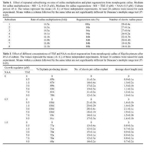
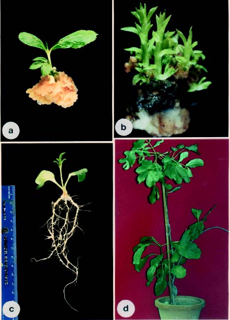
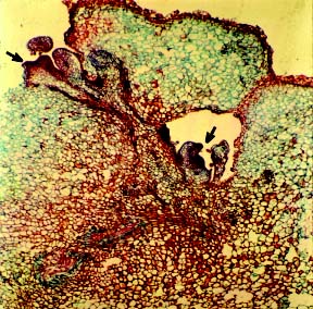
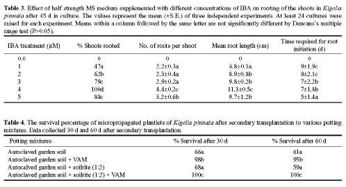
Abstract. An efficient regeneration system was developed for Kigelia pinnata L., a multipurpose tree belonging to the family Bignoniaceae. The nodal segments were cultured in vitro, and the optimum concentrations of plant growth regulators for callus induction were determined. The friable organogenic calli were derived from the basal cut end of the nodal segments. The highest yield of morphogenic callus (100%) was observed when nodal segments were cultured on Murashige and Skoog (MS) medium supplemented with 3 mM 2,4 dichlorophenoxyacetic acid (2, 4-D). The morphogenic callus maintained high regeneration during the first four subcultures in the callus induction medium. The maximum shoots (28/culture) were regenerated at the highest frequency of 100% when 3 mM thidiazuron (N-phenyl N' 1,2,3-thidiazol-5-yl urea) (TDZ) and 0.5 mM naphthaleneacetic acid (NAA) were added to MS medium. The emergence of multiple shoots from the calli was histologically documented. The regenerated shoots showed maximum rooting on ˝ MS medium containing 4 mM indole-3- butyric acid (IBA). The effect of vesicular arbuscular mycorrhizae (VAM) association in averting the transplantation shock was tested and proved to be highly beneficial, giving a 100% survival rate after 60 d of transplantation. This efficient plant regeneration system provides a foundation for generating transgenic plants of this multipurpose tree.

Keywords: Kigelia pinnata L.; Medicinal plant; Plant regeneration; Thidiazuron; Tissue culture; Vesicular arbuscular mycorrhizae.

Abbreviations: 2, 4-D, 2,4 dichlorophenoxyacetic acid; IBA, Indole-3- butyric acid; NAA, Naphthaleneacetic acid; TDZ, thidiazuron (N-phenyl N' 1,2,3-thidiazol-5-yl urea); VAM, vesicular arbuscular mycorrhizae.

Introduction

Forest species predominate in temperate and equatorial zones, and the wood produced by the trees is the most abundant material on the earth's surface (Gammie, 1981). Wood provides fuel for most of the population of the world, particularly in developing countries, and is a leading industrial raw material. Despite its economic importance, the production of wood is threatened by population growth, desertification, industrial development, and attack by numerous parasites. The classical conservation techniques such as crossing, sexual and somatic hybridization, and breeding give a genetic blind mixture. These techniques are limited by the sterility of the descents, the genetic barrier between species, and the long life cycle of certain trees (Sederoff, 1995). Plant tissue culture offers many unconventional techniques for plant improvement. While tissue culture technology has been developed for the mass propagation of several fruit tree species, several other tree species are lagging behind due to a recalcitrant nature to in vitro techniques.

Kigelia pinnata, a fast growing, multipurpose tree used for ornamental and roadside planting, belongs to the fam

ily Bignoniaceae. Several parts of the plant are employed for medicinal purposes by certain aboriginal people. Traditional healers in India have used various parts of this plant to treat a wide range of skin ailments, from relatively mild complaints, such as fungal infections, boils, and psoriasis, to the more serious diseases like leprosy, syphilis, and skin cancer. Other medicinal applications include the treatment of dysentery, ringworm, tapeworm, post-partum haemorrhaging, malaria, diabetes, pneumonia, and toothache. An antimalarial compound known as lapachol has been extracted from the root of K. pinnata (Binutu et al., 1996). Another compound obtained from the wood, quinone, shows antimalarial activity against drug resistant strains of Plasmodium falciparum superior to chloroquine and quinine (Carvalho et al., 1988). Conventionally, K. pinnata reproduces via viable seeds, but the low percentage of seed viability limits its natural propagation. Hence alternative methods like in vitro techniques could be used to propagate this plant and thereby multiply elite genotypes. Micropropagation has many advantages over conventional propagation of fruit trees (Stushnoff and Fear, 1985) and is important for the regeneration following transformation (Ainsley et al., 2000) and cryopreservation (Channuntapipat et al., 2000). Thidiazuron (TDZ), a urea-derived cytokinin, is a potent cytokinin for woody plant tissue culture (Huetteman and

*Corresponding author. Telephone: +91-04822-212316,17; Fax: +91-04822-216313; E-mail: den_thuruthiyil@yahoo.com


Botanical Bulletin of Academia Sinica, Vol. 45, 2004

Preece, 1993) and is extensively used for the induction of shoot regeneration in several plant species (Li et al., 2000; Mohan and Krishnamurthy, 2002; Liu et al., 2003). The present study was carried out to standardize an efficient regeneration system for K. pinnata L. using thidiazuron.

Materials and Methods

Plant Material for Callus Initiation

Callus cultures of K. pinnata were initiated from nodal segments of healthy disease free plants growing near our college campus. The nodal segments were washed in 5% Teepol solution for 10-15 min followed by disinfecting in 0.1% mercuric chloride (HgCl2) solution for 5 min in sterilized autoclaved bottles and finally washed three times with sterile-distilled water. Since the use of sodium hypochlorite and bromine water did not prevent contamination, mercuric chloride was used as sterilizing agent throughout the experiment. Utmost care was taken while using mercuric chloride, and autoclaved hand gloves were employed while sterilizing the explant. The surface sterilized nodal segments were cultured on MS (Murashige and Skoog, 1962) medium supplemented with various concentrations (1-7 mM) of 2, 4-D (Sigma-Aldrich, U.S.A.) in a test for callus induction. Callus was subcultured onto fresh medium (MS + 3 mM 2, 4-D) every 45 d.

Induction and Development of Multiple Shoots

For multiple shoot induction, calli (about 8 mm ´ 8 mm in size) were transferred to MS medium supplemented with various concentrations (0.5 - 9 mM) of TDZ (Sigma-Aldrich, USA) alone or in combination with NAA (Sigma-Aldrich, USA) (0.5 and 1 mM). The % cultures responding, mean shoot length, and the number of shoots per tube were calculated 45 d after callus culture.

Rooting and Transplantation

Shoot clusters with a shoot 1.5 cm or longer were transferred to ˝ MS solid medium supplemented with IBA (Sigma-Aldrich, USA). The rooted plants were taken out of the tubes, and the agar was gently removed under running tap water. The shoots were transferred to glass bottles containing a mixture of autoclaved vermiculate and sand (2:1) moistened with MS basal liquid medium and were grown at 25±2°C under a 16-h photoperiod. This is referred to in the Results and Discussion as primary transplantation. After four weeks the individual plantlets were transferred to plastic pots (25 cm diameter) containing potting mixture and acclimatized under greenhouse conditions for 60 d. Four different potting mixtures were used for acclimatization. They were autoclaved garden soil; autoclaved garden soil and VAM; autoclaved garden soil and soil rite (1:2); autoclaved garden soil, soil rite (1:2) and VAM. These plantlets were transferred to greenhouse with 85% humidity and a temperature of 28±20°C. This is referred to in the Results and Discussion as secondary transplantation.

VAM Inoculation

Spore of Glomus fasciculatum and Glomus macrocarpum were isolated from the soil obtained from the rhizosphere of Leucaena leucocephala growing in the college campus. Sudan grass (Sorghum halepense) grown in flat earthen pots was inoculated with these spores. Both species of Glomus were multiplied and maintained independently. The rhizosphere soil of the 6-month-old pot culture of Sudan grass was used as inoculum of these VAM fungal species. Each gram of this inoculum consisted of 30-40 spores plus hyphae, arbuscules, and vesicles of fungus, identified using the standard staining techniques of Phillips and Hayman (1970), plus root segments of Sudan grass and soil particles. For establishment of mycorrhizal association with plantlets of K. pinnata, about 1 g of inoculum of either G. fasciculatum or G. macrocarpum was applied below the roots, during secondary transplantation (Puthur et al., 1998; Sharmila et al., 2000)

Culture Conditions

All the media were fortified with 20 g/l sucrose (Sigma-Aldrich, USA) and gelled with 0.8% agar (Sigma-Aldrich, USA), and the pH was adjusted to 5.8 after adding the growth regulators. The media were steam sterilized in an autoclave under 1.5 kg/cm2 and 121°C for 15 min. All the cultures were grown at 25±2°C under a 16-h photoperiod supplied by two Philips TL 40W fluorescent tubes. At least twenty-four cultures were raised for each treatment, and all experiments were performed three times. Analysis of variance and Duncan's multiple range test were used for comparison among treatment means.

Histological Studies

The calli with multiple shoots were fixed for 24 h in FAA (formalin + glacial acetic acid + 70% ethanol in a 1:1:18 ratio by volume). Following dehydration in tertiary butanol the material was infiltrated and embedded in paraffin wax (E-Merck) (Johansen, 1940). The paraffin blocks were mounted on wooden stubs, and 8 mm thick sections were cut using a Spencer microtome (USA) attached with a steel knife. The sections were mounted on microslides, dewaxed, and double stained with safranin (1%) and astra blue (1%) (Johansen, 1940).

Results and Discussion

Callus Induction

When nodal segments were cultured on callus induction medium (i.e. 2, 4-D) callus growth was initiated from the basal cut end of the nodal segments in about four d, and rapid growth followed for the majority of the explants. Pale yellow friable morphogenic calli were well developed 45 d after nodal segments culture (Figure 1a). The rate of callus induction and the percentage of cultures responding varied depending up on the 2, 4-D concentrations applied. 2, 4-D in the range of 1-7 mM alone produced pale yellow calli for each concentration tested, but based on


Thomas and Puthur — Regeneration in Kigelia pinnata L.

the rate of callus induction and the percentage of cultures responding, 3 mM 2, 4-D was optimum. Above or below this concentration the rate of callus induction or the percentage of cultures responding decreased (data not shown). Hence further experiments were carried out with 3 mM 2, 4-D.

For detailed experiments, maintaining callus with a high regeneration ability for a prolonged period is essential. Moreover, most of the transformation procedures have relied on inducing long term morphogenic potential after repeated subculture, and this is critical for successful transformation. With this goal in mind the callus was multiplied on 3 mM 2, 4-D and maintained a high regeneration rate from 1-8 subcultures on this medium. The multiplica

tion rate was high with an average 16.3 fold in the first four subcultures and significantly higher than that in subsequent subcultures (Table 1). The multiplication rate was maintained at approximately an 11 fold level for subsequent subcultures. No significant differences in regeneration rates or the number of plants generated per callus piece appeared among the first four subcultures. From the fifth subculture onwards regeneration rates and the number of plants generated per callus piece showed significant differences. Although the callus maintains its regeneration capacity for a longer period, the available literature suggests that prolonged subculturing may lead to a higher frequency of mutants especially in higher concentrations of 2, 4-D (Choi et al., 2001).


Botanical Bulletin of Academia Sinica, Vol. 45, 2004

Shoot Regeneration

The morphogenic callus generated from the nodal segments was transferred to various concentrations of TDZ alone or in combination with NAA (Table 2). Shoot initiation was observed one week after callus culture. Often several shoots developed from one callus mass (Figure 1b). The optimum response in terms of percentage of explants producing shoots and the highest number of shoot buds per explant was recorded on MS medium supplemented with TDZ (3 mM) and NAA (0.5 mM). On this medium 100% cultures responded with an average 28 shoots per culture. The regenerated shoots attained a height of about 2 cm in about 45 d of callus culture. TDZ, a synthetic phenylurea, is considered to be one of the most active cytokinins for shoot induction in plant tissue culture (Huetteman and Preece, 1993; Murthy et al., 1998). TDZ-induced shoot regeneration from different explants of many recalcitrant spe

cies as well as from medicinal plants has been reported (Bhagwat et al., 1996; Seneviratne and Flagmann, 1996; Murthy et al., 1998; Pelah et al., 2002; Schween and Schwenkel, 2002; Liu et al., 2003; Mithila et al., 2003; Thomas, 2003). Several reports suggest that TDZ results in shoot regeneration better than other cytokinins (Barna and Wakhlu, 1995; Thomas, 2003). TDZ-induced morphogenesis probably depends on the levels of endogenous growth regulators, and TDZ modulates the endogenous auxin levels (Murthy et al., 1995; Hutchinson and Saxena, 1996). In the present study TDZ in combination with NAA gave better results than TDZ alone. Hence, the synergistic combination of auxin and cytokinin promoted shoot regeneration. The promoting effect of auxin and cytokinin combinations on organogenic differentiation has been well established in several systems (Lisowska and Wysonkinska, 2000; Pereira et al., 2000; Pretto and Santarém, 2000; Xie and Hong, 2001; Koroch et al., 2002).

Figure 1. Different stages in the shoot organogenesis and complete plant regeneration in Kigelia pinnata. a, A nodal segment culture taken out from MS medium supplemented with 2, 4-D (3 mM) 45 d after culture. Luxuriantly growing morphogenic friable calli formed at the basal cut end of the nodal segment (´1.2); b, High frequency in vitro callus regeneration on MS medium supplemented with TDZ (3 mM) and NAA (0.5 mM). Photographs were taken 45 d after culture. Well developed, healthy shoots emerged from the calli (´3.2); c, Rooting of regenerated shoots ˝ MS medium with 4 mM IBA for 45 d. Note the long healthy roots with root hairs (´1.8); d, A one-year-old acclimatized plant growing in the green house. The plant developed two new branches (´0.1).


Thomas and Puthur — Regeneration in Kigelia pinnata L.

Rooting

Root differentiation was observed after about 1 week of culture. The optimal medium for rooting contained MS + IBA (4 mM), on which a maximum of 100% shoots rooted with an average 4.4 roots per shoot and an average root length of 11.3 cm (Table 3; Figure 1c). Complete plants were obtained 45 d after the regenerated plantlets were transferred to this medium. IBA is the most commonly used auxin for root induction. The superior role of IBA in root induction has been reported in several plants (Fracaro and Echeverrigaray, 2001; Abrie and Van Staden, 2001; Thomas, 2003).

Transplantation and Acclimatization of Regenerated Shoots

Primary transplantation was carried out in order to increase the survival percentage. From earlier trials we found that survival of transplants improved by 20-30% if transplantation was carried out in two steps. In the secondary transplantation, plantlets were transplanted to four different potting mixtures (Table 4). Of these, the potting mixture containing autoclaved garden soil, soil rite, and VAM showed maximum response (100%), in terms of survival percentage after 60 d of transplantation. The medium containing autoclaved garden soil, and VAM was also found suitable for transplantation, with 95% survival (Table 4). After 30 d the emergence of new leaves was observed. The better survival rate of VAM inoculated plants may be due to the beneficial physiological features imparted to the plant by the symbiotically associated VAM fungi. The beneficial physiological features of these plants include, development of a superior root system, increase in water conducting capacity, increase in phosphorous uptake, enhanced nitrogen levels, increased photosynthetic

efficiency, aversion of possible attack by soil born pathogens, enhanced uptake of micro and immobile nutrients, and alleviated environmental stress (Puthur et al., 1998; Sharmila et al., 2000). Plants transferred to the greenhouse have been established well in the soil (Figure 1d).

Histology

The histological analysis of the regenerating calli clearly showed that the shoot buds had emerged from the periph

Figure 2. Histological analysis of the regenerating callus. A section of a 15-d-old callus showing initiation of shoot (arrow marked) from the peripheral meristematic tissue (´40).


Botanical Bulletin of Academia Sinica, Vol. 45, 2004

Koroch, A., H.R. Juliani, J. Kapteyn, and J.E. Simon. 2002. In vitro regeneration of Echinacea purpurea from leaf explants. Plant Cell Tiss. Org. Cult. 69: 79-83.

Li, H., S.J. Murch, and P.K. Saxena. 2000. Thidiazuron induced de novo shoot organogenesis on seedlings, etiolated hypocotyls and stem segments of Huang-qin. Plant Cell Tiss. Org. Cult. 62: 169-173.

Lisowska, K. and H. Wysokinska. 2000. In vitro propagation of Catalpa ovata G. Don. Plant Cell Tiss. Org. Cult. 60: 171-176.

Liu, C.Z., S.J. Murch, E.L. Demerdash, and P.K. Saxena. 2003. Regeneration of the Egyptian medicinal plant Artemisia judaica L. Plant Cell Rep. 21: 525-530.

Mithila, J., J.C. Hall, J.M.R. Victor, and P.K. Saxena. 2003. Thidiazuron induces shoot organogenesis at low concentrations and somatic embryogenesis at high concentrations on leaf and petiole explants of African violet (Saintpaulia ionantha Wendl.). Plant Cell Rep. 21: 408-414.

Mohan, M.L. and K.V. Krishnamurthy. 2002. Somatic embryogenesis and plant regeneration in pigeonpea. Biol. Plant. 45: 19-25.

Murashige, T. and F. Skoog. 1962. A revised medium for rapid growth and bioassays with tobacco tissue culture. Physiol. Plant. 15: 473-497.

Murthy, B.N.S., S.J. Murch, and P.K. Saxena. 1995. Thidiazuron induced somatic embryogenesis in intact seedlings of peanut (Arachis hypogaea L.): endogenous growth regulator levels and significance of cotyledons. Physiol. Plant. 94: 268-276.

Murthy, B.N.S., S.J. Murch, and P.K. Saxena. 1998. Thidiazuron: a potential regulator of in vitro plant morphogenesis. In Vitro Cell. Dev. Biol. - Plant 34: 267-275.

Phillips, J.M. and D.S. Hayman. 1970. Improved procedures for clearing roots and staining parasitic and vesicular arbuscular mycorrhizal fungi for rapid assessment of infection. Trans Brit. Mycol. Soc. 55: 15-162.

Pelah, D., R.A. Kaushik, Y. Mizrahi, and Y. Sitrit. 2002. Organogenesis in the vine cactus Selenicereus megalanthus using thidiazuron. Plant Cell Tiss. Org. Cult. 71: 81-84.

Pereira, A.M., B.W. Bertoni, B. Appezzato-da-Glória, A.R.B. Araujo, A.H. Januário, M.V. Lourenco, and S.C. Franca. 2000. Micropropagation of Pothomorphe umbellate via direct organogenesis from leaf explants. Plant Cell Tiss. Org. Cult. 60: 47-53.

Pretto, F.R. and E.R. Santarém. 2000. Callus formation and regeneration from Hypericum perforatum leaves. Plant Cell Tiss. Org. Cult. 62: 107-113.

Puthur, J.T., K.V. S.K. Prasad, P. Sharmila, and P.P. Saradhi. 1998. Vesicular arbuscular mycorrhizal fungi improves establishment of micropropagated Leucaena leucocephala plantlets. Plant Cell Tiss. Org. Cult. 53: 41-47.

Schween, G. and H.G. Schwenkel. 2002. In vitro regeneration in Primula ssp. via organogenesis. Plant Cell Rep. 20: 1006-1010.

Sederoff, R.R. 1995. Forest trees. In K. Wang., A. Herrera-Estrell and M. Van Montagu (eds.), The Transformation of Plants and Soil Microorganisms. Cambridge University Press, Cambridge, pp. 150-163.

Seneviratne, P. and A. Flagmann. 1996. The effect of thidiazuron on axillary shoot proliferation of Hevea brasiliensis in vitro.

eral nodular structures, which consisted of closely arranged and highly cytoplasmic cells. In some shoots the vascular supply was found to be continuous with the vasculature of the callus (Figure 2).

Conclusion

In conclusion this paper describes a procedure for micropropagation of K. pinnata via callus regeneration and a successful adaptation of plants to field conditions. To the best of our knowledge this is the first report on the micropropagation of K. pinnata. The present study showed that inducing high frequency organogenesis in nodal segments derived calli by using thidiazuron and NAA is possible. This efficient plant regeneration system can play an important role in generating transgenic plants of this multipurpose tree.

Literature Cited

Abrie, A.L. and J. Van Staden. 2001. Micropropagation of the endangered Aloe polyphylla. Plant Growth Regul. 33: 19-23.

Ainsley, P.J., G.G. Collins, and M. Sedgley. 2000. Adventitious shoot regeneration from leaf tissue of almond (Prunus dulcis Mill.). In Vitro Cell. Dev. Biol. Plant 36: 470-474.

Barna, K.S. and A.K. Wakhlu. 1995. Effect of thidiazuron on micropropagation of rose. In Vitro Cell. Dev. Biol. Plant 31: 44-45.

Bhagwat, B., L.G.E. Vieria, and L.R. Erickson. 1996. Stimulation of in vitro benzyladenine and gibberellic acid. Plant Cell Tiss. Org. Cult. 46: 1-7.

Binutu, O.A., K.E. Adesogan, and J.I. Okogun. 1996. Antibacterial and antifungal compounds from Kigelia pinnata. Planta Med. 62: 335-353.

Carvalho, L.H., E.M.M. Rocha, D.S. Raslan, A.B. Oliveira, and A.U. Krettli. 1988. In vitro activity of natural and synthetic naphthoquinones against erythrocytic stages of Plasmodium falciparum. Braz. J. Med. Biol. Res. 21: 485-487.

Choi, H., P.G. Lemaux, and M. Cho. 2001. Selection and osmotic treatment exacerbate cytological aberrations in transformed barley (Hordeum vulgare L.). J. Plant Physiol. 158: 935-943.

Channuntapipat, C., G. Collins, T. Bertozzi, and M. Sedgley. 2000. Cryopreservation of in vitro almond shoot tips by vitrification. J. Hort. Sci. Biotech. 75: 228-232.

Fracaro, F. and S. Echeverrigaray. 2001. Micropropagation of Cunila galioides, a popular medicinal plant of south Brazil. Plant Cell Tiss. Org. Cult. 64: 1-4.

Gammie, J. 1981. World Timber to the Year 2000. The Economist Intelligence Unit Special Report, No. 98, Economist Intelligence Unit Ltd, London.

Huetteman, C.A. and J.E. Preece. 1993. Thidiazuron: a potent cytokinin for woody plant tissue culture. Plant Cell Tiss. Org. Cult. 33: 105-119.

Hutchinson, M.J. and P.K. Saxena. 1996. Acetylsalicylic acid enhances and synchronizes thidiazuron-induced somatic embryogenesis in geranium (Pelargonium × hortorum Bailey) tissue culture. Plant Cell Rep. 15: 512-515.

Johansen, D.A. 1940. Plant Microtechnique, Mc Graw-Hill Company, New York.


Thomas and Puthur — Regeneration in Kigelia pinnata L.

J. Rubber Res. Inst. Srilanka 77: 1-14.

Sharmila, P., J.T. Puthur, and P.P. Saradhi. 2000. Vesicular arbuscular mycorrhizal fungi improves establishment of micropropagated plants. In K. G. Mukerji (ed.), Mycorrhizal Biology. Kluwer Academic/Plenum Publishers, USA, pp. 235-250.

Stushnoff, C. and C. Fear. 1985. The Potential Use of in vitro

Storage for Temperate Fruit Germplasm, A Status Report, IBPGR, Rome.

Thomas, T.D. 2003. Thidiazuron induced multiple shoot induction and plant regeneration from cotyledonay explants of mulberry. Biol. Plant. 46: 529-533.

Xie, D.Y. and Y. Hong. 2001. Regeneration of Acacia mangium through somatic embryogenesis. Plant Cell Rep. 20: 34-40.