Bot. Bull. Acad. Sin. (2005) 46: 43-52

LEE et al. — Sucrose phosphate synthase from rice

Biochemical characterization of rice sucrose phosphate synthase under illumination and osmotic stress

Mu-Ho Lee, Chien-Chi Yang, Jong-Ching Su, and Ping-Du Lee*

Institute of Microbiology and Biochemistry; Department of Biochemical Science and Technology, College of Life Science, National Taiwan University, Taipei 106, TAIWAN

(Received April 6, 2004; Accepted October 5, 2004)

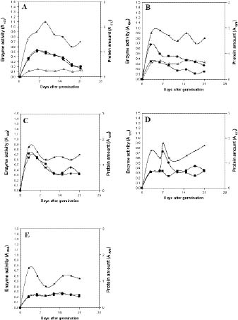
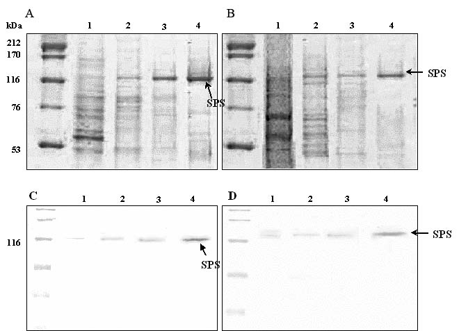
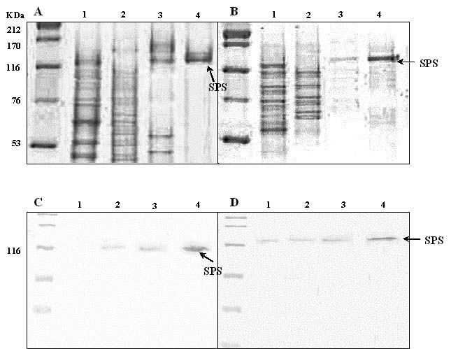
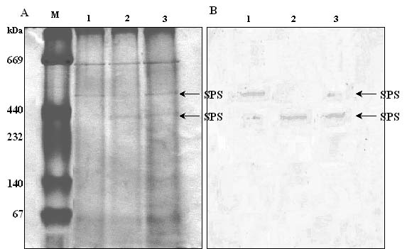
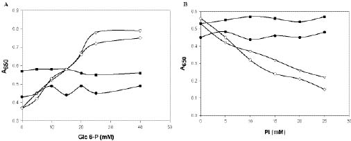
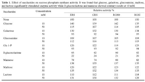
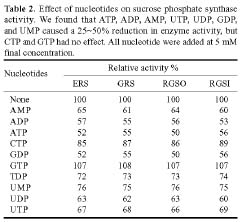
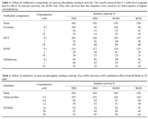
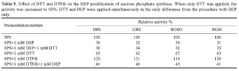
Abstract. Sucrose phosphate synthase (SPS) is one of a number of sucrose-metabolizing enzymes that regulates the sucrose synthesis pathway. SPSs were purified from etiolated rice seedlings (ERS), green rice seedlings (GRS), rice grain suspension cells under osmotic stress (RGSO), and rice grain suspension cells under illumination (RGSI). A native molecular mass of ca. 420 and 520 kDa was found using native-PAGE. The SDS-PAGE analyses revealed SPSs to be homotetramers composed of subunits with a mass of 116-120 kDa. The maximum activity for SPSs was observed on the third day. As far as their biochemical characterization was concerned, the optimum pH of the enzyme reactions lay generally between 6-8, the optimum temperatures between 35-40°C. The ERS and RGSO SPS Km values for Fru 6-P and UDPG were 1.8 and 35 mM, respectively. However, the GRS and RGSI SPS had similar Km values for Fru 6-P and UDPG of 1.5 and 28 mM, respectively. GRS and RGSI SPS activities were allosterically regulated by Glc 6-P (activator) or Pi (inhibitor), but ERS and RGSO SPS had no effect. From their regulations and Km values two enzyme forms (SPS-I and SPS-II) could be discriminated in the rice. SPS-II was induced by illumination, but SPS-I by osmotic stress. All SPSs were activated by Mg2+. The nucleotides AMP, ADP, ATP, UMP, UDP, GDP and UTP inhibited enzyme activity by about 25-50%. Thiol reagents became sensitized to the enzyme activity, but could be restored with DTT or b-ME. Glucose, galactose, glucosamine, maltose, and lactose activated the enzymes and were inhibited by d-gluconolactone and mannose. SPSs were also inhibited by PCMBS, cibacron blue F3G-A, and DEP.

Keywords: Etiolated rice seedlings; Green rice seedlings; Illumination; Isoforms; Osmotic stress; Rice grain suspension cells; Sucrose phosphate synthase.

Abbreviations: DEP, diethylpyrocarbonate; DTNB, 5,5'-dithiobis-2-nitrobenzoic acid; DTT, 1,4-dithiothreitol; ERS, etiolated rice seedlings; GRS, green rice seedlings; RGSI, rice grain suspension cells under illumination; RGSO, rice grain suspension cells under osmotic stress; b-ME, b-mercaptoethanol; PCMBS, p-chloromercuribenzenesulphonic acid; Glc 6-P, glucose 6-phosphate; Fru 6-P, fructose 6-phosphate; Pi, inorganic phosphate; UDPG, uridine 5'-diphosphoglucose.

Introduction

Sucrose is the major stable product of photosynthesis for many plants, and it is the form in which most carbon is transported in phloem vessels from photosynthetic organs into non-photosynthetic organs such as roots, grains, fruits, and tubers (Chris et al., 1999; Rees, 1984). Sucrose accumulates in most plant tissues. This is essential if the plant is to be able to respond effectively to environmental stresses like low temperatures (Guy, 1990), water (Morgan, 1984; Quick et al., 1989; Ramos et al., 1999), and salinity (Balibrea et al., 1997). However, sucrose can also act as a regulator, carbohydrate store, and substrate for biosynthesis and is temporarily preserved in the leaves as a buffer for plants (Chris et al., 1999).

Sucrose synthesis can be catalyzed by the coordination of two enzymes in higher plants: sucrose phosphate

synthase (E.C. 2.4.1.14; SPS) and sucrose phosphate phosphatase (E.C. 3.1.3.24; SPP) (Huber and Huber, 1996). The catalytic reaction is as follows:

SPS is known to be a key regulative enzyme responsible for sucrose synthesis in plants (Stitt et al., 1988; Huber and Huber, 1992; Huber and Huber, 1996). All sucrose-synthesizing organisms contain SPS, which is regulated by several interacting mechanisms, including: (i) Regulation of gene expression (Huber and Huber, 1996), (ii) Covalent modification via reversible phosphorylation (Huber and Huber, 1996), and (iii) Allosteric regulation via metabolites (Doehlert and Huber, 1985).

When purifying SPS in the leaves of plants, it was found that SPS activity changes in coordination with the light cycle (Galtier et al., 1995). In addition, SPS can be activated in spinach leaves under illumination and mannose treatments, but is inhibited under darkness and when inorganic phosphate (Douglas and Huber, 1983) comes in

*Corresponding author. Tel & Fax number: 02-23633610; E-mail: pingdu@ntu.edu.tw


Botanical Bulletin of Academia Sinica, Vol. 46, 2005

contact with the plants. Modification by phosphorylation / dephosphorylation occurs in response to light and darkness (Amir and Preiss, 1982; Cheink and Brenner, 1992). In leaves, illumination induces dephosphorylation, which leads to the enzyme being activated, and darkness results in inactivation because of phosphorylation (McMichael et al., 1993, 1995). SPS is also active in other sucrose synthesizing organs or tissues, including those adapting to cold or drought, fruits, etiolated cotyledons, germinating seeds, sugar cane (Saccharum officinarum) stems, and beet-roots (Beta vulgaris) (Huber and Huber, 1996). However, only a few studies discuss SPS in heterotrophic tissues under illumination, osmotic stress, and darkness.

Our previous experiments found that SPS in sweet potato calli under illumination and osmotic stress had a different biochemical characterization (Lee et al., 2003). The object of this report focused on darkness, illumination, and osmotic stress as regulatory factors affecting the properties of various types of SPSs such as ERS, GRS, RGSO, and RGSI. The effects of these regulatory factors on rice SPSs among the photosynthetic (ERS, GRS) and non-photosynthetic (RGSO, RGSI) tissues were also observed.

Materials and Methods

Plant Materials

Etiolated and green seedlings of rice (Oryza sativa L. cv. Tainong 67) were grown for 14 days under conditions of darkness and illumination. Rice grain suspension cells under illumination and osmotic stress were cultured as previously described (Wang et al., 2000). Briefly, they were subcultured by being transferred twice weekly into a fresh N6 salt medium containing 2,4-D 0.4 µg·mL-1, casamino acid 1 g·µL-1, and sucrose 30 g·L-1. Cells for experiments were harvested at 14 days after routine transfer into the same medium. ERS, GRS, RGSO and RGSI were collected at three-day intervals for a total of 14 days of cultivation and then were used for further analyses.

Extraction and Partial Purification of SPSs

Approximately 100 g of materials from ERS, GRS, RGSO and RGSI were blended and homogenized in 400 mL of ice-cold extraction buffer (50 mM Hepes-KOH, pH 7.5, 10 mM MgCl2, 1 mM EDTA, 2.5 mM DTT, 0.1% [v/v] Triton X-100, 1 mM PMSF and 1% [w/v] PVPP) containing protease inhibitors (PMSF) as above. The crude extracts were centrifuged at 6,000 g for 10 min. The supernatants were decanted and fractionated with polyethylene glycol (PEG) 8000. Proteins in the 30% PEG fraction were dissolved in 20 mL of buffer A (20 mM Tris-HCl, pH 7.5, 1 mM EDTA, and 2 mM DTT) containing protease inhibitors. All procedures to this point were carried out at 4°C. The precipitated protein in 30% PEG fraction from rice was dissolved and applied to a 5 mL DEAE-Sephacel column (Pharmacia), equilibrated with buffer A. Proteins were eluted with a linear salt gradient (1 M NaCl over 50 mL), collecting all fractions of 2 mL per tube. The bulk of the SPS protein was eluted between 280-340 mM NaCl. The corresponding frac

tions were pooled and desalted by passing them through a MonoQ column (HR5/5, Pharmacia/LKB). The Mono Q column was eluted using a fast protein liquid chromatography system (FPLC, Pharmacia/LKB) at a flow rate of 0.5 mL/min, washed with buffer for 20 min, and then with a linear NaCl gradient in buffer (0~1 M NaCl) for 60 min. Fractions of 0.5 mL per tube were collected after 20 min.

Protein Quantitation

Proteins concentrations were determined by the dye-binding method (Bradford, 1976) using bovine serum albumin as a standard.

Assay of SPS

SPSs were assayed under both limiting (Vlimit) and saturated (Vmax) substrate conditions (according to the method of Huber et al., 1989). The enzymes were monitored using the anthrone test during the purification procedure (Huber et al., 1989). In this test, 70 µL of the reaction mixture including the extracts were adjusted to a final concentration of 4 mM Fru 6-P, 20 mM Glc 6-P, 3 mM UDPG, 50 mM Hepes-KOH (pH 7.5), 5 mM MgCl2, and 1 mM EDTA. The mixtures were incubated at 37°C for 15 min before adding 70 µL 30% (w/v) KOH and heated for 10 min at 95°C. To this 1 mL 0.14% (w/v) anthrone in 95% H2SO4 was then added. The mixtures were incubated for 20 min at 37°C, and A650 was measured. Methods of biochemical characterization were used when various nucleotides, thiol reagents, and sugars were tested as activators or inhibitors.

Gel Electrophoresis and Western Blotting

Proteins were separated by 10% SDS-PAGE and 7.5% native-PAGE as described in Laemmli (1970) and either stained with Coomassie Brilliant Blue R-250 or transferred to a nitrocellulose membrane by electroblotting. Protein gel blots were blocked using 5% fat-free powdered milk solution in 20 mM Tris-HCl, pH 7.5, and 500 mM NaCl. Blots were incubated with rabbit antiserum raised against RBE1, glutathione S-transferase (GST)-SPK fusion protein and GST-Suc synthase fusion protein (Urao et al., 1994) in order to detect each protein. Immunodecorated proteins were revealed with horseradish peroxidase-conjugated anti-goat secondary antibody using the Vectastain ABC kit (Vector Laboratories, Burlingame, CA) and DAB substrates (Funakoshi, Tokyo, Japan).

Results

Comparative Analyses of the Enzyme Activty of ERS, GRS, RGSO and RGSI

Rice seedlings (i.e., ERS and GRS) and rice grain suspension cells (i.e., RGSO and RGSI) were collected and employed as primary experimental materials of this study. The rice SPS's activity under different conditions was recorded every three days (Figure 1). The illuminated cells were placed under a constant illumination of 5000 Lux (wavelength of 700 nm) from 06:00 to 21:00 (with no illu


LEE et al. — Sucrose phosphate synthase from rice

Figure 1. SPS activities and protein level of rice varied with different treatment under different growth phase. It was discovered that ERS, GRS, RGSI, and RGSO display increasing SPS enzyme activity with the increase of time. A, ERS; B, GRS; C, RGSO; D, RGSI; E, Rice grain suspension cell (control). The assay condition of Vmax and Vlimt are expressed in the text. SPS Vmax activity (~); SPS Vlimit activity (¢); SPS roots activity (r); Protein content (p).

mination from 21:00 to 05:00) for 14 days (Figure 1D). It was discovered that the activity of SPS from ERS had gone down on the sixth day (Figure 1A) and GRS had dwindled a little on the fourth day (Figure 1B). The highest SPS activity from RGSO was on the third day (Figure 1C), and RGSI beignet surged on the sixth day (Figure 1D), indicating that the modulating forms of SPSs in RGSI and RGSO were different. However, a low SPS activity was found in normally developing suspension cells (Figure 1E) and in the roots from ERS and GRS (Figure 1A-B).

Purification of the Enzymes

ERS, GRS, RGSO and RGSI as materials were put through SPS purification. Analyses of the native-PAGE revealed that the RGSI SPS is a tetramer with a molecular mass of 420 and 520 kDa (Figure 4A-B). The apparent molecular mass of SPSs from ERS and RGSO as revealed in

the native-PAGE is 520 kDa, while SPS from GRS is 420 kDa (data not shown). The 420 kDa bands are thicker in the samples from rice suspension cells under illumination for 3, 6 and 12 days. This implied that the 420 kDa protein can be induced by light. The SDS-PAGE analyses showed a molecular mass of 116-120 kDa as a monomer (Figures 2,3). From immuno-staining patterns of SPSs on native-PAGE analyses, we propose that two isozymes exist in RGSI (Figure 4B).

Biochemical Characterization of Rice SPSs

SPS optimum and stability pH values lay between 6-8. When the temperature was higher than 40°C, SPS activity in ERS, GRS, RGSO and RGSI would go down and become unstable (data not shown). Therefore, in the process of purification and analyses, the enzymes are best preserved in ice water so as to maintain their activity. The optimum


Botanical Bulletin of Academia Sinica, Vol. 46, 2005

Figure 2. CBR staining and immuno-staining patterns of SPS fraction on 10% SDS-PAGE from ERS (A, C) and RGSI (B, D) separated after the different purification steps. Lane 1, crude extract; lane 2, PEG 30%; lane 3, DEAE-Sephacel column; lane 4, FPLC Mono Q column. It is estimated that the molecular mass of subunit is around 116~120 kDa.

Figure 3. CBR staining and immuno-staining patterns of SPS fraction on 10% SDS-PAGE from RGSO (A, C) and GRS (B, D) separated after the different purification steps. Lane 1, crude extract; lane 2, PEG 30%; lane 3, DEAE-Sephacel column; lane 4, FPLC Mono Q column. It is estimated that the molecular mass of subunit is around 116 to 120 kDa.


LEE et al. — Sucrose phosphate synthase from rice

Effect of Sugars and Sugar Phosphates

Different sugars influenced rice SPS activity (Reimholz et al., 1994). It was found that glucose, galactose, glucosamine, maltose, and lactose significantly stimulated enzyme activity while d-gluconolactone and mannose showed contrary results at 10 mM (Table 1). However, the activity of SPS was enhanced by 50 mM mannose. The weaker inhibition of 10 mM mannose to SPS may be due to the experimental error. The structure of lactone in d-gluconolactone is half-chair, similar to the carbonium-oxonium ion, which may occupy the glucosyl transfer site of SPS. The transition state is likely to be a glucose-enzyme complex in which the glucosyl unit has the half-chair conformation. This reaction occurs via the formation of an unstable glucose-enzyme complex that leads to the inactivation of the enzyme (Reimholz et al., 1994).

temperature should be between 35-40°C. From GRS and RGSI, SPS was allosterically regulated by Glc 6-P as an activator and Pi as an inhibitor (Figure 5). It had the highest activity at 30 mM (Glc 6-P). Pi at 5 mM inhibited its activity, and 25 mM Pi exhibited the maximum inhibition effect. In contrast, ERS and RGSO were not activated by Glc 6-P (Figure 5A) or inhibited by Pi (Figure 5B). In fact, the enzymes were activated by Mg2+ at 7.5-12.5 mM (data not shown).

Kinetic Characteristics of Rice SPSs

SPSs from various rice materials in different concentrations of substrates, Fru 6-P and UDPG, did not demonstrate the same results. The Km values for Fru 6-P and UDPG were calculated. Similar Km values for UDPG and Fru 6-P from ERS, RGSO and GRS, RGSI SPSs were observed.

Figure 4. CBR-staining (A) and immuno-staining (B) patterns of RGSI SPS on 7.5% native-PAGE. This research also verified that two isozymes exist in rice grain suspension cells. Lane 1, 3 days under illumination; lane 2, 6 days under illumination; lane 3, 12 days under illumination.

Figure 5. Effects of Glc 6-P and Pi on the activity of SPS from ERS, GRS, RGSI and RGSO. A, Effect of Glc 6-P on SPS from rice; B, Effect of inorganic phosphate on SPS from rice. SPS from etiolated rice seedlings and rice grain suspension cells with osmotic stress treatment was not regulated by Glc 6-P or Pi. ERS (~); GRS (s); RGSI (¯); RGSO (¢).


Botanical Bulletin of Academia Sinica, Vol. 46, 2005

Effect of Nucleotides

The effects of various nucleotides on enzyme activity were summarized in Table 2. We found that ATP, ADP, AMP, UTP, UDP, GDP, and UMP caused a 25~50% reduction in enzyme activity, but CTP and GTP had no effect (Table 2). It is possible that ATP, ADP, AMP, UTP, UDP, and UMP competed with the UDPG binding site of SPSs.

Effect of Thiol Reagents

SPS activity is subjected to the influence of the thiol group within the protein sequences (Doehlert et al., 1985). It was tested with different thiol reagents to determine how the -SH group affected its activity in the amino acid sequence of rice SPSs. The results showed that 0.1 mM thiol reagents had no effect on enzyme activity, but b-ME can enhance SPS activity (Table 3). However, the SPS activity was inhibited as the concentration was highly raised. They also showed that the enzymes were sensitive to thiol regents at higher concentrations (Table 3).

Effect of Inhibitors

PCMBS was repressive to the sulfhydryl groups complex (Slabnik et al., 1968). It was found that PCMBS increasingly repressed SPS when its concentration was heightened. Cibacron blue F3G-A, similar to the substrate UDPG, was demonstrated to be a high-affinity competitive inhibitor (Hatzfeld et al., 1995). The results obtained in Table 4 indicate a 50% inhibition effect for rice SPSs from PCMBS at 20-50 mM. In wheat seedlings (Salerno and Pontis, 1978), maize leaves (Doehlert and Huber, 1985), potato tuberous roots (Slabnik et al., 1968), and spinach leaves (Amir and Preiss, 1982), PCMBS completely represses SPS activity, which can be restored by DTT.

Modifying Effect of DEP

As a chemical modification effecter, DEP modifies the histidine in protein by producing an N-carbethoxyhistidyl derivation. This derivate absorbance was measured at 230~250 nm (Sinha et al., 1998). The aim of this study was primarily to understand whether histidine affects SPS activity. The reactions were carried out at 25°C with different incubation times. Twenty mM histidine was used to stop the reaction. The results indicate that the inhibition of SPS became apparent in the presence of increasing concentrations of DEP (Figure 6). It was inhibited 33%


LEE et al. — Sucrose phosphate synthase from rice

Figure 6. Time course of the activity of DEP-modified SPS. DEP modifies a protein by converting His residues to N-carbethobxyhistidyl derivative. A, Etiolated rice seedlings; B, Green rice seedlings; C, Rice grain suspension cell under osmotic stress; D, Rice grain suspension cell under illumination. 0 mM (~); 0.5 mM (™); 1.0 mM (q); 5.0 mM (s); 10 mM (¢)


Botanical Bulletin of Academia Sinica, Vol. 46, 2005

by 1 mM DEP at 25°C, resulting in a 10-min reaction. When only DTT was applied, the activity was decreased to 60%. No synergetic effects were apparent when DTT and DEP were added together as compared to DEP alone (Table 5). The influence of DTNB described in Table 5 showed that the enzymes were activated by 1 mM DTNB.

Discussion

Nothing in the literature yet describes the biochemical characterization of SPSs in rice under different treatments. This report finds that SPSs underwent different isoforms and activities depending on whether they were affected in ERS, GRS, RGSO or RGSI. Darkness, illumination, and osmotic stress were then used to examine the change of SPS activity among photosynthetic and non-photosynthetic tissues under various environmental treatments for rice. Different biochemical characterizations between SPSs were observed in ERS, RGSO and GRS, RGSI. This was verified by the following results, which distinguished two SPS isoforms in rice: First, SPSs activity from GRS and RGSI were subjected to allosteric regulation by Glc 6-P (activator) and Pi (inhibitor) (Figure 5) (Douglas and Huber, 1983). Both of them had similar Km values for UDPG and Fru 6-P. Secondly, Glc 6-P and Pi did not affect the SPSs activity from ERS and RGSO. Both SPSs have a similar Km to UDPG and Fru 6-P. Furthermore, the apparent molecular mass of SPS-I was estimated to be 520 kDa in the native-PAGE. Its expression in rice grain tissue cultures increased in response to the osmotic stress. On the other hand, SPS-II, estimated at 420 kDa on native-PAGE, increased in response to the illumination. No band existed in native-PAGE without light treatment. From 7.5% native-PAGE and immuno-staining patterns of RGSI SPSs, two enzyme isoforms (SPS-I and SPS-II) (Figure 4) resulted. The results also indicated that light could induce SPS isoforms both in photosynthetic (GRS) and non-photosynthetic (RGSI) tissues.

Two enzyme isoforms had similar results when affected by saccharides, nucleotides, sulfhydryl compounds, and inhibitors. They were activated by glucose, galactose, glucosamine, maltose, and lactose but were inhibited by d-gluconolactone and mannose (Table 1). Glc 1-P increased enzyme activity at 10 mM, but not at 50 mM (Table 1). AMP, ADP, ATP, UMP, UDP, UTP, and TDP possibly

played the role of competitive inhibitors toward their UDPG-binding site.

SPSs in spinach, seed of Pisum sativum and leaf of Prosopis juliflora were affected by the thiol reagent concentrations (Harbron et al., 1981; Lunn and Rees, 1990; Sinha et al., 1997). The -SH group may be directly involved in catalysis or may be also required to maintain an appropriate enzyme conformation. Therefore, sulfhydryl compounds may also play a peculiar role in controlling rice SPSs activity; the results did not indicate an absolute requirement of free -SH groups for enzyme activity (Table 3). Usually, the allosteric sites of most enzymes contain the thiol group, and the substrate molecules do not. However, this was not necessary for the catalytic site area (Sinha et al., 1997). Most SPS amino acid sequences contain 10 cysteine residues. Possibly one or more of these ten exist in the catalytic site.

SPSs from ERS, GRS, RGSO and RGSI were inhibited by PCMBS and cibacron blue F3G-A, suggesting the presence of the sulfhydryl group is essential for enzyme activity. The effects of DTT, DTNB and DEP on rice SPSs indicated that the DEP function was not affected by the -SH group (Harbron et al., 1981; Lunn and Rees, 1990), but rice SPS activity was inhibited by histidine (Table 5). It is believed that the effect of DEP is on the phosphorylate enzyme area. The phosphate carries a negative charge while the histidine is positively charged, and the modifying of DEP in histidine in protein breaks the counter charges, making an electrical balance which protects the active site. So DEP modified the histidine, thus affecting the binding between substrate and binding site.

According to biochemical characterization and enzyme activity analyses, we conclude that at least two enzyme isoforms, SPS-I and SPS-II, exist in rice. They can be induced by illumination both in the photosynthetic and non-photosynthetic tissues. SPS-I was found in ERS and RGSO, and activity was regulated by osmotic stress and darkness while SPS-II was induced and activated by illumination in GRS and RGSI. But, active sites of SPS-I and SPS-II show a similar modification pattern by chemical reagents. This feature must be important in causing at least some of the metabolic and physiological effects observed in this study, and it is further shown that SPS in rice is a enzyme in source-sink relationships in response to environmental factors, e.g. light, osmotic stress, and darkness.


LEE et al. — Sucrose phosphate synthase from rice

Acknowledgements. The authors would like to thank the National Science Council in Taiwan for financially supporting this research (under Grant NSC No. 91-2313-B-002-313). We appreciate Dr. Dong-Jiann Huang for generously providing the SPS antibody.

Literature Cited

Amir, J. and J. Preiss. 1982. Kinetic characterization of spinach leaf sucrose-phosphate synthase. Plant Physiol. 69: 1027-1030

Balibrea, M.E., A.M. Rus-Alvarez, M.C. Bolarin, and F. Perez-Alfocea. 1997. Fast changes in soluble carbohydrates and proline contents in tomato seedlings in response to ionic and nonionic iso-osmotic stresses. J. Plant Physiol. 151: 221-226.

Bradford, M.M. 1976. A rapid and sensitive method of the quantitation of microgram quantities of protein utilizing the principle of protein-dye binding. Anal. Biochem. 72: 248-254.

Cheink, N. and M.L. Brenner. 1992. Effect of dark and light conditions and role of gibberellins and abscisic acid. Plant Physiol. 100: 1230-1237.

Chris, P., A. Cairns, J. Gallagher, J. Farrar, D. Tomos, and O. Koroleva. 1999. Sweetness and Light: the Role of Sucrose in Higher Plants. Iger innovations 6-9.

Doehlert, D.C. and S.C. Huber. 1985. The role of sulhydryl groups in the regulation of spinach leaf sucrose phosphate synthase. Biochim. Biophys. Acta 830: 267-273.

Douglas, C.D. and S.C. Huber. 1983. Regulation of spinach leaf sucrose phosphate synthase by glucose-6-phosphate, inorganic phosphate, and pH. Plant Physiol. 73: 989-994.

Galtier, N., C.H. Foyer, E. Murchie, R. Alred, and P. Quick. 1995. Effects of light and atmospheric carbon dioxide enrichment on photosynthesis and carbon partitioning in the leaves of tomato (Lycopersicon esculentum L.) plant over-expressing sucrose phosphate synthase. J. Exp. Bot. 46: 1335-1344.

Guy, C.L. 1990. Cold acclimation and freezing stress tolerance: role of protein metabolism. Annu. Rev. Plant Physiol. Plant Mol. Biol. 41: 187-223.

Harbron, S., C. Foyer, and D. Walker. 1981. The purification and properties of sucrose-phosphate synthase from spinach leaves: the involvement of this enzyme and fructose bisphosphatase in the regulation of sucrose biosynthesis. Arch. Biochem. Biophys. 212: 237-246.

Hatzfeld, W.D. and E. Moesinger. 1995. Active-site-directed inhibition of sucrose-phosphate synthase by Cibacron blue F3G-A and 1-deoxynojirimycin. Planta 197: 296-300.

Huber, J.L.A., S.C. Huber, and T.H. Nielsen. 1989. Protein phosphorylation as a mechanism for regulation of spinach leaf sucrose-phosphate synthase activity. Arch. Biochem. Biophy. 270: 681-690.

Huber, S.C and J.L.A. Huber. 1992. Role of sucrose-phosphate synthase in sucrose metabolism in leaves. Plant Physiol. 99: 1275-1278.

Huber, S.C. and J.L. Huber. 1996. Role and regulation of sucrose-phosphate synthase in higher plants. Annu. Rev. Plant Physiol. Plant Mol. Biol. 17: 431-444.

Laemmli, U.K. 1970. Cleavage of structural proteins during the assembly of the head of bacteriophage T4. Nature 227: 680-685.

Lee, M.H., C.C. Yang, H.L. Wang, and P.D. Lee. 2003. Regulation of sucrose phosphate synthase of the sweet potato callus is related to illumination and osmotic stress. Bot. Bull. Acad. Sin. 44: 257-265.

Lunn, J.E. and A. Rees. 1990. Purification and properties of sucrose phosphate synthase from seeds of Pisum sativum. Phytochemistry 29: 1057-1063.

McMichael, R.W.Jr., R.R. Klein, M.E. Salvucci, and S.C. Huber. 1993. Identification of the major regulation phosphorylation site in sucrose phosphate synthase. Arch. Biochem. Biophys. 307: 248-252.

McMichael, R.W.Jr., M. Bachman, and S.C. Huber. 1995. Spinach leaf sucrose-phosphate synthase and nitrate reductase are phosphorylated/inactivated by multiple protein kinase in vitro. Plant Physiol. 108: 1077-1082.

Morgan, J.M. 1984. Osmoregulation and water stress in higher plants. Ann. Rev. Plant Physiol. 35: 299-319.

Quick, P., G. Siegl, E. Neuhaus, R. Feil, and M. Stitt. 1989. Short-term water stress leads to a stimulation of sucrose synthesis by activating sucrose-phosphate synthase. Planta 177: 536-546.

Ramos, M.L.G., A.J. Gordon, F.R. Minchin, J.I. Sprent, and R. Parsons. 1999. Effect of water stress on nodule physiology and biochemistry of a drought tolerant cultivar of common bean (Phaseolus vulgaris L.). Ann. Bot. 83: 57-63.

Rees, A. 1984. Sucrose metabolism. In Storage Carbohydrates In Vascular Plants. Cambridge University Press. London, pp. 53-57.

Reimholz, R., P. Geigenberger, and M. Stitt. 1994. Sucrose phosphate synthase is regulated via metabolites and protein phosphorylation in potato tubers, in a manner analogous to the enzyme in leaves. Planta 192: 480-488.

Salerno, G.L. and H.G. Pontis. 1978. Sucrose phosphate synthase. Separation from sucrose synthtase and a study of its properties from wheat. Planta 142: 41-48.

Slabnik, E., R.B. Frydman, and C.E. Cardini. 1968. Some properties of potato tuber UDPG: D-fructose-2-glucosyltransferase and UDPG: D-fructose-6-phosphate-2- glucosyltransferase. Plant Physiol. 43: 1063-1068.

Sinha, A.K., U.V. Pathre, and P.V. Sane. 1997. Purification and characterization of sucrose- phosphate synthase from Prosopis juliflora. Phytochemistry 46: 441-447.

Sinha, A.K.,U. Pathre, and P.V. Sane. 1998. Essential histidyl residues at the active site of sucrose-phosphate synthase from Prosopis juliflora. Biochim. Biophys. Acta. 1388: 397-404.

Stitt, M., I. Wilke, R. Feil, and H.W. Heldt. 1988. Coarse control of sucrose -phosphate synthase in leaves. Planta 174: 217-230.

Urao, T, T. Katagiri, T. Mizoguchi, K.Yamaguchi-Shinozaki, N. Hayashida, and K. Shinozaki. 1994. An Arabidopsis thaliana cDNA encoding Ca2+-dependent protein kinase. Plant Physiol. 105: 1461-1462.

Wang, H.L., P.D. Lee, W.L. Chen, D.J. Huang, and J.C. Su. 2000. Osmotic stress-induced changes of sucrose metabolism in cultured sweet potato cells. J. Exp. Bot. 51: 1991-1999.


Botanical Bulletin of Academia Sinica, Vol. 46, 2005