Bot. Bull. Acad. Sin. (2005) 46: 143-149

HSU et al. — Anatomical characteristics of the secondary phloem in branches of Zelkova serrata

Anatomical characteristics of the secondary phloem in branches of Zelkova serrata Makino

Ying-Shan HSU1, Shiang-Jiuun CHEN1, Chin-Mei LEE2, and Ling-Long KUO-HUANG1,*

1Department of Life Science; Institute of Ecology and Evolutionary Biology, National Taiwan University, Taipei, Taiwan, Republic of China

2Division of Forest Utilization, Taiwan Forestry Research Institute, Taipei, Taiwan, Republic of China

(Received June 21, 2004; Accepted December 23, 2004)

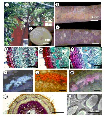
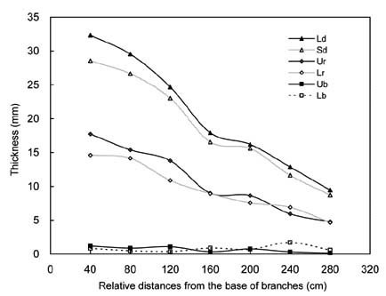
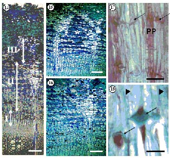
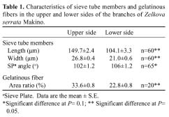
Abstract. The properties and tissue compositions of reaction wood in the leaning trunks or branches of trees have been extensively investigated, but studies on the influence of the related reaction on the secondary phloem are few and incomplete. In this work, formation of the vascular and cork cambium and the secondary phloem in the upper and lower sides of branches of Zelkova serrata Makino were studied. Vascular cambium is formed first in the second internode of shoot. In the third internode, cork cambium began to initiate subepidermally, meanwhile two to four layers of grouped gelatinous fibers were first found in the cortex outside of all the primary phloem. Gelatinous fibers were also found in both the secondary xylem and secondary phloem. The leaning branches exhibited pronounced radial secondary growth promotion to the upper side, and the reaction wood formed eccentrically. The secondary phloem formed layers of conducting sieve elements alternately with gelatinous fibers. Comparing the secondary phloem in the upper side (reaction phloem) of the branches with that in the lower side (opposite phloem), there is no obvious difference in thickness. Nevertheless, it was found that in the cross sections, the gelatinous fibers formed earlier, and there were more continuous cell seriates and a much larger area ratio in the upper side. Besides, the sieve tubes in the upper side of secondary phloem were longer and wider and possessed a very horizontally-orientated sieve plate between two sieve elements. These features may imply that, in branches of Zelkova serrata, the secondary phloem of the upper side (reaction phloem) may have a higher translocation efficiency.

Keywords: Branches; Gelatinous fibers; Reaction phloem; Secondary phloem; Zelkova serrata.

Introduction

With continued secondary growth, a tree's vascular cambium pertinently divides and produces secondary xylem inward and secondary phloem outward. The inclined trunk and branches frequently display eccentric growth and show specific structural changes in the cell layers, cell morphology, and cell wall constitution during the formation of secondary tissues (Fahn, 1990; Jourez et al., 2001). The timber which secondary growth slants toward one side is called reaction wood (Wilson and Archer, 1977). Generally, reaction wood is classified as compression wood or tension wood. Compression wood occurs in most gymnosperms, and tension wood forms mostly in the woody angiosperms (Wilson and Archer, 1977; Timell, 1986). Compression wood forms in the lower side of leaning trunks and branches and manifests its effect by slowly pushing the stem while tension wood forms in the upper side of leaning stems and functions by pulling the stems to the needed orientation. A tree, in effect, ingeniously adjusts its growth stress to the requirements of its environment, thereby satisfying its physical needs.

It is reasonable to expect the inductive causes of reaction wood formation to also affect the derivatives of the vascular cambium on the correlative phloem side. Scurfield and Wardrop (1962) first coined the term reaction phloem. The terms reaction, compression, and tension barks were coined by Timell (1986). The different morphology and structure of barks on the lower and the upper sides of branches or on the leaning stems of conifers have been mentioned by several investigators. In Chamaecyparis obtuse and Cryptomeria japonica, Onaka (1949) found radial growth in the bark to be larger on the lower, compression wood side than on the upper side although the difference was less than in the wood. As a consequence of the larger radial growth of the compression wood, outer bark on the lower side tended to split and fall off, and compression bark as a result was thin. Kutscha et al. (1975) observed that in compression phloem of Abies balsamea the bands of tannin cells were discontinuous instead of continuous as in normal phloem. Thick, unlignified wall layers have been observed in gelatinous fibers present in tension phloem, for example in Eucalyptus (Dadswell and Wardrop, 1955), Tilia cordata (Böhlmann, 1971), and Populus euramericana (Nanko et al., 1982).

Many studies have explored the structural, physiological, and mechanical properties of reaction wood (Wilson and Archer, 1977; Timell, 1986). However, the for

*Corresponding author. Tel: (02) 33662510; Fax: (02) 23673374; E-mail: linglong@ntu.edu.tw


Botanical Bulletin of Academia Sinica, Vol. 46, 2005

mation and regulation mechanisms are still not well understood. It has been reported that the formation of reaction wood is closely related to the distribution of plant hormones (Little and Eklund, 1998; Yoshida et al., 1999). It is certain that the secondary phloem in the stem is very important not only because of its translocation of specific plant hormones but also as the nutrients sink of trees. Comparing works on wood properties, studies on secondary phloem, whether of gymnosperms or angiosperms, are limited, especially studies on reaction phloem.

Zelkova is a highly-prized hardwood with a beautiful grain, fine texture, and high strength properties. In Taiwan, the plantation area of Zelkova serrata covers over six thousand hectares. This is a species known to have a very high level of growth stress. During sawing, timber yield invariably decreases as a result of heart crack and distortion of board owing to the release of growth stress (Sasaki et al., 1978; Huang et al., 2004). Investigations into the reaction phloem of Zelkova serrata may help us to understand the mechanism of reaction formation and the trees' adaptation in physiology.

Materials and Methods

Branches of Zelkova serrata Makino were collected from the trees planted on the campus of National Taiwan University or in the timberland of Taiwan Forestry Research Institute. Before branches were cut at the base (Figure 1), their upper sides were marked. The transverse sections of the leaning branches exhibited radial promotion to the upper side (Figure 2). The tension wood showed a silvery white metallic shine, and its color was much lighter than the wood opposite. The color of the outer bark on the upper side of the branches was light brown (Figure 3) while that on the lower side is usually dark brown (Figure 4).

In order to understand when and where the vascular cambium and cork cambium were initiated, transverse 10-20 µm paraffin sections (Kuo-Huang et al., 2002) were made from the youngest to the fifth internode of shoots. From the upper and the lower side of the basal part of branches with obviously eccentric growth, small slices of bark with thin sap wood were directly sectioned by free-hand or by using the Erma sliding microtome. The paraffin sections and the free-hand sections were stained with safranin O and fast green. Some slices of bark were carefully cut into small pieces (1mm × 1 mm × 2 mm), double fixed with 2.5% glutaraldehyde and 1% OsO4, and then dehydrated and embedded in spurr's resin. With ultramicrotome (Reichert Ultracut E), 1 µm sections were made and stained with toluidine blue. Materials for scanning electron microscopy were prepared following the methods by Sheue et al. (2003). For a histochemical test of the cell wall to identify the distribution of gelatinous fibers in the tissues, suitable transverse free-hand sections were made and stained with phloroglucinal-HCl (Gahan, 1984), Herzberg reagent, or toluidine blue. Materials were examined on a Leica Diaplan light microscope or Hitachi S-520 SEM. Sixty to 65 sieve tube elements from the upper and lower sides of branches

were randomly chosen for measuring the length, width, and the sieve plate angles. The area percentages of phloem fibers were also counted. Mean values and standard deviations were calculated.

Results

Initiation of Vascular Cambium and Cork Cambium

In all cross sections of the young shoots, many trichomes were found extended from the epidermal cells. In the second internode, vascular cambium was first found between the primary xylem and phloem (Figure 5) while in the third internode, cork cambium was initiated subepidermally (Figure 6). In the transverse sections of the fourth internode (Figure 7), vascular cambium was found to divide periclinally several times while cork cambium formed only one layer of phelloderm inwardly and 1-2 layers of phellem outwardly.

Two to four layers of gelatinous fibers were found in the cortex of the transverse sections from the third to fifth internodes (Figures 6-11). They occurred in groups directly outside of the primary phloem and were found to be connected almost as a ring (Figure 11). Histochemical tests of the cell wall of gelatinous fibers showed that the inner gelatinous wall layer was nonlignified and contained mainly cellulose (Figures 9-10). The cortical cells outside the gelatinous fibers were irregularly enlarged and later formed the stone cells (Figures 10-11). Unlike the gelatinous fiber occurring in phloem, the xylary fibers, in the first to the fifth internodes, contained only the lignified secondary wall and a distinct cell lumen.

Outer Bark and Secondary Phloem

Figure 13 shows changes in the long and short diameters of the branches, the thickness measurements of the tension wood and opposite wood and their related barks with the relative distances from the base to the tip of the branches. The leaning branches exhibited pronounced radial secondary growth promotion to the upper side, and the reaction wood formed eccentrically. The basal part the branches was more as ellipical. Nevertheless, there were no obvious differences in the thickness of barks between the upper and lower sides of the branches.

The outer bark consisted of periderm and cortex with conspicuous tangential bands of stone cells. The inner bark was composed of the secondary phloem. In the outer part of the secondary phloem, ray cells divided anticlinally and formed expansion rays (Figures 14-16). Several times in a growing season, tangential bands of two to four layers of conducting sieve elements accompanied the parenchymatous cells, and then one to four layers of phloem fibers formed alternately (Figures 15 and 16). All of the mature phloem fibers contained a gelatinous layer in the inner cell wall (Figure 12). Staining demonstrated that the inner cell wall was not lignified and consisted mainly of cellulose. These multi-layered gelatinous phloem fibers were ob


HSU et al. — Anatomical characteristics of the secondary phloem in branches of Zelkova serrata

Figures 1-12. Figure 1. The branch used in this study. It is 2.5 m in height from the ground; Figure 2. Transection of the basal of the sampling branch; Figure 3. The upper side of the sampling branch; Figure 4. The lower side of the sampling branch; Figure 5. Cross section of the second internode of young branch. Vascular cambium (arrow) formed between primary xylem and phloem. (Bar = 100 µm); Figure 6. Cross section of the third internode of young branch. The origin of phellogen (long arrow) located in subepidermal cells. Gelatinous fibers (GF) formed outside of the primary phloem. Some vascular cambium cells divided (short arrow). (Bar = 100 µm); Figure 7. Cross section of the fourth internode of young branch. Phellogen cells divided 1~2 times (arrow). (Bar = 100 µm); Figure 8. Stained by Toluidine blue, gelatinous fibers show a bright red color and are surrounded by lignified cell wall, which appears sky blue under polarizing microscope. (Bar = 100 µm); Figure 9. Stained by Herzberg reagent, gelatinous fibers are surrounded by a light orange color. (Bar = 100 µm); Figure 10. Stained by phloroglucin-HCL, gelatinous fibers are surrounded by the purplish red color that is lignin rich. (Bar = 100 µm); Figure 11. Cross section of the fifth internode of young branch. Gelatinous fibers (arrows) almost form a continuous ring in the outer phloem. Lignin is stained pink by phloroglucinol-HCL, especially in xylem tissue. (Bar = 25 µm); Figure 12. SEM of gelatinous fiber in phloem shows the thick non-lignified inner layer. (Bar = 3.0 µm).


Botanical Bulletin of Academia Sinica, Vol. 46, 2005

Figure 13. The long and short diameters and the thickness of xylem and phloem at the upper and lower sides of the branch in different distances from the trunk. Ld: Long diameters; Sd: Short diameters; Ur: Radiuses of upper sides; Lr: Radiuses of lower sides; Ub: Thickness of barks of upper sides; Lb: Thickness of barks of lower sides.

Figures 14-18. Figure 14. Cross section of the bark. The secondary phloem could be divided into three parts: the inner one consisting of phloem cells just divided from vascular cambium (I); the middle part being the mature phloem (II); and the outer phloem composing of collapsed cells (III). (Bar = 50 µm); Figure 15. Cross section of the secondary phloem of upper side showing layers of sieve elements (light-stained) and gelatinous fibers (dark-stained; *) arranged alternately. (Bar = 100 µm); Figure 16. Cross section of the secondary phloem of lower side showing layers of sieve elements (light-stained) and gelatinous fibers (dark-stained; *) arranged alternately. (Bar = 100 µm); Figure 17. Radial longitudinal section of secondary phloem with sieve tube elements (arrows), sieve plate, and phloem parenchymatous cells (PP). (Bar = 100 µm); Figure 18. Radial longitudinal section of secondary phloem, showing the sieve plates (arrows) between adjacent sieve elements and the companion cells (short arrows). (Bar = 50 µm).


HSU et al. — Anatomical characteristics of the secondary phloem in branches of Zelkova serrata

served on both sides of the branches (Figures 15 and 16). The distribution of such gelatinous phloem fibers approximately corresponded to that of the gelatinous fibers in the cortex outside of the primary phloem and in the tension wood.

Comparing the secondary phloem on the upper side of the branches with that on the lower side (Figures 15 and 16), it was found that the gelatinous phloem fibers formed earlier and more continuous cell seriates. Addtionally, they showed slightly more rows of cells in the radial extent, and their area ratio percentages were higher (Table 1).

The newly formed functional phloem consisted of the sieve elements, companion cells, and parenchymatous cells, but no gelatinous fibers had been formed yet. As seen in the longitudinal sections, a series of sieve tube members were connected end-to-end by the sieve plates (Figures 17 and 18). The cells on the upper side differentiated and renewed faster than those on the lower side. The sieve tube members of the secondary phloem on the upper side were longer and wider than those on the opposite side, and the inclination of the sieve plates tended to be more horizontal (Table 1).

Discussion

Comparing the outer morphology of the upper and lower sides of branch barks of Zelkova serrat, considerable differences in color emerge, i.e. the lower bark is dark brown while the upper one is usually light brown. As Timell (1986) suggested, variations in growth rate, amounts of illumination, and exposure to rain could account for these differences. It may not necessarily be relevant to inner growth stress.

The phellogen of Zelkova serrata originated subepidermally and formed almost simultaneously with, or just shortly after, the formation of vascular cambium. Waisel et al. (1967) suggested that the activity of vascular cambium may make the stem robust, and therefore repress the outward tissues to promote the phellogen's initiation and production of periderm. Kandelia candel (L.) Druce contains thicker periderm in the reaction side of leaning stems (Chang, 1984). However, in the case of Zelkova serrata, the thickness of the reaction bark did not show the variations that occurred in the reaction wood. The cell layers of periderm contributing to barks were limited, and the outer non-functional secondary phloem was often compressed to collapse and did not accumulate their thickness as secondary xylem tissue did. This may be why the bark of Zelkova serrata did not show apparent difference in thickness between the upper and lower sides.

In the third internode of the branches of Zelkova serrata, gelatinous fibers were first observed in the cortex outside of the primary phloem. However, no gelatinous fibers were found in the primary xylem. Therefore, reception of the stimulation of stress may be expressed in the phloem earlier than in the xylem. The formation of reaction tissue (gelatinous fibers) is not limited to secondary growth and may be present as early as primary growth. In

the primary tissues of the seedlings of Acer (Kang and Soh, 1992) and Ricinus (Kang and Soh, 1994), gelatinous fibers were also found. Gelatinous fibers occur in Acer's first internode and only in xylem, but Ricinus resembles Zelkova serrata in that the gelatinous fibers occur in the phloem earlier. In Zelkova serrata, the gelatinous fibers were distributed around the entire transverse section as was observed outside of phloem in stems of Gnetum gnemon (Tomlinson, 2001) and roots of Juniperus communis (Timell, 1986).

The formation of tension wood is generally associated with growth eccentricity. However, this phenomenon occurring on phloem tissue is reported very rarely. Metzger, as early as 1908, suggested that any change in the formation of reaction xylem should be accompanied by changes in the phloem as well (Krishnamurthy et al., 1997). Several species, such as Eucalyptus, Quercus alba, Q. falcate, Tilia cordata (Timell, 1986), and Populus euramericana (Nanko et al., 1982), have been reported to possess gelatinous fibers in the secondary phloem. These gelatinous fibers can be observed only on the upper side of leaning or horizontally positioned branches and are independently formed or related to the reaction wood. In the branches of Zelkova serrata, gelatinous fibers are distributed in both the secondary xylem and the phloem of the upper and lower sides although these fibers still occur in higher area percentages on the upper side.

The sieve tubes are the cells responsible for translocating nutrients in plants. In the branches of Zelkova serrata, the sieve tubes in the secondary phloem of the upper side are wider and longer. Besides, their ends have smaller obtuse angles, i.e. the sieve tubes possess a highly horizontally-orientated sieve plate between two sieve elements. As a whole, in the branches of Zelkova serrata the secondary phloem of the upper side is supposed to have higher translocation efficiency.

The anatomical characteristics of tension wood generated by plant hormones, like GA3 and auxin, have been investigated (Baba et al., 1995; Yoshida et al., 1999; Kwon et al., 2001). GA3, for example, is transported mainly by phloem. We have supposed that on the upper side of the branches the translocation efficiency is higher. As a re


Botanical Bulletin of Academia Sinica, Vol. 46, 2005

sult the reaction side tissue would receive more accounts of GA3 than the opposite side. In this situation, reaction phloem undoubtedly plays an important role in the formation of reaction wood. However, further studies should be undertaken to clarify this.

Acknowledgements. This work was financially supported by the National Science Council of Taiwan, ROC. (NSC 91-2815-C-002-063-B). The authors thank Dr. Y. S. Huang for the valuable discussion and C. Y. Lin and C. Y. Tong (Electron Microscope Laboratory of the College of Science, National Taiwan University) for their technical assistance with Scanning electron microscopy.

Literature Cited

Baba, K., K. Adachi, T. Take, T. Yokoyama, T. Itoh, and T. Nakamura. 1995. Induction of tension wood in GA3-treated branches of the weeping type of Japanese cherry, Prunns spachiana. Plant Cell Physiol. 36: 983-988.

Böhlmann, D. 1971. Zugbast bei Tilia cordata Mill. Holzforschung 25: 1-4.

Chang, C.L. 1984. The origin of the first periderm in stem, with special reference to periderm formation in Kandelia candel (L.) Druce. Master thesis of National Taiwan University.

Dadswell, H. and A.B. Wardrop. 1955. The nature of reaction wood IV. Variations in cell wall organization of tension wood fibers. Aust. J. Bot. 8: 177-189.

Fahn, A. 1990. Plant Anatomy. 4th ed. Pergamon Press. Oxford, pp. 366-369.

Gahan, P.B. 1984. Plant Histochemistry and Cytochemistry: An introduction. Academic Press, London.

Huang, Y.S., S.S. Chen, L.L. Huang-Kuo, and M.C. Lee. 2004. Growth stress of Zelkova serrata and its reduction by heat treatment. Forest Prod. J.

Jourez, B., A. Riboux, and A. Leclercq. 2001. Anatomical characteristics of tension wood and opposite wood in young inclined stems of poplar (Populus euramericana cv `Ghoy'). IAWA J. 22: 133-157.

Kang, K.D. and W.Y. Soh. 1992. Differentiation of reaction tissues in the first internode of Acer saccharinum L. seedling positioned horizontally. Korean J. Bot. 35: 211-217.

Kang, K.D. and W.Y. Soh. 1994. Development of reaction tissues in the hypocotyls of Ricinus communis L. seedling grown horizontally. Phytomorphology 44: 207-221.

Krishnamurthy, K.V., N. Venugopal, V. Nandagopalan, Y.

Hariharan, and A. Sivakumari 1997. Tension phloem in some legumes. J. Plant Anat. Morph. 7: 20-23.

Kuo-Huang, L.-L., T.-B. Yen, and S.-C. Chen. 2002. Formation of calcium oxalate and calcium carbonate depositions in the leaves of Ficus pumila L. var. awkeotsang (Makino) Corner. Taiwania 47: 1-10.

Kutscha, N.P., F. Hyland, and J.M. Schwarzmann. 1975. Certain seaconal changes in balsam fir cambium and its derivatives. Wood Sci. Technol. 9: 175-188.

Kwon, M., D.L. Bedgar, W. Piastuch, L.B. Davin, and N.G. Lewis. 2001. Induced compression wood formation in Douglas fir (Pseudotsuga menziesii) in microgravity. Phytochemistry 57: 847-857.

Little, C.H.A. and L. Eklund.1998. Ethylene in relation to compression wood formation in Abies balsamea shoots. Trees 13: 173-177.

Metzger, K. 1908. Über das Konstructionsprinzip des Sekundaren Holzkörpers. Naturw. Z. Forst. Landwirtsch. 6: 249.

Nanko, H., H. Saiki, and H. Harada. 1982. Structural modification of secondary phloem fibers in the reaction phloem of Populus euramericana. Mokuzai Gakkaishi 28: 202-207.

Onaka, F. 1949. Studies on Compression and Tension Wood. Mokuzai Kenkyo Wood Res. Inst. Kyoto Univ., 1. 88 pp.

Sasaki, Y., T. Okuyama, and Y. Kikata. 1978. The evolution process of the growth stress in the tree: the surface stresses on the tree. Mokuzai Gakkaishi 24: 149-157.

Scurfield, G. and A.B. Wardrop. 1962. The nature of reaction wood VI. The reaction anatomy of seedlings of woody perennials. Aust. J. Bot. 10: 93-105.

Sheue, C.-R., Y.-P. Yang, and L.-L. Kuo-Huang. 2003. Altitudinal variation of resin ducts in Pinus taiwanensis Hayata (Pinaceae) needles. Bot. Bull. Acad. Sin. 44: 305-313.

Timell, T.E. 1986. Compression Wood in Gymnosperms. Vol. I. Berlin: Springer-Verlag, pp. 259-262.

Tomlinson, P.B. 2001. Reaction tissues in Gnetum gnemon a preliminary report. IAWA J. 22: 401-413.

Waisel, Y., N. Liphschitz, and T. Arzee. 1967. Phellogen activity in Robinia pseudacadia L. New Phytol. 66: 331-335.

Wilson, B.F. and R.R. Archer. 1977. Reaction wood: induction and mechanical action. Annual Rev. Plant Physiol. 28: 23-43.

Yoshida, M., T. Nakamura, H. Yamamoto, and T. Okuyama. 1999. Negative gravitropism and growth stress in GA3-treated branches of Prunus spachiana Kitamura f. spachiana cv. Plenarosea. J. Inst. Wood Sci. 45: 368-372.


HSU et al. — Anatomical characteristics of the secondary phloem in branches of Zelkova serrata