Bot. Bull. Acad. Sin. (2005) 46: 189-196

KOKILADEVI et al. — Characterization of a-amylase inhibitor in Vigna sublobata

Characterization of Alpha-amylase inhibitor in Vigna sublobata

E. KOKILADEVI*, A. MANICKAM, and B. THAYUMANAVAN

Center for Plant Molecular Biology, Tamil Nadu Agricultural University, Coimbatore 641 003, Tamil Nadu, India

(Received August 30, 2004; Accepted January 14, 2005)

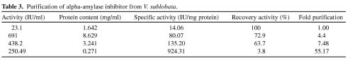
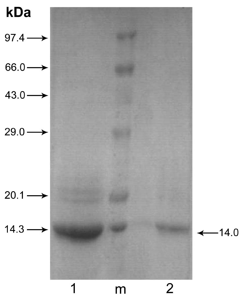
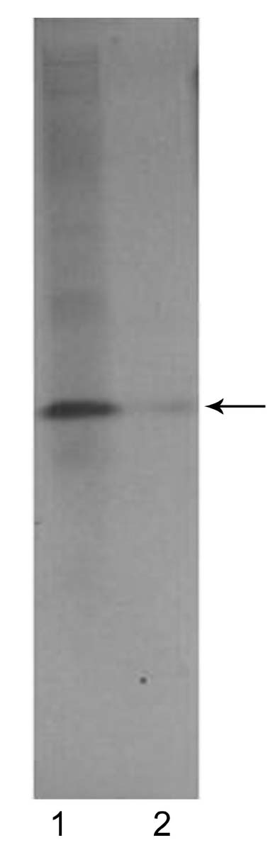
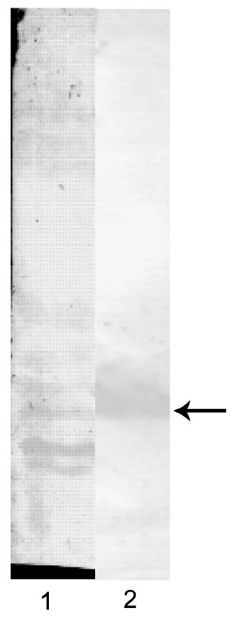
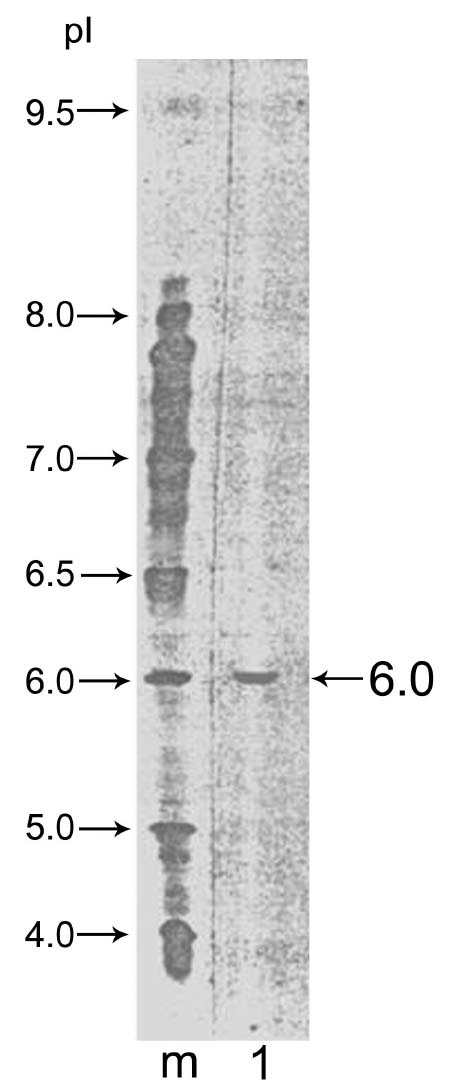
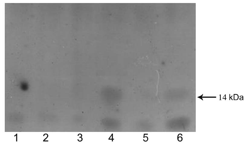
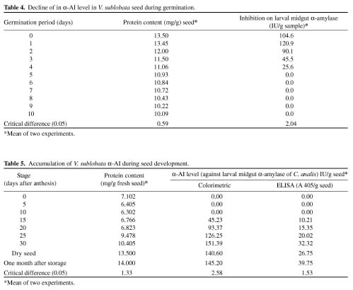
Abstract. Alpha-amylase inhibitor protein, which inhibits the activity of insect (Callosobruchus analis) a-amylase, was characterized from V. sublobata. The molecular weight of purified inhibitor protein was 14 kDa by SDS-PAGE. The inhibitor is non-glycosylated protein and its N-terminal sequence is similar (A P S P V…) to Phaseolus vulgaris a-AI-1. Its pI value is 6.0 and largely localised in cotyledons. The inhibitory activity decreased during germination from days one to five. In the developmental stages of seed formation from anthesis to 30 days the inhibitor content increased.

Keywords: a-Amylase; Alpha-amylase inhibitor; Callosobruchus analis; Vigna sublobata.

Introduction

Many edible plant seeds contain substances that inhibit enzymes, especially hydrolases. Most of these compounds are proteins by nature, which specifically inhibit enzymes by forming complexes that block the active site or alter enzyme conformation, ultimately reducing the catalytic function. Substances present in the seeds of P. vulgaris suggested to play a role in insect resistance include heteropolysaccharides, lectins, and protease- and amylase inhibitors (Applebaum et al., 1969). It has also been suggested that the various forms of resistance are mainly due to the presence of genes of the lectin family (Chrispeels and Raikhel, 1991).

Among the storage pests, the genus- Callosobruchus (Coleoptera: Bruchidae) is the most serious pest on mungbean and other Vigna species (Talekar and Lin, 1981). Alpha-amylase inhibitors have been studied in several common beans (Marshall and Lauda, 1975) maize (Blanco-Labra and Iturbe-Chinas, 1981), sorghum (Kutty and Pattabiraman, 1986), wheat (Warchalewski, 1977), and barley (Mundy and Rogers, 1986). Seed alpha-amylase inhibitor (a-AI) in several cultivars of the common bean plays a protective role against bruchid pests (Ishimoto and Kitamura, 1989). The a-AI strongly inhibited the larval midgut a-amylase activities of adzuki bean weevil (C. chinensis L.) and cowpea weevil (C. maculatus), non-pest species of the common bean. The gene encoding an a-AI was isolated from common bean and transferred to pea (Pisum sativum), and transgenic peas were produced that expressed the transgene (Shade et al., 1994). Bean a-AI-1 in transgenic peas provided complete protection from pea weevil (Bruchus pisorum) under field conditions (Morton et al., 2000). Earlier studies from our laboratory also re

ported that the presence of a-AI is one among the possible strong factors for bruchid resistance in the wild relatives of mungbean, V. sublobata (Sahu, 1996). In the present study an a-AI from V. sublobata has been isolated from seed and characterized.

Materials and Methods

Source of Seed Materials

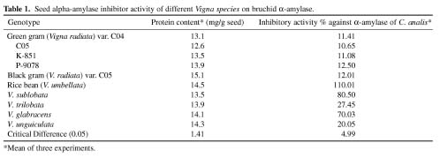
Seeds of Vigna genotypes, including viz., green gram (var. CO 4, CO 5, K-851, P-9078), black gram (var. CO 5), V. glabracens, V. sublobata, V. trilobata and V. umbellata obtained from the Department of Pulses, Center for Plant Breeding and Genetics, Tamil Nadu Agricultural University, Coimbatore were used.

Extraction of Seed Proteins

The extraction of proteins from seed flour was done following the method of Mirkov et al. (1994) for electrophoresis (SDS-PAGE) and immunological analysis. One hundred mg of finely ground seed flour was extracted in the extraction buffer (10 mM Tris-HCl [pH 7.5], 500 mM NaCl, 1% 2-mercapto-ethanol, 0.1% triton-X-100, 2 mM phenylmethylsulphonyl fluoride [PMSF] [1 ml/mg sample]) by homogenization followed by incubation at 4°C for 1 h. It was then centrifuged at 15,000 rpm for 15 min at 4°C. The supernatant was collected and stored frozen in aliquots. The protein content in the extract was quantified following Bradford's (1976) method.

a-AI Assay

The a-AI was assayed by quantifying the reducing sugar (maltose equivalent) liberated under the assay conditions. The enzyme inhibitory activity is expressed as the decrease in units of maltose liberated. A modified dinitrosalicylic acid (DNS) method of Bernfeld (1955) was

*Corresponding author. E-mail: cmkokila@yahoo.com


Botanical Bulletin of Academia Sinica, Vol. 46, 2005

followed to estimate the maltose content. Two hundred microlitres of a-AI extract was pre-incubated with 50 µl of the larval amylase for 30 min, and 1 ml (1% w/v) starch solution was added before further incubating at 37°C for 10 min. The reaction was stopped by adding 1 ml DNS reagent (1 g DNS, 200 mg crystalline phenol, and 50 mg sodium sulphite dissolved in 1% NaOH [w/v] [prepared fresh]), and then the contents were heated in a boiling-water bath for 5 min. One ml potassium sodium tartrate (40% [w/v]) solution was added while the contents were still warm and volume in each tube was made up to 10 ml. A blank was set without a-AI extract, and another one without amylase enzyme, replaced by equal quantities of extraction buffer. The absorbance was measured at 530 nm. The reducing sugar released from starch was estimated as maltose (1 mg/ml) equivalent from the standard graph.

Purification of a-AI

Finely ground seed flour (250 g) of V. sublobata was extracted in 1l of extraction buffer (10 mM Tris-HCl (pH 7.5), 500 mM NaCl, 1% 2-mercapto-ethanol, 0.1% Triton-X-100, 2 mM phenylmethylsulphonyl fluoride, PMSF). The crude protein extract was subjected to ammonium sulphate precipitation (80-100% saturation) at room temperature. The precipitate dissolved in extraction buffer and was dialyzed against water in batches. The dialyzed material was lyophilized and stored at -20°C until further analysis.

The a-AI was fractionated by repeated size exclusion chromatography on a Sephadex G-50 column (26 × 1.2 cm) with 20 mM phosphate buffer (pH 7.0) and then by reversed phased high performance liquid chromatography (RP-HPLC) on C18 column (5 µ column 250 × 4.6 mm). Mobile phase used was acetonitrile : water (70:30) containing 0.1% trifluoroacetic acid (TFA).

Extraction of Alpha-Amylase from Larvae of C. analis

The procedure adopted is a modification of the method used by Bernfeld et al. (1955). Infested greengram seeds were dissected, and the active larvae (100 no.) were homogenized in 2 ml extraction buffer (20 mM sodium phosphate [pH 6.9], 300 mM sodiumchloride, 2 mM calcium

chloride) followed by centrifugation at 15,000 rpm for 30 min at 4°C. The supernatant was distributed into 0.5 ml aliquots and stored at -20°C. This was used as the enzyme extract without any dilution.

Electrophoresis (SDS-PAGE) of Proteins and Immunoblotting

The polypeptides in the samples were fractionated in (15%) polyacrylamide gel in a discontinuous buffer system as described by Laemmli (1970). Polypeptide markers (medium range) were used to estimate the molecular weight of resolved polypeptides. Western blotting and immunostaining of a-AI were carried out as described (Bio-Rad technical bulletin). Pea a-AI antiserum (gift from Dr. Willy Peumans, Belgium) was used.

N-Terminal Amino Acid Sequencing

The N-terminal partial amino acid sequence of the purified a-AI of V. sublobata was determined by Edman degradation using the National Facility available at the Department of Biochemistry, Indian Institute of Science, Bangalore.

Results and Discussion

Among the genotypes screened by conducting preference and non-preference studies of the bruchid pest, C. analis, Vigna sublobata, and V. umbellate, showed resistance to the bruchid attack. Crude protein extracts from the seeds of ten Vigna genotypes were assayed for inhibitory activity against the larval a-amylase of C. analis. Vigna umbellata, V. sublobata, and V. glabracens showed higher levels of inhibitory activity. Vigna trilobata showed a moderate level of inhibition while other Vigna species showed lower inhibitory activity (Table 1). Intervarietal variation in the content of a-AIs in seeds is not uncommon and has been reported in crops such as common bean (Ishimoto and Kitamura, 1991; Ishimoto et al., 1995; Baker et al., 1991; Sanchez-Monge et al., 1996), cowpea (Prasad et al., 1996), barley (Jarrett et al., 1997), and rye (Taeufel et al., 1997). The a-AI content in chickpea varieties ranged from 11.6 to 84.4 units/g seed (Mullimani et al., 1994).


KOKILADEVI et al. — Characterization of a-amylase inhibitor in Vigna sublobata

Purification of a-AI

Crude protein extract of V. sublobata seeds was precipitated using ammonium sulphate at different levels of saturation. Greater a-amylase inhibitory activity (85%) was observed in the fraction precipitated between 80 and 100 percent ammonium sulphate saturation. This fraction was re-suspended in phosphate buffer (pH 7.0) and subsequently applied onto a Sephadex G-50 column. The chromotograph of V. sublobata (Figure 1) showed a minor peak followed by a major peak. The fractions of major peak (fractions 10-14; 25 ml) that showed inhibitory activity were pooled.

RP-HPLC

Sephadex G-50 column-eluted fraction was further fractionated on a C18 column (BDS, Shimadzu). The profile showed six major peaks and five minor peaks (Figure 2). Assay of major peaks (Table 2) revealed that the only peak with retention time 5.2 min of V. sublobata strongly inhibited the C. analis larval a-amylase while others did not.

The protein content of V. sublobata was estimated at 13.5 mg/g seed. For the a-AI purification process 3375 mg crude protein was extracted from 250 g seed flour. Based on the colorimetric assay, it was found that the specific activity of the inhibitor at this stage was very low. In the present study, the seeds of V. sublobata contained 0.8-1.0 percent of the a-AI and showed resistance to bruchids. Subsequent chromatographic separation including RP-HPLC removed another small percentage of the total proteins. The procedure used to purify this inhibitor gave a pure protein, the characteristics of which were closely related to other inhibitors previously reported in different beans.

Ho and Whitaker (1993) achieved an 18.5-fold increase in the specific activity of white kidney bean a-AI from the aqueous crude extract by heat denaturation, ethanol fractionation, and subsequent chromatographic separations on DEAE cellulose column. Transgenic pea seeds, which completely resisted bruchid attack, contained 1-2 percent of the a-AI (Marshall and Lauda, 1975; Blanco-Labra and Iturbe-Chinas, 1981). Fold purification of alpha-amylase inhibitor from V. sublobata is summarized in Table 3.

Molecular Weight

The purified a-AI fraction from V. sublobata showed a molecular weight of 14 kDa (approximately) as estimated

Figure 1. Gel exclusion chromatograph of V. sublobata a-AI on Sephadex G-50 column. Lyophilized sample (2 mg/ml) in equilibration buffer (0.02 M sodium phosphate, pH 6.9 containing 0.3 M NaCl) was applied on to Sephadex G-50 column (26 × 1.2 cm) bed volume 30 ml.

Figure 2. HPLC profile of V. sublobata crude protein extract from seed. The fraction obtained from Sephadex G-50 column was subjected to RP-HPLC (Hypersil BDS C18, 5 µ column [250 × 4.6 mm]). Fifty microlitre of fraction was injected into the system. The proteins were eluted using a linear gradient of 70% acetonitrile containing 0.1% TFA.


Botanical Bulletin of Academia Sinica, Vol. 46, 2005

by Sephadex G-50 gel filtration in relation to standard proteins (aprotinin, cytochrome-C, carbonic anhydrase, ovalbumin, bovine serum albumin and phosphorylase b) chromatographed, separately. The analysis of the purified protein on SDS-PAGE confirmed the above observation. The a-AI showed a molecular weight of 14 kDa on a 15% PAGE containing SDS (Figure 3). Ho and Whitaker (1993) found that the molecular weight of white kidney bean a-AI (WKB 858 A) was 42, 40 and 38 kDa on FPLC superose gel filtration chromatography, Sephadex G-75 gel filtration chromatography, and native gel electrophoresis, respectively. a-AIs form a large family of proteins with varying molecular masses. Insect-specific albumins are low molecular weight proteins of 12-24 kDa (Feng et al., 1996). Rice a-AIs are relatively small proteins of around 14 kDa (Feng et al., 1991).

Native Gel Analysis

The purified a-AI protein from V. sublobata was subjected to electrophoresis under non-denaturing and non-

reducing conditions. The results showed a single band after silver staining revealing the (near) homogeneity of the purified fraction (Figure 4).

Western Blot Patterns

Western blot analysis of V. sublobata using French bean a-AI antiserum revealed an array of at least five bands in the low molecular weight region. The weak signals seen in the high molecular weight range may be due to other proteins, such as arcelin and phytohaemagglutinins (PHA), antigenically related to a-AI. The Western blot analysis of purified proteins from V. sublobata using French bean a-AI antiserum clearly showed a single band in the 14 kDa region (Figure 5). The French bean a-AI antiserum used in the present study is polyclonal in nature and hence capable of producing many immunosignals. The proteins from other crop seeds have also been reported to have molecular weights in the range of 14-20 kDa. In addition, many different types of a-AI do occur. The result of the present study is in accordance with that of the report by

Figure 3. Molecular weight of a-AI from V. sublobata seed. Crude proteins (»20 mg) were fractionated on 15% SDS-PAGE. Lane 1, 80-100% ammonium sulphate precipitated proteins; lane 2, Sephadex G-50 column eluted protein; and lane m, molecular weight marker.

Figure 4. Purified a-AI from V. sublobata seed on a native gel. Protein (»5 mg) was electrophoresed on 15% gel under non-reducing and non-denaturing conditions and silver stained. Lane 1, crude extract and lane 2, purified protein.


KOKILADEVI et al. — Characterization of a-amylase inhibitor in Vigna sublobata

Iguti and Lajolo (1991), wherein isoforms of a-AI have been reported in French bean.

N-Terminal Sequence

The purified protein of V. sublobata was transferred onto PVDF membrane and subjected to sequencing. The first five amino acids in the N-terminus are: Ala-Pro-Ser-Pro-Val (A P S P V). Furthermore, the protein is not N-terminally blocked. The first five amino acid residues of V. sublobata a-AI are similar to those of P. vulgaris a-AI-1 (Blanco-Labra et al., 1996). The N-terminus of V. sublobata inhibitor showed less homology with the already characterized legume and cereal a-AIs. French bean a-AI antiserum cross-reacted with V. sublobata has an amino acid sequence more or less similar to P. vulgaris a-AI-1.

pI of the Protein

The a-AI protein from V. sublobata was focused at pI 6.0 (Figure 6). Common bean a-AI-1 showed wider pI values (4.5-6.5). The pI value for tepary bean is 4.7 (Blanco-Labra et al., 1996). Most of the reported a-AI proteins showed pI between 4.5 and 9.0. Blanco Labra et al. (1996) reported the a-AI of tepary bean (Phaseolus acutifolius) to be pH 4.7.

Localisation of a-AI

The protein is mainly localized in the cotyledons and embryonic axes as examined by SDS-PAGE followed by silver staining (Figure 7). Dot blot analysis with French bean

Figure 5. Protein blot analysis. Immunoblot of SDS-PAGE separated polypeptides from V. sublobata (lane 1); purified protein (5 µg) on immunoblot was probed with French bean a-AI antiserum (lane 2).

(a)

(b)

Figure 7. (a) Localization of a-AI protein from V. sublobata. Protein (5 µg) were fractionated on 12% SDS-PAGE and silver-stained. Lane 1, leaf; lane 2, stem; lane 3, seed coat; lane 4, cotyledon; lane 5, root and lane 6, embryonic axis. (b) Dot blot analysis for the localization of a-AI in different tissues of V. sublobata. Crude extract (20 mg protein) were probed with French bean a-AI antiserum. Dot 1, seed coat; dot 2, leaf; dot 3, stem; dot 4, cotyledon and dot 5, embryonic axis.

Figure 6. Isoelectric focusing of the purified a-AI. Five µg of the purified a-AI protein from V. sublobata was subjected to isoelectric focusing. Lane m, pI markers; lane 1, V. sublobata.


Botanical Bulletin of Academia Sinica, Vol. 46, 2005

a-AI antiserum reinforced the above observation (Figure 7). Earlier studies by Moreno et al. (1989) showed that in common bean the a-AI was detected only in the cotyledons and embryonic axis.

a-AI in V. sublobata During Germination and Seed Development

The fate of a-AI during seed germination was followed by assaying the inhibition of C. anlis amylase using the extracts prepared at 24-h intervals from the time of soaking to the 10th day of germination. The inhibitory activity decreased from day one through five. No activity was observed during subsequent days of germination (Table 4). SDS-PAGE also showed that the a-AI (14 kDa) polypeptide band disappeared beyond 8 days of germination (Data not shown).

During seed development, the inhibitor accumulated in the seeds at levels detectable by ELISA within 15 days of seed development (Table 5). The inhibitory activity was nearly or completely undetectable by both colorimetric and

ELISA until day 10 of seed development. The level increased up to 30 days of seed formation. Thereafter, the inhibitor level remained unaltered or slightly decreased during the drying of the seed.

As the germination progressed, the seed inhibitor content decreased. The inhibitor was undetectable in the cotyledons by day 5 of germination (Table 3). This result is in accordance with the findings of Baumgartner and Chrispeels (1976) in mungbean. Moreno et al. (1989) showed that in bean seeds the a-AI appeared in detectable amounts seventeen days. The characterisation of new inhibitors, such as the one isolated in the present work, is important considering that inhibitors obtained from different sources differ in characteristics. In this work the V. sublobata a-AI demonstrated activity against C. analis that attack mungbean (V. radiata). Considering that V. sublobata can hybridize with V. radiata and V. mungo, improving this crop may be possible through traditional breeding programs, with the aim of obtaining materials of better specific resistance characteristics to these insect pests.


KOKILADEVI et al. — Characterization of a-amylase inhibitor in Vigna sublobata

Literature Cited

Applebaum, S.W., U. Macro, and Y. Birk. 1969. Saponins as possible factors of resistance of legume seeds to the attack of insects. J. Agric. Food Chem. 17: 618-622.

Baker, J.E., S.M. Woo, J.E. Thorne, and P.L. Finney. 1991. Correlation of alpha-amylase inhibitor content in Eastern soft wheats with development parameters of the rice weevil. (Coleoptera, Curculionidae). Env. Entomol. 20: 53-60.

Baumgartner, B. and M.J. Chrispeels. 1976. Partial characterization of a protease inhibitor which inhibits the major endopeptidase present in the cotyledons of mung beans. Plant Physiol. 58: 1-6.

Bernfeld, P. 1955. Amylases, a and b. In S.P. Colowick and N.O. Kaplan (eds.), Methods in Enzymology. Academic Press, New York, pp. 149-158.

Blanco-Labra, A. and F.A. Iturbe-Chinas. 1981. Purification and characterization of glycoprotein alpha-amylase inhibitor from tepary bean seeds (Phaseolus acutifolius A. Gray). J. Food Biochem. 5: 1-17.

Blanco-Labra, A., L.S. Cardoso, E.M. Olaya, S.V. Rodriguez, and M.G. Lopez. 1996. Purification and characterization of glycoprotein alpha-amylase inhibitor from tepary bean seeds (Phaseolus acutifolius A. Gray). J. Plant Physiol. 149: 650-656.

Bradford, M.M. 1976. A rapid and sensitive method for the quantification of microgram quantities of protein utilizing the principle of protein dye-binding. Anal. Biochem. 72: 248-254.

Chrispeels, M.J. and N.V. Raikhel. 1991. Lectins lectin genes, and their role in plant defense. Plant Cell. 3: 1-9.

Feng, G.H., M.S. Chen, K.J. Kramer, and G.R. Reeck. 1991. Alpha-amylase inhibitors from rice: fractionation and selectivity toward insect, mammalian, and bacterial alpha-amylases. Cereal Chem. 68: 516-521.

Feng, G.H., N.T. Richardson, M.S. Chen, K.J. Kramer, T.D. Morgan, and G.R. Reeck. 1996. Alpha-amylase inhibitors from wheat: Amino acid sequences and patterns of inhibition of insect and human alpha-amylases. Insect Biochem. Mol. Biol. 26: 419-426.

Ho, M.F. and J.R. Whitaker. 1993. Purification and partial characterization of white kidney bean (Phaseolus vulgaris) alpha-amylase inhibitors from two experimental cultivars. J. Food Biochem. 17: 15-33.

Iguti, A.M. and F.M. Lajolo. 1991. Occurrence and purification of alpha-amylase isoinhibitor in bean (Phaseolus vulgaris L.) varieties. J. Agric. Food Chem. 39: 2131-2136.

Ishimoto, M. and K. Kitamura. 1989. Growth inhibitory effects of an a-amylase inhibitor from the kidney bean, Phaseolus vulgaris (L.) on three species of bruchids (Coleoptera: Bruchidae). Appl. Ent. Zool. 24: 281-284.

Ishimoto, M. and K. Kitamura. 1991. Effect of absence of seed alpha-amylase inhibitor on the growth inhibitory activity to adzuki bean weevil (C. chinensis) in common bean (Phaseolus vulgaris L.). Jap. J. Breed. 41: 231-240.

Ishimoto, M., K. Suzuki, M. Iwanaga, F. Kikuchi, and K. Kitamura. 1995. Variation of seed alpha-amylase inhibitors in the common bean. Theor. Appl. Genet. 90: 425-429.

Jarrett, S.J., R.J. Marschke, M.H. Symons, C.E. Gibson, R.J. Henry, and G.P. Fox. 1997. Alpha-amylase/subtilisin inhibitor levels in Australian barleys. J. Cereal Sci. 25: 261-266.

Kutty, A.V.M. and T.N. Pattabiraman. 1986. Isolation and characterization of an amylase inhibitor from sorghum seeds, specific for human enzymes. J. Agric. Food Chem. 34: 552-557.

Laemmli, E.K. 1970. Cleavage of structural proteins during the assembly of the head of bacteriophage T4. Nature 227: 680-685.

Marshall, J.J. and C.M. Lauda. 1975. Purification and properties of phaseolamin, an inhibitor of alpha-amylase from the kidney bean, Phaseolus vulgaris. J. Biol. Chem. 250: 8030-8037.

Mirkov, T.E., J.M. Wahlstrom, K. Hagiwara, F. Finardi-Filho, S. Kjemtrup, and M.J. Chrispeels. 1994. Evolutionary relationship among proteins in the phytohemagglutinin-arcelin-alpha-amylase inhibitor family of the common bean and its relatives. Plant Mol. Biol. 26: 1103-1113.

Moreno, J., T. Altabella, and M.T. Chrispeels. 1989. Characterization of alpha-amylase inhibitor, a lectin-like protein in the seeds of Phaseolus vulgaris. Plant Physiol. 92: 703-709.

Morton, R.L., H.E. Schroeder, K.S. Bateman, M.J. Chrispeels, E. Armstrong, and T.J. Higgins. 2000. Bean alpha-amylase inhibitor 1 in transgenic peas (Pisum sativum) provides complete protection from pea weevil (Bruchus pisorum) under field conditions. Proc. Nat. Acad. Sci. USA 97: 3820-3825.

Mullimani, V.H., G. Rudrappa, and D. Supriya. 1994. Alpha-amylase inhibitors in chickpea (Cicer arietinum L.). J. Sci. Food Agric. 64: 413-415.

Mundy, J. and J.C. Rogers. 1986. Selective expression of a probable alpha-amylase/subtilisin inhibitor. Planta 169: 51-63.

Prasad, D.T., N.S. Umapathy, and R. Veeranna. 1996. Genotypic variation in cowpea (Vigna unguiculata) cultivars in relation to insect resistance. J. Plant Biochem. Biotech. 5: 47-49.

Sahu, R.K. 1996. Alpha-Amylase Inhibitor and Bruchid Resistance in Grain Legumes. M.Sc. (Biotech) Thesis. Tamil Nadu Agricultural University, Coimbatore.

Sanchez-Monge, R., G. Garcia-Casado, J.M. Malpika, and G. Salcedo. 1996. Inhibitory activities against heterologous alpha-amylases and in vitro allergenic reactivity of Einkorn wheats. Theor. Appl. Genet. 93: 745-750.

Shade, R.E., H.E. Schroeder, J.J. Peuyo, L.M. Murdock, T.J. Higgins, and M.J. Chrispeels. 1994. Transgenic pea seeds expressing the alpha-amylase inhibitor of the common bean are resistant to bruchid beetles. Biotechnol. 12: 793-796.

Taeufel, A., H. Bohm, and W. Flamme. 1997. Protein inhibitors of alpha-amylase in mature and germinating grain of rye (Secale cereal). J. Cereal Sci. 25: 267-273.

Talekar, N.S. and Y.H. Lin. 1981. Two sources with differing modes of resistance to Callosobruchus chinensis in mungbean. J. Econ. Entomol. 74: 634-642.

Warchalewski, J.R. 1977. Isolation and purification of native alpha-amylase inhibitors from winter wheat. Bull. Acad. Polon Sci. Ser. Sci. Biol. 25: 731-735.


Botanical Bulletin of Academia Sinica, Vol. 46, 2005