Bot. Bull. Acad. Sin. (2005) 46: 217-222

WANG et al. — Nuclear transplantation in Antrodia cinnamomea

Variation and segregation following nuclear transplantation in Antrodia cinnamomea

Wei-Ming WANG1, Rey-Yuh WU1, and Wen-Hsiung KO2,*

1Development Center for Biotechnology, 101 Lane 169, Kang Ning St., Hsi Chih, Taipei, Taiwan

2Department of Plant and Environmental Protection Sciences, University of Hawaii at Manoa, Hilo, Hawaii 96720, USA

(Received July 29, 2004; Accepted January 27, 2005)

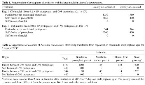
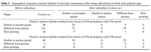
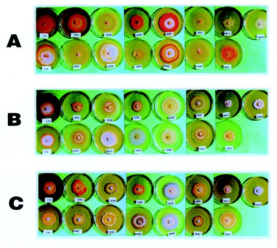
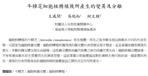
Abstract. Intraspecific fusion of protoplasts with nuclei in Antrodia cinnamomea lengthened the time for development of colonies on regeneration medium and reduced the number of protoplasts capable of regeneration. When 2.1 × 10⁵ C98 protoplasts were fused with C96 nuclei, 1750 colonies appeared on the regeneration medium, and 742 of them were putative nuclear hybrids as their colonies were different from that of the protoplast parent. Without the presence of C96 nuclei, 3160 colonies appeared on the regeneration medium and 400 of them tested were similar to the protoplast parent in colony appearance. No colonies appeared when self-fused nuclei were plated on regeneration medium. The rate of successful transfer was 23%. The success rate was 2.4% for reciprocal transfer. About 77 to 82% of nuclear hybrids were slow in growth. These isolates resumed normal growth upon subculture. All the nuclear hybrids obtained were unstable. Segregation occurred during growth of mycelia, resulting in the formation of colonies different from those of the originals in subcultures. After five consecutive subcultures, 13 stable nuclear hybrids were obtained. These isolates differed greatly from the parental isolates and from each other in pigmentation, growth rate, and colony pattern.

Keywords: Antrodia cinnamomea; Colony morphology; Nuclear hybrid; Nuclear transplantation.

Introduction

Antrodia cinnamomea T. T. Chang & W. N. Chou is a rare and extremely valuable medicinal fungus in Taiwan (Chang and Chou, 1995, 2004). The basidiomes of the fungus are well known locally for their effectiveness in treating various human illnesses, including liver cancer, kidney problems, diabetes, and food poisoning (Chen et al., 2001b). New compounds with different medicinal properties have been isolated from these basidiomes in recent years (Chen and Yang, 1995; Cherng and Chiang, 1995; Cherng et al., 1995; Chiang et al., 1995). Antrodia cinnamomea is indigenous to Taiwan, and the protected precious endemic tree species Cinnamomum kanehirai Hayata is its only known host (Chang and Chou, 1995). Since it is still impossible to produce fruiting bodies of A. cinnamomea in cultures, the basidiomes of the fungus collected from natural forests by the natives are in great demand and have been sold for about U.S. $15,000 per kg (Wu et al., 1997). Some medicinal effects have been detected in liquid as well as solid cultures of A. cinnamomea (Chen et al., 2001a; 2001b). Isolates of this fungus with the ability to produce large quantities of medicinally ac

tive compounds are, therefore, urgently needed. Unfortunately, the number of isolates available for selection is very limited because of the plant's rarity in nature.

Recently, hybrids of Phytophthora resulting from nuclear transplantation have been shown to display increased variation in both physiological and morphological characteristics (Gu and Ko, 1998; 2000; 2001). A project was, therefore, initiated to develop a protocol for the nuclear transplantation of A. cinnamomea to generate variation for selection of strains capable of producing large quantities of medicinal compounds. Reports on nuclear transplantation in fungi are few. Successful application of nuclear transplantation requires a selective method for isolation of nuclear hybrids. Initially, complementary auxotrophic mutants were used, and prototrophic strains capable of growing on minimal medium subsequent to nuclear transplantation were selected as nuclear hybrids (Ferency and Pesti, 1982: Sivan et al., 1990; Vagvolgyi and Ferenczy, 1992). Recently, two mutants, each resistant to a different fungicide, were used, and strains capable of growing on medium containing both fungicides were selected as nuclear hybrids (Gu and Ko, 1998; 2000; 2001). The generation of auxotrophic mutants and induction of fungicide resistant mutants are tedious and time consuming tasks. In this study we developed a method for detection of nuclear hybrids based on a comparison of colony morphology with parental types.

*Corresponding author. Tel: +1-808-974-4105; Fax: +1-808-974-4110; E-mail: kowh@hawaii.edu


Botanical Bulletin of Academia Sinica, Vol. 46, 2005

Materials and Methods

Cultures

Isolates C96 (CCRC 35396) and C98 (CCRC 35398) of A. cinnamomea were obtained from Culture Collection and Research Center of Food Industry Research and Development Institute, Hsinchu, Taiwan. They were cultured on malt peptone agar consisting of 2% malt extract, 2% glucose, 0.1% peptone, and 2% agar. After incubation at 28°C for 21 days on this medium, C96 formed a pink colony with a whitish center while C98 formed a lightly yellow colony with a yellow center. These two isolates were very stable. Their colony morphology did not change over repeated subcultures.

Isolation of Protoplasts

The existing methods (Gu and Ko, 1998; Stasz et al., 1988) were modified for protoplast isolation. Eight culture blocks (ca 4 × 4 × 2 mm) obtained from the advancing margin of a colony were cut into tiny pieces with a scalpel and used to inoculate 10 ml of malt peptone broth consisting of 2% malt extract, 2% glucose, and 0.1% malt peptone in a 125 ml flask. After incubation on a rotary shaker at 28°C for 3 days, young mycelia were collected on Miracloth (Calbiochem, La Jolla, CA) and washed twice each with 200 ml of a stabilizer solution consisting of 0.6 M mannitol, 0.01 M CaCl2, and 0.01 M Tris-HCl, pH 7.5. Mycelia were transferred to a 50-ml centrifuge tube and incubated with 5 ml Novozyme 234 (Novo Laboratories, Wilton, CT) at the concentration of 10 mg/ml stabilizing solution for 3-4 h at 28°C with gentle shaking to release protoplasts. Protoplasts were separated from non-digested mycelia by filtration through Miracloth and 150 µm nylon sieve and washed twice each with 10 ml stabilizer solution by centrifugation at 100 g for 5 min. The entire procedure was performed under aseptic conditions. The number of nuclei in protoplasts obtained in this study ranged from 0 to 2 per protoplast. The concentration of protoplasts was determined with a microliter pipette (P20, West Coast Scientific, Oakland CA) (Ko et al., 1973).

Nuclear Isolation

The method by Sivan et al. (1990) was modified for isolation and transplantation of nuclei. Protoplasts in 0.5 ml suspension were pelleted by centrifugation at 1,000 g for 15 min, and resuspended in 0.2 ml extraction buffer consisting of 10 mM Tris-HCI (pH 7.0), 4 mM spermidine hydrochloride, 1 mM spermine hydrochloride, 0.1 M KCl, 10 mM EDTA, 0.5 M sucrose, 1 mM phenylmethylsulfonyl flouoride, and 0.1% 2-mercaptoethanol. The suspension was mixed with 50 mg fine quartz sand adjusted to 5 ml with extraction buffer and homogenized for 3 min in a Tenbroeck tissue grinder (Corning Glass Works, Corning, NY) to rupture the protoplasts. The homogenized suspension was filtered sequentially through polycarbonate membranes (Nuclepore Corp., Pleasanton, CA) with pore sizes of 20, 12, 10, 5, and 2 µm to remove residual protoplasts and centrifuged at 1,500 g for 5 min for sedimentation of

nuclei but not mitochondria (Bhargava and Halvorson, 1971). The nuclear pellet was suspended in 1 ml extraction buffer. All the processes of nuclear isolation were performed at 4°C. To assure the purity of the preparations, drops of nuclear suspension were stained with DAPI (4´, 6-diamidino-2-phyenylindole) (Stasz et al., 1988) or Rhodamine 123 (Weiss and Chen, 1984) to detect the presence of nuclei and mitochondria, respectively. After being stained with DAPI, isolated nuclei of C96 and C98 appeared as round bodies and irregular masses, respectively. No intact protoplasts were detected. Rhodamine staining also did not detect mitochondria, thus confirming the purity of the preparations.

Nuclear Transplantation

For nuclear transplantation, 1 ml nuclear suspension from 1 ml protoplast suspension was gently added to the 0.5 ml protoplast suspension in a centrifuge tube. After incubation at 30°C for 5 min, 100 µl fusion solution consisting of 60 (w/v) PEG 3350, 10 mM CaCl2 and 10 mM Tris-HCl (pH 7.5) was gently mixed with the protoplast-nucleus suspension. A second and third 250 µl fusion solution were added and mixed at 5 min intervals. The nuclei and protoplasts were then forced together by centrifugation at 1,000 rpm for 5 min. This pellet was incubated for an additional 5 min and suspended in 2 ml stabilizer solution. The diluted mixture was centrifuged at 1,000 rpm for 5 min, and the supernatant was replaced with 1.2 ml stabilizer solution. The whole protoplast-nucleus suspension was spread on six plates (0.2 ml/plate) of regeneration medium consisting of malt peptone agar plus 10% sucrose. For controls, the same amount of nuclei and protoplasts was separately processed through the same fusion procedure and suspended in 0.8 ml stabilizer solution. Each suspension was spread on four plates of regeneration medium.

A marking pen was used to mark 100 dots on the bottom of each malt peptone agar plate (Ho and Ko, 1997). After regeneration at 28°C for 100 h, all the colonies regenerated from fusion products were each isolated and placed on the surface of the agar medium above one of the dots. The number of colonies regenerated from self fusion protoplasts was estimated by counting colonies in one-eighth of each plate. After counting, 400 colonies were isolated as described above for each experiment.

Segregation of Putative Nuclear Hybrids

For observation of colony characteristics, each colony from initial isolation plates was transferred to the center of a malt peptone agar plate and incubated at 28°C for 7 days. For the study of segregation, a 10-mm culture disk was obtained from the advancing margin of a putative nuclear hybrid with a cork borer, placed in the center of the malt peptone agar plate, and incubated at 28°C for 7 days. For comparison of the colony characteristics between stable putative nuclear hybrids and the parental isolates, each isolate was grown on malt peptone agar, potato dextrose agar (Difco Lab., Michigan), and malt extract agar (Difco Lab., Michigan) for 28 days at 28°C in darkness.


WANG et al. — Nuclear transplantation in Antrodia cinnamomea

protoplasts. When the same amount of C96 protoplasts were fused with C98 nuclei, only 344 colonies appeared on the regeneration plates (Table 1). Nuclear fusion caused an approximate 97% reduction in number of C96 protoplasts capable of regeneration.

Colony Characteristics of Products of Nuclear Transplantation

Among the 1750 isolates regenerated from C98 protoplasts following fusion with C96 nuclei, 1008 isolates were similar to the protoplast parent and 742 isolates were different from the protoplast parent (Table 2). The latter were considered putative nuclear hybrids, as all of the 400 isolates regenerated from self-fused C98 protoplasts were similar to the protoplast parent. The putative hybrids included 38 isolates similar to the nuclear parent, 134 isolates different from both parents, and 570 isolates slow in growth (Table 2).

Among the 344 isolates regenerated from C96 protoplasts following fusion with C98 nuclei, 98 isolates were similar to the protoplast parent and 246 isolates were different (Table 2). The latter were considered putative nuclear hybrids, as all the 400 isolates regenerated from self-fused C96 protoplasts were similar to the protoplast parent. The nuclear hybrids included 40 isolates similar to the nuclear parent, 4 isolates different from both parents, and 202 isolates slow in growth (Table 2).

Segregation of Putative Nuclear Hybrids

When colonies of putative nuclear hybrids were subcultured by transferring a small piece of agar culture from

Results

Protoplast Regeneration after Nuclear Transplantation

Fusion of protoplasts with nuclei lengthened the regeneration time and reduced the regeneration frequency of the protoplasts. Numerous tiny colonies appeared in all four regeneration plates containing self-fused C98 protoplasts in 52 h. During the same period of time, three out of six regeneration plates containing C98 protoplasts fused with C96 nuclei showed a scant number of tiny colonies. None of the other plates had any colonies. It took 100 h for all six plates to become full of tiny colonies. No colonies appeared on the regeneration plates containing self-fused nuclei during the experiment. After 100 h, approximately 3160 colonies appeared on the regeneration plates containing self-fused C98 protoplasts. When the same number of C98 protoplasts were fused with C96 nuclei, only 1750 colonies appeared on the regeneration plates (Table 1). Nuclear fusion caused about a 50% reduction in the number of C98 protoplasts capable of regeneration.

All four regeneration plates containing self-fused C96 protoplasts were also full of tiny colonies in 52 h, and only one out of six regeneration plates containing C96 protoplasts fused with C98 nuclei showed some tiny colonies during the same time. None of the other five plates had any colonies. It took 100 h for tiny colonies to appear on all six plates. No colonies appeared on the regeneration plates containing self-fused C98 nuclei during the experiment. After 100 h, about 10360 colonies appeared on the regeneration plates containing self-fused C96


Botanical Bulletin of Academia Sinica, Vol. 46, 2005

the advancing margin to fresh malt peptone agar, all of them developed colonies different from their origins in appearance (Table 3). For putative nuclear hybrids resulting from fusion of C98 protoplasts with C96 nuclei, 38 isolates similar to the nuclear parent, 134 isolates different from both parents, and 10 slow growing isolates were tested. Among the 38 isolates similar to the nuclear parent, 29 isolates reverted and became similar to the protoplast parent while 9 became different from both parents as a result of segregation during mycelial growth. Of the 134 isolates different from both parents, 75 reverted and became similar to the protoplast parent. All of the 10 slow growing isolates tested reverted to normal growth following segregation. Nine of them became similar to the protoplast parent while the remaining one became different from both parents.

For putative nuclear hybrids resulting from fusion of C96 protoplasts with C98 nuclei, 40 isolates similar to nuclear parent, 4 isolates different from both parents, and 10 slow growing isolates were tested. Among the 40 isolates similar to the nuclear parent, 18 isolates reverted and became similar to the protoplast parent while 22 isolates became different from both parents as a result of segregation during mycelial growth (Table 3). All 4 isolates different from the parents reverted and became similar to the protoplast parent. All 10 slow growing isolates tested also became normal in growth following segregation. Eight of them reverted and became similar to the protoplast parent while the other two became different from the parent.

All isolates that were different from both parents after subculture (Table 3) were subcultured once every 14 days by transferring a small piece of agar culture from the advancing margin to fresh malt peptone agar. Thirteen stable nuclear hybrids were obtained, after five consecutive subcultures. The 10 isolates from the fusion of C96 nuclei with C98 protoplasts were designated S68A to S68J while the other 3 isolates from the fusion of C98 nuclei with C96 protoplasts were designated S86A and S86C. These stable nuclear hybrids differed from the parental C98 and C96 isolates in color, size, and pattern of the colony on the same medium, and in response to change of culture medium (Figure 1).

Discussion

When protoplasts of C98 or C96 isolates of A. cinnamomea were processed through fusion treatment, the regenerated colonies were similar to their respective parent in colony appearance, and the same type of colony appeared on subsequent subculture. However, when these protoplasts were fused with nuclei from different isolates, 42 and 72% of the regenerated colonies were different from their respective parents in the reciprocal experiments. All these cultures displayed segregation during the growth of mycelia, suggesting that these isolates were hybrids resulting from nuclear transplantation. Our result demonstrates the possibility of detecting nuclear hybrids based on comparison of colony morphology. Although this novel method requires the isolation of a large number of colonies for comparison, it obviates the tedious and time consuming process of generating auxotrophic or fungicide-resistant mutants.

When 2.1 × 10⁵ C98 protoplasts were fused with C96 nuclei, 1750 colonies appeared on the regeneration medium (Table 1), and 742 of them were putative nuclear hybrids (Table 2). Without the presence of C96 nuclei, 3160 colonies regenerated on the regeneration medium. The rate of successful transfer was 23%. The successful rate was 2.4% for reciprocal transfer. The rates of successful nuclear transplantation obtained in this study were higher than those reported previously (Ferenczy and Pesti, 1982; Gu and Ko, 2000; 2001; Sivan et al., 1990; Vagvolgyi and Ferenczy, 1992). The isolates used in this study were wild types while those used by others were mutants. It is conceivable that mutation treatment of the test organisms might have weakened the regeneration ability of their protoplasts following nuclear transplantation.

Nuclear hybrids obtained in this study were unstable. Sector formation was very common, and all of their subcultures showed different colony morphology from their origins. A parasexual cycle is known to occur following nuclear transplantation (Gu and Ko, 1998). It is possible that in the nuclear hybrids, heterozygous diploid nuclei may result from spontaneous nuclear fusion. Aneuploid nuclei and recombinant haploid nuclei may also result from


WANG et al. — Nuclear transplantation in Antrodia cinnamomea

Figure 1. Colony characteristics of parental isolates and nuclear hybrids of Antrodia cinnamomea on malt peptone agar (A), malt extract agar (B), and potato dextrose agar (C) after incubation at 28°C in darkness for 28 days. C98 and C96 were parental isolates. S68A to S68J were hybrids resulted from fusion of C96 nuclei with C98 protoplasts while S86A to S86C were from fusion of C98 nuclei with C96 protoplasts.

chromosome nondisjunction and mitotic crossing over (Brody and Williams, 1974). Segregation of these different kinds of nuclei may occur during mycelial growth resulting in the formation of sectors and the appearance of different kinds of colony morphology during consecutive subcultures.

About 77 to 82% of nuclear hybrids were slow in growth. However, these colonies resumed normal growth upon subculture. An inserted foreign nucleus is apparently inhibitory to growth of hybrid cells. After some adjustment, hybrid cells probably could overcome the inhibitory effect by suppression or by elimination of the inhibitory factor. Nuclear transfer of Trichoderma harzianum (Sivan et al., 1990) and Aspergillus nidulans (Vagvolgyi and Ferenczy, 1992) also caused slow growth of regenerated colonies. Increased growth rates were also observed among single-

spore isolates derived from slow-growing nuclear hybrids of T. harzianum (Sivan et al., 1990). Growth inhibition of nuclear hybrids by inserted nucleus, and the subsequent reversion during subculture appear to be unique to haploid fungi. No similar phenomenon was observed in the nuclear hybrid of diploid Phytophthora (Gu and Ko, 1998; 2000). In fact, most of the intraspecific and interspecific nuclear hybrids of Phytophthora parasitica and P. capsici exhibited greater growth rates than their parental isolates (Gu and Ko, 2001).

The stable nuclear hybrids obtained in this study differed greatly from the parental isolates and from each other in pigmentation, growth rate, and colony pattern (Figure 1). Whether these nuclear hybrids can produce larger quantity of medicinally active compounds than the parental isolates remains to be tested.


Botanical Bulletin of Academia Sinica, Vol. 46, 2005

Literature Cited

Bhargava, M.M. and H.O. Halvorson. 1971. Isolation of nuclei from yeast. J. Cell Biol. 49: 423-429.

Brody, T. and K.L. Williams. 1974. Cytological analysis of the parasexual cycle in Dictyostelium discorideum. J. Gen. Microbiol. 82: 371-383.

Chang. T.T. and W.N. Chou. 1995. Antrodia cinnamomea sp. nov. on Cinnamomum kanehirai in Taiwan. Mycol. Res. 99: 756-758.

Chang, T.T. and W.N. Chou. 2004. Antrodia cinnamomea reconsidered and A. salmonea sp. nov. on Cunninghamia konishii in Taiwan. Bot. Bull. Acad. Sin. 45: 347-352.

Chen, C.H. and S.W. Yang. 1995. New steroid acids from Antrodia cinnamomea, a fungal parasite of Cinnamomum micranthum. J. Nat. Prod. 58: 1655-1661.

Chen, C.J., C.H. Su, and M.H. Lan. 2001a. Study on solid cultivation and bioactivity of Antrodia camphorata. Fung. Sci. 16: 65-72.

Chen, C.J., W.H. Lin, C.N. Chen, S.J. Sheu, S.T. Huang, and Y.L. Chen. 2001b. Development of Antrodia camphorata mycelium with submerge culture. Fung. Sci. 16: 7-22.

Cherng, I.H. and H.C. Chiang. 1995. Three new triterpenoids from Antrodia cinnamomea. J. Nat. Prod. 58: 365-371.

Cherng, I.H., D.P. Wu, and H.C. Chiang. 1995. Triterpenoids from Antrodia cinnamomea. Phytochemistry 41: 263-267.

Chiang, H.C., D.P. Wu, I.W. Cherng, and C.H. Ueng. 1995. A sesquiterpenelactone, phenyl and biphenyl compounds from Antrodia cinnamomea. Phytochmistry 39: 613-616.

Ferenczy, L. and M. Pesti. 1982. Transfer of isolated nuclei into protoplasts of Saccaromyces cerevisiae. Curr. Microbiol. 7: 157-160.

Gu, Y.H. and W.H. Ko. 1998. Occurrence of a parasexual cycle following the transfer of isolated nuclei into protoplasts of Phytophthora parasitica. Curr. Genet. 134: 120-123.

Gu, Y.H. and W.H. Ko. 2000. Segregation following interspecific transfer of isolated nuclei between Phytophthora parasitica and P. capsici. Can. J. Microbiol. 46: 410-416.

Gu, Y.H. and W.H. Ko. 2001. Creation of hybrid vigor through nuclear transplantation in Phytophthora parasitica. Can. J. Microbiol. 47: 662-666.

Ho, W.C. and W.H. Ko. 1997. A simple method for obtaining single-spore isolates of fungi. Bot. Bull. Acad. Sin. 38: 41-44.

Ko, W.H., L.L. Chase, and R.K. Kunimoto. 1973. A microsyringe method for determining concentration of fungal propagules. Phytopathology 63: 1206-1207.

Sivan, A., G.E. Harman, and T.E. Stasz. 1990. Transfer of isolated nuclei into protoplasts of Trichoderma harzianum. Appl. Environ. Microbiol. 56: 2404-2409.

Stasz, T.E., G.E. Harman, and N.F. Weeden. 1998. Protoplast preparation and fusion in two biocontrol strains of Trichoderma harzianum. Mycologia 80: 141-150.

Vagvolgyi, C. and L. Ferenczy. 1992. Transfer of isolated nuclei into protoplasts of Aspergillus nidulans. Experientia 48: 271-272.

Weiss, M.J. and L.B. Chen. 1984. Rhodamine 123: a lipophilic, cationic mitochondrial-specific vital dye. Kodak Lab. Chem. Bull. 55: 1-4.

Wu, S.H., L. Ryvarden, and T.T. Chang. 1997. Antrodia camphorata - ("niu-chang-chih"), new combination of a medicinal fungus in Taiwan. Bot. Bull. Acad. Sin. 38: 273-275.