Bot. Bull. Acad. Sin. (2005) 46: 315-324

CHEN et al. — Duplex telomeric DNA-binding proteins in Arabidopsis

Functional redundancy of the duplex telomeric DNA-binding proteins in Arabidopsis

Chung-Mong CHEN1,*, Chi-Ting WANG1, Yu-Hsin KAO1, Geen-Dong CHANG2, Chia-Hsing HO2, Feng-Ming LEE3, and Ming-Jhy HSEU4

1Institute of Plant and Microbial Biology, Academia Sinica, Taipei, Taiwan 115, Republic of China

2Institute of Biochemical Sciences, National Taiwan University, Taipei, Taiwan 106, Republic of China

3Institute of Plant Biology, National Taiwan University, Taipei, Taiwan 106, Republic of China

4Institute of Biological Chemistry, Academia Sinica, Taipei, Taiwan 115, Republic of China

(Received May 23, 2005; Accepted July 22, 2005)

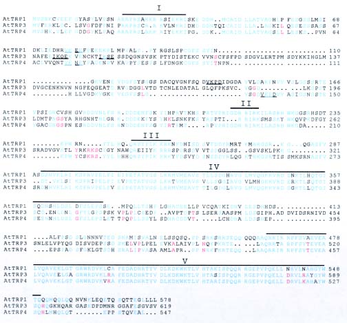
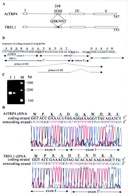
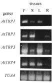
Abstract. AtTRP1 is an Arabidopsis protein that binds duplex telomeric DNA in vitro. Here we showed that knockout of AtTRP1 did not change significantly the telomere length in plant. This implies that either AtTRP1 does not participate in the regulation of telomere length or the Arabidopsis genome contains other genes functionally redundant to AtTRP1. Sequence analysis of Arabidopsis genome together with molecular cloning enabled us to identify two additional genes AtTRP3 and AtTRP4 and the corresponding cDNA clones encoding AtTRP1-like proteins. The C-terminal regions of both AtTRP3 and AtTRP4 proteins bind specifically duplex telomeric DNA in vitro. The amino acid sequence of AtTRP4 is identical to that of another Arabidopsis protein TRFL1 except with an internal deletion of six amino acids, suggesting that AtTRP4 and TRFL1 may be derived from the same gene by alternative splicing. This speculation was further confirmed by DNA sequence analysis of RT-PCR products specific for AtTRP4 and TRFL1 transcripts. Our data together with reports from other researchers revealed that Arabidopsis contains at least seven different duplex telomeric DNA-binding proteins encoded by a six-member gene family, named AtTRP. We proposed that some members of the AtTRP family may be functionally redundant in the regulation of telomere length in Arabidopsis.

Keywords: Alternative splicing; Arabidopsis thaliana; Functional redundancy; Knockout mutant; Telomere length; Telomeric DNA-binding protein.

Introduction

Telomeres are unique structures that are found at the ends of chromosomes in most eukaryotes and essential for the maintenance of the integrity of those chromosomes and for genome stability (Blackburn, 2001). Telomeric DNA consists of short DNA repeats, which are tandem arrayed and terminated with a single-stranded 3´ G-rich overhang. The synthesis of telomeric DNA at the chromosome end is primarily catalyzed by the telomerase. However, the access to telomerase is regulated by various factors, including the duplex telomeric DNA-binding proteins such as Rap1p in budding yeast, Taz1p in fission yeast, and TRF1 and TRF2 in human cells (Smogorzewska and de Lange, 2004; Vega et al., 2003).

The protein Rap1p negatively regulates telomere length (Marcand et al., 1997) in addition to controlling the transcription of multiple genes in budding yeast (Shore, 1994).

The C-terminal protein-interaction domain of Rap1p is required for the regulation of telomere length (Kyrion et al., 1992; Marcand et al., 1997) and for telomere clustering (Levy and Blackburn, 2004). It has been proposed that the recruitment of other proteins to telomere by the C-terminal domain of Rap1p can prompt the telomere to form a high-order structure inaccessible to telomerase (Levy and Blackburn, 2004).

Taz1p contains a Myb DNA-binding domain at its C-terminus and deletion of this C-terminus results in telomere lengthening (Cooper et al., 1997), indicating that Taz1p plays a negative role in the maintenance of telomere length in fission yeast. Although Taz1p was predicted to have a dimerization domain at N-terminus (Fairall et al., 2001), gel filtration analysis of purified Taz1p revealed that the native form of Taz1p can be as big as hexamers (Tomaska et al., 2004). Incubation of artificial telomeres with purified Taz1p prompted telomeric DNA to form a t-loop structure on which the associated Taz1p particles were also estimated to be hexamers (Tomaska et al., 2004). This observation implied that binding of Taz1p to telomeric DNA as oligomers may be important for t-loop formation and the regulation of telomere length in fission yeast.

*Corresponding author. Email: chen922@gate.sinica.edu.tw; Tel: (+886)-2-27899590 ext. 325; Fax: (+886)-2-27827954.

GenBank accession no.: AY181997 and AY395985.


Botanical Bulletin of Academia Sinica, Vol. 46, 2005

Human telomeric proteins TRF1 and TRF2 also contain a Myb DNA-binding domain at the C-terminus and a protein-dimerization domain in the central region of each protein (Chong et al., 1995; Bilaud et al., 1997; Broccoli et al., 1997; Fairall et al., 2001). TRF1 negatively regulates telomere length in human cells (van Steensel and de Lange, 1997). Binding of TRF1 induces the conformational change of telomeric DNA which may become inaccessible to telomerase (Bianchi et al., 1997; Griffith et al., 1998). Human TRF2 promotes telomeres to form a t-loop structure which is thought to protect telomeres from end-to-end fusions (van Steensel et al., 1998; Griffith et al., 1999).

We have previously described an Arabidopsis protein AtTRP1 which binds duplex telomeric DNA in vitro (Chen et al., 2001). This protein contains a single Myb-like DNA-binding domain at the C-terminus. Proteins highly homologous to AtTRP1 have been identified in other plants such as maize (Lugert and Werr, 1994), parsley (da Costa e Silva et al., 1993), rice (Yu et al., 2000) and tobacco (Yang et al., 2003), indicating that AtTRP1-like proteins are widely distributed in both monocotyledonous and dicotyledonous plants. Moreover, recent papers reported that the Arabidopsis genome contains six genes which encode a family of duplex telomeric DNA-binding proteins including AtTBP1, AtTRP1, TRFL1 and TRFL9 (Hwang et al., 2001; Karamysheva et al., 2004). Here we show that knockout of AtTRP1 did not cause a significant change in telomere length in Arabidopsis. Analysis of genome sequence along with molecular cloning enabled us to isolate two additional genes, AtTRP3 and AtTRP4, which encode proteins not only highly homologous to AtTRP1 but that also bind telomeric DNA in vitro. The protein AtTRP3 is identical to TRFL9, while AtTRP4 and TRFL1 are derived from the same gene by alternative splicing. Our results together with another report (Karamysheva et al., 2004) reveal that the Arabidopsis genome contains six genes encoding at least seven duplex telomeric DNA-binding proteins. Some of these proteins may be functionally redundant with each other.

Materials and Methods

Plant Growth

Arabidopsis thaliana plants were grown in an environmental growth chamber at 22°C with a 16-h photoperiod. To identify and characterize the knockout mutants, seeds were surface-sterilized and germinated on solid medium containing half-strength MS salt (Murashige and Skoog, 1962) with or without 50 µg/ml kanamycin.

Oligonucleotides

The primers described in this paper are listed in Table 1.

Isolation of AtTRP1 Knockout Mutant

The pooled DNA samples from T-DNA insertion lines of the Ws ecotype, generated by the Arabidopsis Knock

out Facility at The University of Wisconsin (Krysan et al., 1999), were screened by the PCR-based method (McKinney et al., 1995) to identify mutants with T-DNA insertion in AtTRP1. The PCR products were subjected to Southern blot analysis using an AtTRP1-specific probe, and the DNA fragments detected by the probe were purified and sequenced to define the site in AtTRP1 where the T-DNA was inserted. PCR conditions were 3 min at 94°C, followed by 40 cycles (94°C, 45 s; 65°C, 1 min; 72°C, 2 min) and a final extension at 72oC for 7 min. One plant heterozygous for a mutation in the AtTRP1 locus, named attrp1-1, was identified and self-pollinated to produce homozygous plants, which were maintained by self-pollination for subsequent experiments.

Characterization of attrp1-1 Knockout Mutant

Analysis of the AtTRP1 transcript in both wild-type plants and mutant plants homozygous for attrp1-1 was performed by reverse transcriptase-mediated PCR (RT-PCR). To measure telomere length, terminal restriction fragment (TRF) analysis was performed (Allshire et al., 1989). Genomic DNA was extracted from pooled 16-day-old seedlings using the DNeasy Plant Kits (Qiagen, Nalencia, CA). The DNA was digested with MseI, fractionated by electrophoresis in a 0.5% agarose gel, Southern transferred to a nylon membrane and probed with (TTTAGGG)4 end-labeled with digoxigenin (DIG)-11-ddUTP (Roche Applied Sci., Germany). Hybridization and detection conditions were described previously (Chen et al., 1997).

Isolation and Characterization of cDNA Clones Encoding AtTRP1 Homologs

An NCBI BLASTP search of the A. thaliana genome identified five putative genes, At1g07540, At3g12560, At3g46590, At3g53790 and At5g13820, which encode AtTRP1 homologs. The cDNAs corresponding to At1g07540, At3g53790 and At5g13820 have been cloned and shown to encode a duplex telomeric DNA-binding protein (Hwang et al., 2001; Karamysheva et al., 2004). To clone the cDNA corresponding to At3g12560, fragments a and b were amplified from the CD4-15 library provided by the Arabidopsis Biological Resource Center (ABRC) using primer pair AtTRP3F and T7, and primer pair AtTRP3R and T3, respectively. Fragment a was cut with XbaI plus HindIII, and fragment b was cut with BamHI plus XbaI. The largest fragments recovered from both digestions were ligated to pUC18 cut with BamHI plus HindIII to obtain a 2.3 kb cDNA clone, named AtTRP3 (GenBank accession number AY181997).

To clone the cDNA corresponding to At3g46590, fragments c and d were amplified from a Clontech library (Chen et al., 2001) using primer pair AtTRP4F and GADP2, and primer pair AtTRP4R and GADP1, respectively. Both PCR products were digested with EcoRI, and the largest fragments recovered from both digestions were ligated to pUC18 cut with EcoRI to obtain a 2.2 kb cDNA clone, named AtTRP4 (GenBank accession number AY395985).


CHEN et al. — Duplex telomeric DNA-binding proteins in Arabidopsis

for RT-PCR were annealed to sequences in exons on both sides of an intron. The primer TUA4R was annealed to exon 2 of the tubulin 4a (TUA4) gene of Arabidopsis (Kopczak et al., 1992). RT-PCR conditions were 30 min at 50°C to synthesize the initial cDNA, followed by 2 min at 94°C, 40 cycles (94°C, 30 s; 60°C, 30 s; 68°C, 2 min) and a final extension at 68°C for 7 min.

Results

Identification of attrp1-1 Allele

To understand the biological function of AtTRP1, a plant heterozygous for attrp1-1 was identified from a collection of 60,480 T-DNA insertion lines using a PCR-based procedure. The heterozygous mutant was allowed to self-pollinate, and the progeny were tested for kanamycin resistance carried by the inserted T-DNA and analyzed by PCR for the isolation of mutants homozygous for attrp1-1. The T-DNA insert in the heterozygous mutant was inherited in a Mendelian manner, and one-fourth of the progeny were found to be homozygous for attrp1-1 (26/106). PCR analysis using the T-DNA left border primer JL202 along with AtTRP1F or AtTRP1R generated frag

To find out whether proteins AtTRP3 and AtTRP4 bind plant telomeric DNA, the cDNA regions encoding the potential DNA-binding domains of both proteins were amplified from the corresponding full-length cDNA clones by PCR and subcloned into the XhoI and BamHI sites of plasmid pET15b (Novagen, Madison, WI). Plasmids were transformed into Escherichia coli BL21 (DE3) to overexpress the corresponding proteins (Chen et al., 2001). Crude bacterial extracts were prepared and 1 µl of each extract containing 5-10 µg protein was examined for telomeric DNA-binding activity by gel-shift assay. Primer pairs AtTRP3cF2 and AtTRP3cR1, and AtTRP4cF2 and AtTRP4cR1 were used, respectively, for subcloning and expression of AtTRP3502-619 and AtTRP4437-547 peptides in bacteria.

Reverse Transcription-Polymerase Chain Reaction (RT-PCR)

RNA was isolated either from various tissues or from whole seedlings of Ws ecotype using the RNeasy Mini Kit (Qiagen). Total RNA (1 µg) from each sample was used for the synthesis of cDNA using the Titan One Tube RT-PCR Kit (Roche). All of the primers except TUA4R used


Botanical Bulletin of Academia Sinica, Vol. 46, 2005

ments of 1.6-kb or 0.9-kb, respectively, from the genomic DNA of plants carrying attrp1-1 (Figure 1A). A 2.5-kb fragment was amplified by PCR from the genomic DNA of wild-type plants and heterozygous mutants but not from the genomic DNA of mutants homozygous for attrp1-1 using the oligonucleotides AtTRP1F and AtTRP1R as primers (Figure 1A). Sequence analysis of the 1.6-kb and 0.9-kb mentioned above revealed that attrp1-1 contained an inverted T-DNA repeat at the junction of exon 7 and intron 7 of AtTRP1 (Figure 1A).

The nature of attrp1-1 was studied further by RT-PCR. AtTRP1 mRNA-specific primers (Table 1, Figure 1A) were used in various combinations to amplify the AtTRP1 transcript from the wild-type Ws plants and the attrp1-1 homozygous mutants (Figure 1B). Coupling the forward primer e2/3F with any one of the reverse primers can amplify DNA fragments with expected sizes from the total RNA of wild-type plants (Figure 1B, lanes 1-3). A DNA fragment was amplified from the total RNA of the attrp1-1 mutant in the reaction containing primers e2/3F and e7/8R (Figure 1B, lane 5) but not in those containing primers e2/3F and e8/9R or e9/10R (Figure 1B, lanes 4 and 6), suggesting that the transcript of attrp1-1 is deficient in exons 8 to 10 of AtTRP1. Based on PCR and RT-PCR analysis, the structure of the peptide encoded by attrp1-1 was predicted and shown schematically in Figure 1C.

No Significant Change in Telomere Length in Plants Carrying attrp1-1

Plants homozygous for attrp1-1 did not exhibit obvious morphological changes even after they were maintained for nine generations by self-pollination. TRF analysis of DNA from a pool of mutant seedlings from each generation revealed that telomeres generally became shorter, but with occasional lengthening, during the first five generations. From the sixth generation (T6) on, the telomeres gradually became stable, but were only slightly shorter than those of wild-type plants (Figure 1D). Since a variation in telomere length was also observed among individual plants of wild-type Arabidopsis (Shakirav and Shippen, 2004), a slight shortening in telomere length can not be regarded as the result of a single mutation in AtTRP1.

Multiple Genes in the Arabidopsis Genome Encoding Duplex telomeric DNA-Binding Proteins

Mutants homozygous for attrp1-1 displayed only a slight decrease in telomere length after long-term maintenance (Figure 1D), raising the possibility that the Arabidopsis genome may contain other AtTRP1 homologs which can maintain telomere length in the absence of AtTRP1. Searching the Arabidopsis genome sequence followed by molecular cloning enabled us to identify two cDNA clones, AtTRP3 and AtTRP4, which encode proteins highly homologous to AtTRP1 (Figure 2). We consider these proteins, therefore, to be members of the AtTRP family. At least five regions along these protein sequences were predicted or confirmed to have biological functions

(Figures 1 and 2). The sequences in regions I, II and III are rich in basic residues and have been predicted previously to contain nuclear localization signals (NLS) (Chen et al., 2001). The N-terminal region of the tobacco telomeric DNA-binding protein NgTRF1 also contains sequences similar to those in regions I-III of the AtTRP family (Yang et al., 2003). Fusion of this N-terminal peptide of NgTRF1 to a green fluorescent protein (GFP) enabled the GFP to be transported into plant nuclei (Yang et al., 2003). The sequence in region IV is homologous to ubiquitin (Buchberger, 2002) and is defined as the ubiquitin domain. Region V in AtTRP1 has been confirmed to be essential for specific telomeric DNA-binding activity (Chen et al., 2001). In addition, several short sequences in all of these proteins were found to be the consensus binding motif of the small ubiquitin-like modifier (SUMO), jKXE(D), where j is a large hydrophobic amino acid, K is lysine to which SUMO is conjugated, X is any amino acid, and E and D are glutamic acid and aspartic acid, respectively (Melchior, 2000).

During the preparation of this manuscript, several duplex telomeric DNA-binding proteins, including TRFL1 and TRFL9 in Arabidopsis were reported (Karamysheva et al., 2004). Sequence comparison revealed that the amino acid sequence of AtTRP3 was identical to that of TRFL9 (Karamysheva et al., 2004), demonstrating that they are the same clone (data not shown). The amino acid sequence of AtTRP4 was also identical to that of TRFL1 except that an internal deletion of six amino acids was observed in AtTRP4 (Figure 3A), implying that they may be derived from the same gene by alternative splicing. To verify this speculation, the forward primers e3/4f1 and e3/4f2, specific for AtTRP4 and TRFL1, respectively, were coupled separately with a common reverse primer e4/5R for RT-PCR analysis of the total RNA from leaves (Table 1 and Figure 3B). A DNA fragment with an expected size of around 150 bp was amplified from each reaction (Figure 3C). Sequence analysis of each DNA fragment revealed that they were derived from different parts of At3g46590 that shared a common sequence (Figure 3B and D), confirming that both cDNA clones originated from At3g46590 by alternative splicing. The amount of RT-PCR product for AtTRP4 (Figure 3C, lane 1) was less than that for TRFL1 (Figure 3C, lane 2), implying that the mRNA of AtTRP4 is quantitatively different from that of TRFL1 in Arabidopsis leaves.

The C-terminal regions of AtTRP3 and AtTRP4, which correspond to the DNA-binding domain of AtTRP1 (Chen et al., 2001), were produced in E. coli and assayed for telomeric DNA-binding activity (Figure 4A and B). As expected, the bacterial extract containing each peptide formed complexes with the double-stranded DNA probes containing plant telomeric repeats. This complex formation can be prevented effectively by the duplex oligonucleotides containing the Arabidopsis telomeric sequence (G2T3AG)4 but not by duplex oligonucleotides containing the human telomeric sequence (T2AG3)4, indicating that both proteins bind specifically to plant telomeric DNA in vitro.


CHEN et al. — Duplex telomeric DNA-binding proteins in Arabidopsis

Figure 1. Identification and TRF analysis of a mutant homozygous for attrp1-1. A, Schematic representation of AtTRP1 and attrp1-1. The T-DNA from the plasmid pD991 was integrated as an inverted repeat after the nucleotide 2626 of AtTRP1 to create attrp1-1. Nucleotides 2627 to 2637 of AtTRP1 were deleted in attrp1-1. The positions of the primers, used in the PCR reactions for the identification of the mutant, or in RT-PCR reactions for the characterization of the gene transcript, are shown on the map. Open boxes represent the exons and solid boxes stand for exons encoding the DNA-binding domain; B, RT-PCR analysis of the transcripts of AtTRP1 and attrp1-1. Total RNA isolated from the seedlings of wild-type plants (lanes 1-3) or mutant plants homozygous for attrp1-1 (lanes 4-6) was RT-PCR amplified using mRNA-specific oligonucleotides as primers (shown above each lane). M, the molecular weight markers; C, Schematic description of the structural relationship between AtTRP1 and attrp1-1 proteins. Open boxes represent predicted or confirmed domains. Domains I to III are potential nuclear localization signals (NLS) (Chen et al., 2001). Domain IV is homologous to ubiquitin (Buchberger, 2002), and domain V is required for telomeric DNA-binding activity (Chen et al., 2001); D, TRF analysis of DNA from wild-type plants and mutant plants homozygous for attrp1-1. Genomic DNA from a pool of seedlings from each generation of the mutant or from wild-type seedlings (Ws) was subjected to TRF analysis. T1-T9 indicates different generations.


Botanical Bulletin of Academia Sinica, Vol. 46, 2005

Figure 2. Alignment among the AtTRP1, AtTRP3, and AtTRP4. Residues in proteins identical or similar to those in AtTRP1 are in blue. Residues in proteins identical or similar to AtTRP4 are in red. SUMO binding sites are underlined. Homologous regions with potential or confirmed function are labeled with Roman numbers I to V.

Co-expression of AtTRP Members in Different Tissues

RT-PCR was applied to analyze the expression patterns of AtTRP members among different tissues (Figure 5). While the amount of the transcript of the control gene TUA4 was the same in different tissues, the amount of the transcript of each AtTRP member varied during plant development. For instance, the amount of the AtTRP3 transcript in rosette leaves and floral buds was much higher than in roots and stems while the amount of the AtTRP1 transcript was low in rosette leaves, stems and floral buds and undetectable in roots. Similarly, the AtTBP1 transcript was detected in roots, stems, and floral buds but not in

rosette leaves, and the AtTRP4 transcript was lower in roots than in rosette leaves, stems and floral buds. In addition, RT-PCR analysis also indicated that both AtTRP4 and TRFL1 transcripts coexisted in rosette leaves, stems, and floral buds (data not shown). Generally speaking, this result indicates that at least three AtTRP members are transcribed in each tissue examined.

Discussion

Although AtTRP1 was shown to bind duplex telomeric DNA in vitro (Chen et al., 2001), the mutant plant of attrp1-1 did not display a significant change in telomere length


CHEN et al. — Duplex telomeric DNA-binding proteins in Arabidopsis

Figure 3. Sequence analysis of RT-PCR products specific for AtTRP4 and TRFL1. A, Schematic representation of the domain structure of AtTRP4 and TRFL1 proteins and sequence of the six amino acid insert in TRFL1; B, Partial sequence of At3g46590 encompassing the region encoding the six amino acid insert in TRFL1. The nucleotide sequence encompassing the junction of exon 3 and intron 3 and that of intron 3 and exon 4 of both AtTRP4 and TRFL1 is presented. The positions for the RT-PCR primers e3/4f1 and e3/4f2 are indicated as long and broken arrows interrupted with dotted lines. The conserved dinucleotides GT for the 5´ and AG for the 3´ boundaries of the introns are in bold and underlined; C, Fractionation of RT-PCR products specific for AtTRP4 (lane 1) and TRFL1 (lane 2) by electrophoresis on a 2% agarose gel; D, Sequence analysis of RT-PCR products using the oligonucleotide e4/5R as the sequencing primer. The sequence of the noncoding strand around the junction of exons 3 or 3´ and 4 of AtTRP4/TRFL1 is decoded. The sequences for the complementary coding strand and for the corresponding peptides are deduced from the sequence of noncoding strand and shown above each sequence profile. The boundary between two neighboring exons is indicated underneath each sequence profile.


Botanical Bulletin of Academia Sinica, Vol. 46, 2005

Figure 4. Gel-shift assay of the DNA-binding domains of AtTRP3 and AtTRP4. Bacterial extracts containing the polypeptides of (A) AtTRP3502-619 and (B) AtTRP4437-547 were incubated with 1 pmol of duplex oligonucleotide probe DIG-(G2T3AG)4 (plant telomeric sequence) in the absence or presence of double-stranded (ds) competitors at various concentrations, and then analyzed on a 6% native polyacrylamide gel as described (Chen et al., 2001). C and F stand for, respectively, the DNA-protein complexes and the free probe.

(Figure 1). This suggests that either AtTRP1 is not involved in the regulation of telomere length or the Arabidopsis genome contains other genes functionally redundant to AtTRP1. However, the identification of multiple AtTRP members in Arabidopsis genome (Figure 2; Karamysheva et al., 2004) suggests that these genes may more likely be functionally redundant in the regulation of telomere length. Perhaps, Arabidopsis plants with mutation(s) in one or some of these genes may not display an obviously aberrant telomere phenotype. This speculation needs to be clarified by studying plants with mutations in multiple members of this gene family.

Sequence analysis of AtTRP1, AtTRP3, AtTRP4 (Figure 2), and the remaining members of AtTRP family (data not shown) revealed that each contains a ubiquitin-like domain (domain IV) and at least 1-2 SUMO binding sites. In human cells, free TRF1 is ubiquitinated and degraded by the proteasomes (Chang et al., 2003). In both budding and fission yeast (Tanaka et al., 1999; Zho and Blobel, 2005), disruption of a SUMO gene resulted in a slight increase of telomere length. Whether the protein sumoylation or ubiquitination is involved in the metabolism of AtTRP proteins and the regulation of telomere length needs to be investigated further.

Some discrepancy in the expression patterns of members of the AtTRP family was observed between the data presented here (Figure 5) and those from other investigators (Hwang et al., 2001; Karamysheva et al., 2004). For instance, the transcript of AtTBP1 was detected in rosette leaves by Northern blotting (Hwang et al., 2001), but it was not detectable in the same tissue by RT-PCR in this study (Figure 5). Since the 3´ end of each gene encoding the

Figure 5. RT-PCR analysis of the expression patterns of the AtTRP members among plant tissues. Total RNA was isolated from various tissues and analyzed by RT-PCR using primer pairs specific for the transcript of each gene. The primer pairs, TRP1e1/2F with e9/10R, AtTBP1e1/2F with AtTBP1e8/9R, TRP3e1/2F with TRP3e8/9R, and TRP4e1/2F with TRP4e9/10R, were used, respectively, for the amplification of the transcript of AtTRP1, AtTBP1, AtTRP3 and AtTRP4. The primer pair for the amplification of the control gene tubulin 4a (TUA4) was TUA4F and TUA4R. The letters R, L, S and F represent, respectively, root, rosette leaf, stem and floral bud.

DNA-binding domain is highly homologous among the members of AtTRP family, detection of AtTBP1 transcript in leaves by Northern blotting may be the result of a cross-hybridization between AtTBP1 probe and mRNA from other members of the AtTRP family. Karamysheva et al. (2004) reported that each member of the AtTRP family expressed


CHEN et al. — Duplex telomeric DNA-binding proteins in Arabidopsis

repeats in vitro. J. Biol. Chem. 276: 16511-16519.

Chen, C.M., C.T. Wang, C.J. Wang, C.H. Ho, Y.Y. Kao, and C.C. Chen. 1997. Two tandemly repeated telomere-associated sequences in Nicotiana plumbaginifolia. Chromosome Res. 5: 561-568.

Chong, L., B. van Steensel, D. Broccoli, H. Erdjument-Bromage, J. Hanish, P. Tempst, and T. de Lange. 1995. A human telomeric protein. Science 270: 1663-1667.

Cooper, J.P., E.R. Nimmo, R.C. Allshire, and T. R. Cech. 1997. Regulation of telomere length and function by a Myb-domain protein in fission yeast. Nature 385: 744-747.

Cooper, J.P., Y. Watanabe, and P. Nurse. 1998. Fission yeast Taz1 protein is required for meiotic telomere clustering and recombination. Nature 392: 828-831.

Da Costa e Silva, O., L. Klein, E. Schmelzer, G.F. Trezzini, and K. Hahlbrock. 1993. BPF-1, a pathogen-induced DNA-binding protein involved in the plant defense response. Plant J. 4: 125-135.

Fairal, L., L. Chapman, H. Moss, T. de Lange, and D. Rhodes. 2001. Structure of the TRFH dimerization domain of the human telomeric proteins TRF1 and TRF2. Mol. Cell 8: 351-361.

Griffith, J., A. Bianchi, and T. de Lange. 1998. TRF1 promotes parallel pairing of telomeric tracts in vitro. J. Mol. Biol. 278: 79-88.

Griffith, J.D., L. Comeau, S. Rosenfield, R.M. Stansel, A. Bianchi, H. Moss, and T. de Lange. 1999. Mammalian telomeres end in a large duplex loop. Cell 97: 503-514.

Hwang, M.G., I.K. Chung, B.G. Kang, and M.H. Cho. 2001. Sequence-specific binding property of Arabidopsis thaliana telomeric DNA binding protein 1 (AtTBP1). FEBS Lett. 503: 35-40.

Karamysheva, Z.N., Y. V. Surovtseva, L. Vespa, E.V. Shakirov, and D.E. Shippen. 2004. A C-terminal Myb extension domain defines a novel family of double-strand telomeric DNA-binding proteins in Arabidopsis. J. Biol. Chem. 279: 47799-47807.

Kishi, S., X.Z. Zhou, Y. Ziv, C. Khoo, D.E. Hill, Y. Shiloh, and K.P. Lu. 2001. Telomeric protein Pin2/TRF1 as an important ATM target in response to double strand DNA breaks. J. Biol. Chem. 276: 29282-29291.

König, P., R. Giraldo, L. Chapman, and D. Rhodes. 1996. The crystal structure of the DNA-binding domain of yeast RAP1 in complex with telomeric DNA. Cell 85: 125-136.

Kopczak, S.D., N.A. Haas, P. J. Hussey, C.D. Silflow, and D.P. Snustad. 1992. The small genome of Arabidopsis contains at least six expressed a-tubulin genes. Plant Cell 4: 539-547.

Kyrion, G., K.A. Boakye, and A.J. Lustig. 1992. C-terminal truncation of RAP1 results in the deregulation of telomere size, stability, and function in Saccharomyces cerevisiae. Mol. Cell. Biol. 12: 5159-5173.

Krysan, P.J., J.C. Young, and M.R. Sussman. 1999. T-DNA as an insertional mutagen in Arabidopsis. Plant Cell 11: 2283-2290.

Levy, D.L. and E.H. Blackburn. 2004. Counting of Rif1p and Rif2p on Saccharomyces cerevisiae telomeres regulates telomere length. Mol. Cell. Biol. 24: 10857-10867.

Lugert, T. and W. Werr. 1994. A novel DNA-binding domain in the Shrunken initiator-binding protein (IBP1). Plant Mol. Biol. 25: 493-506.

at a similar level in all of the tissues tested. However, our data revealed that a variation at the RNA level existed, not only for the same gene among different tissues, but also for different members of the AtTRP family in the same tissue (Figure 5). It is difficult to make a comment on the result presented by Karamysheva et al. (2004) since no control gene was included in their RT-PCR experiment.

In human cells, the duplex telomeric DNA-binding proteins TRF1 and Pin2 are derived from the same gene by alternative splicing, and the amino acid sequence of Pin2 is identical to that of TRF1, apart from an internal deletion of 20 amino acids (Shien et al., 1997). Both TRF1 and Pin2 can form homodimer and heterodimer (Shien et al., 1997; Fairal et al., 2001) and are involved in the cellular response to double strand DNA breaks and telomere metabolism (Kishi et al., 2001; van Steensel and de Lange, 1997). Here, we showed that Arabidopsis also uses the mechanism of alternative gene splicing to generate duplex telomeric DNA-binding proteins AtTRP4 and TRFL1 from the same gene. Since different members of AtTRP family were shown to interact each other in vitro (Karamysheva et al., 2004), it is likely that AtTRP4 and TRFL1, which differs from each other by only six amino acids, can interact to form both homodimer and heterodimer. On the other hand, whether the six additional residues in TRFL1 can make it different from AtTRP4 both in high-order structure and in function is another issue to be investigated. In conclusion, our results reveal that the Arabidopsis cells contain multiple distinct duplex telomeric DNA-binding proteins, some of which are probably redundant in function.

Acknowledgments. This work was supported by grant no. NSC 91-2311-B-001-132 and NSC 92-2311-B-001-059 from the National Science Council and Academia Sinica in the Republic of China.

Literature Cited

Allshire, R.C., M. Dempster, and N.D. Hastie. 1989. Human telomeres contain at least three types of G-rich repeats distributed non-randomly. Nucleic Acids Res. 17: 4611-4627.

Bianchi, A., S. Smith, L. Chong, P. Elias, and T. de Lange. 1997. TRF1 is a dimer and bends telomeric DNA. EMBO J. 16: 1785-1794.

Bilaud, T., C. Brun, K. Ancelin, C. E. Koering, T. Laroche, and E. Gilson. 1997. Telomeric localization of TRF2, a novel human telobox protein. Nat. Genet. 17: 236-239.

Blackburn, E.H. 2001. Switching and signaling at the telomere. Cell 106: 661-673.

Broccoli, D., A. Smogorzewska, L. Chong, and T. de Lange. 1997. Human telomeres contain two distinct Myb-related proteins, TRF1 and TRF2. Nat. Genet. 17: 231-235.

Buchberger, A. 2002. From UBA to UBX: new words in the ubiquitin vocabulary. Tends Cell Biol. 12: 216-221.

Chang, W., J.N. Dynek, and S. Smith. 2003. TRF1 is degraded by ubiquitin-mediated proteolysis after release from telomeres. Gene. Dev. 17: 1328-1333.

Chen, C.M., C.T. Wang, and C.H. Ho. 2001. A plant gene encoding a Myb-like protein that binds telomeric GGTTTAG


Botanical Bulletin of Academia Sinica, Vol. 46, 2005

Marcand, S., E. Gilson, and D. Shore. 1997. A protein-counting mechanism for telomere length regulation in yeast. Science 275: 986-990.

McKinney, E.C., N. Ali, A. Traut, K.A. Feldmann, D.A. Belostotsky, J.M. McDowell, and R.B. Meagher. 1995. Sequence-based identification of T-DNA insertion mutations in Arabidopsis: actin mutants act2-1 and act4-1. Plant J. 8: 613-622.

Melchior, F. 2000. SUMO-nonclassical ubiquitin. Ann. Rev. Cell Dev. Biol. 16: 591-626.

Murashige, T. and F. Skoog. 1962. A revised medium for the growth and bioassay with tobacco tissue culture. Physiol. Plant. 15: 473-497.

Shakirov, E.V. and D.E. Shippen. 2004. Length regulation and dynamics of individual telomere tracts in wild-type Arabidopsis. Plant Cell 16: 1959-1967.

Shien, M., C. Haggblom, M. Vogt, T. Hunter, and K.P. Lu. 1997. Characterization and cell cycle regulation of the related human telomeric proteins Pin2 and TRF1 suggest a role in mitosis. Proc. Nat. Acad. Sci. USA 94: 13618-13623.

Smogorzewska, A. and T. de Lange. 2004. Regulation of telomerase by telomeric proteins. Ann. Rev. Biochem. 73: 177-208.

Tanaka, K., J. Nishide, K. Okazaki, H. Kato, O. Niwa, T. Nakagawa, H. Matsuda, M. Kawamukai, and Y. Murakami. 1999. Characterization of a fission yeast SUMO-1 homologue, Pmt3p, required for multiple nuclear events, in

cluding the control of telomere length and chromosome segregation. Mol. Cell. Biol. 19: 8660-8672.

Tomaska, L., S. Willcox, J. Slezakova, J. Nosek, and J. D. Griffith. 2004. Taz1 binding to a fission yeast model telomere. J. Biol. Chem. 279: 50764-50772.

Van Steensel, B. and T. de Lange. 1997. Control of telomere length by the human telomeric protein TRF1. Nature 385: 740-743.

Van Steensel, B., A. Smogorzewska, and T. de Lange. 1998. TRF2 protects human telomeres from end-to-end fusion. Cell 92: 401-413.

Vega, L. R., M.K. Mateyak, and V.A. Zakian. 2003. Getting to the end: telomerase access in yeast and humans. Nat. Rev. Mol. Cell. Biol. 4: 948-959.

Yang, S.W., D.H. Kim, J.J. Lee, Y.J. Chun, J.H. Lee, Y.J. Kim, I.K. Chung, and W.T. Kim. 2003. Expression of the telomeric repeat binding factor gene NgTRF1 is closely coordinated with the cell division program in tobacco BY-2 suspension culture cells. J. Biol. Chem. 278: 21395-21407.

Yu, E.Y., S.E. Kim, J. H. Kim, J.H. Ko, M.H. Cho, and I.K. Chung. 2000. Sequence-specific DNA recognition by the Myb-like domain of plant telomeric protein RTBP1. J. Biol. Chem. 275: 24208-24214.

Zhao, X. and G. Blobel. 2005. A SUMO ligase is part of a nuclear multiprotein complex that affects DNA repair and chromosomal organization. Proc. Nat. Acad. Sci. USA 102: 4777-4782.