Bot. Bull. Acad. Sin. (2005) 46: 325-331

YAN et al. — Regulation of camptothecin and 10-hydroxycamptothecin levels

Tissue-specific and developmental regulation of camptothecin and 10-hydroxycamptothecin levels in Camptotheca acuminata

Xiu-Feng YAN1,*, Yang WANG1, Yu-Hong ZHANG2, Tao YU1, Mei-Fang MA1, Shi-Jie JU1, and Sixue CHEN3

1College of Life Sciences, Northeast Forestry University, Harbin 150040, P. R. China

2Open Research Laboratory of Forest Plant Ecology, Northeast Forestry University, Harbin 150040, P. R. China

3Donald Danforth Plant Science Center, St. Louis, MO 63146, U.S.A.

(Received March 26, 2004; Accepted June 24, 2005)

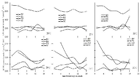
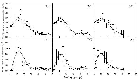
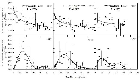
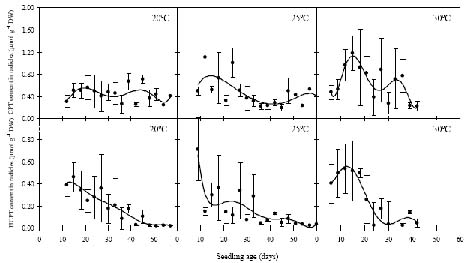
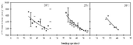
Abstract. Camptothecin (CPT) and its analogue 10-hydroxycamptothecin (HCPT) are two naturally occurring monoterpene indole alkaloids in Camptotheca acuminata. They both show potential efficacy as anti-cancer compounds as well as act as defensive chemicals in plants. Here we report the regulation of CPT and HCPT contents in C. acuminata during seed imbibition and seedling development. The levels of CPT in endosperm was 2 to 3 fold higher than in other tissues, and clearly decreased during the imbibition period. In other tissues, CPT contents exhibited slight fluctuations. In comparison, HCPT contents in all the tissues were much lower. HCPT was less abundant in cotyledon and embryonal axis than in endosperm and seed coat. During seedling development, CPT and HCPT contents in cotyledons reached maximum at 9-15 days stage. In embryonal axis, CPT contents decreased logarithmically, while HCPT content followed the same pattern as in cotyledons. CPT and HCPT levels in radicles were very low, with CPT levels remaining constant and HCPT levels decreasing during development. In leaves, the contents of CPT showed linear reduction during seedling growth. These results demonstrate that the levels of CPT and HCPT are tissue-specifically and developmentally regulated. The physiological implications and significance of the regulation are discussed.

Keywords: Camptotheca acuminata; Camptothecin; 10-hydroxycamptothecin; Regulation.

Introduction

Plant secondary metabolites are naturally occurring compounds which play important roles in various biological functions. For example, certain groups of plant secondary metabolites act as deterrents against pathogens and herbivores or attract pollinators via flower color or scent. A good example of this are alkaloids, which are a diverse group of low-molecular-weight, nitrogen- containing compounds that traditionally have been of interest due to their pronounced and diverse physiological activities in animals (Facchini, 2001). Many alkaloids are known to be poisonous to mammals and have demonstrated functions in plant defense against herbivores and pathogens (Wink, 1998).

Camptothecin (CPT), a monoterpene indole alkaloid produced by a native Chinese tree Camptotheca acuminata Decne., was isolated by Wall and co-workers (Wall et al., 1966) 39 years ago. Because of its high anti-tumor activity in the L1210 mouse leukemia assay, CPT attracted immediate attention for use as a potential cancer chemotherapeutic agent (Geran et al., 1972). The development of CPT as an anti-tumor drug stalled, due to a variety of unacceptable side effects on humans (Giovanella et al., 1989), until

inhibitor of topoisomerase I was discovered by Liu and co-workers (Hsiang et al., 1985) in 1985. Among CPT's many analogues, which have been investigated over more than 30 years, irintecan (Sawada et al., 1991; Masuda et al., 1992; Abigerges et al., 1995; Bleiberg, 1999) and topotecan (Kingsbury et al., 1991; Lilenbaum et al., 1995; Romanelli et al., 1998; Clements et al., 1999) have been approved by United States Food and Drug Administration (FDA) for application to colon/rectum and ovarian cancer. Two other related drugs, 9-aminocaptothecin and 9-nitrocamptothecin (Wani et al., 1986), are currently undergoing extensive clinical trials (Giovanella, 1997; Jeha et al., 1998; Stevenson et al., 1999). 10-hydroxycamptothecin (HCPT), a natural derivative of CPT in C. acuminata and the precursor of irinotecan and topotecan, has shown efficacy against lung, breast, and uterine cervical cancer (Zhang et al., 1998) through the same mechanism of inhibition of topoisomerase I.

Despite the importance of CPT and HCPT in medical applications, research has been conducted to study their distribution in C. acuminata (Lopez-Meyer et al., 1994; Liu and Adams, 1996; Yan et al., 2003), the induction of biosynthesis by environmental factors such as light and water (Liu et al., 1997, 1998), and the effect of seed source variations (Liu and Adams, 1998). However, little work has been done analyzing CPT and HCPT levels in different tissues during the seed imbibition or seedling development

*Corresponding author. E-mail: xfyan@mail.hl.cn; Fax: +86-451-82190052; Tel: +86-451-82192185.


Botanical Bulletin of Academia Sinica, Vol. 46, 2005

tracted at 60°C for 1 h in an ultrasonic bath (Branson Cleaning Equipment Co., U.S.A.). After cooling to room temperature, 60% ethanol was added to equalize volumes. One ml of the extract was centrifuged at 12,500 g for 10 min at 20°C, and the supernatant was used for CPT and HCPT analysis.

Analysis of Alkaloids

CPT and HCPT were separated and quantified by a high-performance liquid chromatography (HPLC) system (JASCO Inc., Japan), which consisted of two pumps (Model 1580), a Techsphere ODS C18 column (25 cm × 4.6 mm, 5 µm, HPLC Technology, U.K.), a Model 1575 UV detector, and a 10 µl sample loop. The elution program was as follows: 24% acetonitrile/water increasing linearly to 40% acetonitrile/water in the first 20 min, followed by 40% acetonitrile/water decreasing linearly to 24% acetonitrile/water in 2.0 min; at 25.0 min, the program stops, and the machine is ready for next injection. The flow rate was 1 ml/min. The detection wavelength was: 266 nm in the first 11.0 min for detection of HCPT, switched to 254 nm for detection of CPT, and then resumed to 266 nm for the next injection. The CPT standard was a kind gift from The Stehlin Foundation for Cancer Research (Houston, TX, USA). The HCPT standard was purchased from Guizhou Hanfang Pharmaceuticals Co., Ltd. (P. R. China). Retention time for HCPT and CPT was 8.6 min and 14.5 min, respectively. CPT and HCPT content was expressed as µmol g-1 dry weight.

Results

CPT and HCPT Levels in Various Seed Tissues During Imbibition

On the second day of imbibition, the concentration of CPT in endosperm was about 7 µmol g-1 dry weight, 2 to 3 times higher than in other tissues. Thereafter, CPT content decreased slightly in endosperm, fluctuated in embryonal axis and seed coat, and was relatively stable in cotyledon (Figure 1 upper). In comparison to CPT levels, the levels of HCPT were much lower in the tissues examined. HCPT content was higher in endosperm and seed coat than in cotyledon and embryonal axis. In both endosperm and seed coat, HCPT levels declined during the first 13 days of treatment and then stabilized while in cotyledon and embryonal axis, they fluctuated slightly during the entire imbibition period (Figure 1 lower). It is interesting to note that different temperature treatments had no significant influence on overall CPT or HCPT content in seeds (Figure 1).

CPT and HCPT Levels in Different Tissues of Seedlings During Development

In cotyledons after germination, CPT and HCPT contents increased in the first 15 days, and then plunged in the next 15 to 20 days. Interestingly, CPT levels only doubled while HCPT content shot up by approximately 300 fold in the first 15 days. The maximum HCPT content was

periods. In the present study, we analyze in detail the distribution of CPT and HCPT contents in various parts of seeds during imbibition and the dynamic variations of CPT and HCPT levels in different organs in the course of seedling development, under three different temperature regimes. The tissue-specific and developmental regulations of CPT and HCPT levels in C. acuminata may suggest physiological and biological significance for these compounds.

Materials and Methods

Plant Growth

Camptotheca acuminata seeds were obtained from Jintang (30.72° N, 104.61° E), Sichuan Province, China in January 2002. Seed aliquots were buried in sand for imbibition in incubators at 20°C, 25°C and 30°C, respectively, in April 2001 and were watered daily. After 16 days of sand burial, germination was easily identified. Seedlings from 3 continuous days of sand burial were transplanted to soil in plastic boxes without supplemental fertilizers and grown in a greenhouse with a photoperiod of 16 h light (45000 lx)/8 h dark, and relative humidity of 20-40% at 20°C, 25°C and 30°C, respectively. Seedlings were planted approximately 6 cm apart. Seedlings taken from the same temperature the same day were looked on as one group, and nine groups of seedlings were pooled.

Sample Collection

To determine the levels of CPT and HCPT in seeds, ten imbibiting seeds treated under different temperatures were sampled every three days from the second day of sand-burying treatment. Each seed was dissected into four different parts, i.e. endosperm, embryonal axis, cotyledon, and seed coat. The same organ of all ten seeds treated under the same conditions was pooled and treated as one sample.

To investigate the production of CPT and HCPT during seedling development, three seedlings from each of the nine groups were sampled every three days. Seedlings of three groups transplanted on different days at the same temperature were collected as replicates. Twenty samples were collected from seedlings geminated and grown at 20°C and 25°C while 13 samples were collected from 30°C samples since seeds germinated at 30°C exhibited a much lower germination rate. As seedlings grew, embryonal axis gradually developed to young stem and root (radicle). Seedlings at an early stage with no distinct embryonal axis (stem) or radicle were only separated into cotyledons and embryonal axises. Seedlings at subsequent stages were dissected into cotyledons, embryonal axises, radicles, and leaves.

Extraction of Alkaloids

Samples were dried at 60°C to a constant weight, followed by grinding in a mortar and storing in a desiccator. Dried material was weighed, transferred to a 5 ml flask, and then 4 ml of 60% ethanol was added. The mixture was ex


YAN et al. — Regulation of camptothecin and 10-hydroxycamptothecin levels

Figure 1. Variations of CPT and HCPT contents in different tissues of seeds of C. acuminata during imbibition. EN, endosperm; EA, embryonal axis; CO, cotyledon; SC, seed coat.

about 6 µmol g-1 dry weight, 1.5 times the CPT content in cotyledons (Figure 2). Seedlings from seeds germinated at 20°C had the lowest initial CPT and HCPT contents while those at 30°C showed the greatest (Figure 2).

In the embryonal axis, CPT contents were generally lower than in cotyledons. During development, CPT levels slowly decreased to about 2 to 4 times that of the initial level over a 50-day period, exhibiting a logarithmical relationship to the seedlings age. In this instance temperature had a marked effect, in that, the higher the temperature, the lower the initial contents (Figure 3 upper). In contrast to the changes of CPT levels, HCPT contents followed a similar single-peak-curve as observed in cotyledons, except that the highest concentration of HCPT in embryonal axis was about 3 to 4 times less in 12-15 day old

seedlings. In this case, higher temperatures tended to decrease the highest concentration peak (Figure 3 lower).

In radicles, the levels of both CPT and HCPT were very low during the 60-day growth. CPT contents fluctuated between 0.2 µmol g-1 dry weight and 1.2 µmol g-1 dry weight. On the other hand, HCPT concentration decreased almost to zero (Figure 4).

The first couple of leaves contained about 5-6 µmol g-1 dry weight CPT after they emerged. Then CPT levels decreased linearly to about 1-2 µmol g-1 dry weight during seedling development (Figure 5). Seedlings from seeds germinated at 20°C showed the highest initial CPT content. It was also the highest CPT content observed in all seedling samples examined. Under the described conditions, HCPT contents were too low to be detected.

Figure 2. Variations of CPT and HCPT contents in cotyledons of C. acuminata during seedlings development. Vertical bars originating from each data point represent the standard error of the mean (n=3).


Botanical Bulletin of Academia Sinica, Vol. 46, 2005

Figure 3. Levels of CPT and HCPT in embryonal axis (stem) of C. acuminata during seedling development. Vertical bars originating from each data point represent the standard error of the mean (n=3).

Figure 4. Levels of CPT and HCPT in radicles of C. acuminata during seedlings development. Vertical bars originating from each data point represent the standard error of the mean (n=3).

Figure 5. CPT contents in leaves of C. acuminata during seedling development. Vertical bars originating from each data point represent the standard error of the mean (n=3).


YAN et al. — Regulation of camptothecin and 10-hydroxycamptothecin levels

We have also determined CPT and HCPT contents in the endosperm and seed coat during the initial seedling development. CPT content in endosperm was 3.4-5.6 µmol g-1 dry weight, lower than those in ungerminated seeds, and in seed coat it was 1.2-2.7 µmol g-1 dry weight, similar to that in seeds. HCPT contents were similar to the low levels measured in seeds (data not shown).

Discussion

Alkaloids are naturally occurring low molecular weight, nitrogen-containing compounds found in approximately 20% of plant species. The diverse biological activities of alkaloids have led to their exploitation as stimulants, pharmaceuticals, narcotics, and poisons to only mention a few. In planta, many of the alkaloids have been ascribed functions in plant pathogen resistance (Wink and Schimmer, 1999; Facchini, 2001). The alkaloids CPT and HCPT are two unique quinoline alkaloids produced by C. acuminata. In this paper, tissue-specific distribution and developmental regulation of CPT and HCPT was studied in great detail by quantitative analysis of the two compounds using reversed-phase HPLC. The results obtained not only demonstrate differential regulation of the CPT and HCPT but also suggest some interesting features pertaining to de novo biosynthesis, conversion, transport, and degradation.

Interestingly, in cotyledons CPT levels only increased about onefold while HCPT levels surged about 300 fold (Figure 2). This indicates CPT-10-hydroxylase catalyzing the production of HCPT from CPT was dramatically up regulated after germination, and that the de novo biosynthesis of CPT must also have been stimulated. Furthermore, embryonal axis CPT levels decreased logarithmically while HCPT levels increased and peaked by day 15 (Figure 3). The net decrease of CPT contents does not appear to account for the increase of HCPT levels unless a low level of CPT de novo biosynthesis was taking place. Therefore, in a tissue where CPT-10-hydroxylase was active, CPT biosynthesis and conversion to HCPT need to be considered as two closely related dynamic processes.

During seed imbibition, the decrease of CPT levels in endosperm and HCPT levels in endosperm and seed coat (Figure 1) may be due to degradation or conversion to other derivatives. At the seedling stage, a decrease of CPT and HCPT levels in several tissues (Figure 2-4) from 15 day to 25 day could be explained by translocation to the leave growth apex, degradation and/or conversion to other derivatives. From 25 days on the decrease demonstrated in the amounts of these two compounds can most likely not be attributed to transport since the levels observed in other tissues showed no increase. Further studies using metabolic profiling and transcriptomic and proteomic tools are needed to elucidate the mechanisms underlying CPT and HCPT metabolism and translocation in plants.

In general, seeds, buds and young tissues tend to sequester or synthesize large amounts of defense chemicals

during vulnerable stages of the plant life cycle (Wink and Schimmer, 1999). Young leaves have been shown to contain much higher amounts of CPT than older leaves (Lopez-Meyer et al., 1994; Liu et al., 1998; Yan et al., 2002). Endosperm, the nutritive part of the seed, contains the highest levels of the animal toxin CPT. Furthermore, by 15 days of seedling growth the production of CPT and its analog HCPT in cotyledon as well as HCPT in embryonal axis reach their maximums. The accumulation of these secondary metabolites at this vulnerable stage may imply an important role for these two defensive compounds in juvenile stages of development. Camptotheca acuminata has CPT in all organs throughout its life cycle (Lin et al., 1977; Liu and Adams, 1996) but produces little HCPT except in cotyledon and embryonal axis of 9-15 day old seedlings (Lopez-Meyer et al., 1994). One can speculate that these two compounds have different roles in plant growth and defense.

In summary, CPT content was the highest in endosperm and young leaves, followed by cotyledons, other parts of the imbibitted seeds, and radicles of seedlings. HCPT content was very high in cotyledons and embryonal axis of 9-15 day old seedlings, and very low in other tissues at different stages. The changes of CPT and HCPT levels in different tissues during development may have nutritional and defensive significance. In addition, the data provided within will prove useful for selecting tissues and developmental stages for extraction of CPT and/or HCPT for anticancer drug synthesis.

Acknowledgments. We thank Dr. Brian J. Green (Biology Department, University of Pennsylvania) for his critical reading of the manuscript. This research was supported by the National Natural Science Foundation of China (No. 30070086) and Science Foundation for Distinguished Young Scholars, Heilongjiang Province, P. R. China (No. JC-02-11).

Literature Cited

Abigerges, D., G.G. Chabot, J.P. Armand, P. Herait, A. Gouyette, and D. Gandia. 1995. Phase I and pharmacologic studies of the camptothecin analog irinotecan administered every 3 weeks in cancer patients. J. Clin. Oncol. 13: 210-221.

Bleiberg, H. 1999. CPT-11 in gastrointestinal cancer. Eur. J. Cancer 35: 371-379.

Clements, M.K., C.B. Jones, M. Cumming, and S.S. Daoud. 1999. Antiangiogenic potential of camptothecin and topotecan. Cancer Chemother. Pharmacol. 44: 411-416.

Facchini, P.J. 2001. Alkaloid biosynthesis in plants: Biochemistry, Cell Biology, Molecular Regulation, and Metabolic Engineering Applications. Annu. Rev. Plant Physiol. Plant Mol. Biol. 52: 29-66.

Geran, R., N. Greenberg, M. MacDonaald, A. Schumacher, and B. Abbott. 1972. Protocols for screening chemical agents and natural products against animal tumors and other biological systems. Cancer Chemother. Rep. 3: 1-63.

Giovanella, B., J. Stehlin, M. Wall, M. Wani, A. Nicholas, L. Liu, R. Silber, and M. Potmesil. 1989. DNA topoisomerase I-targeted chemotherapy of human colon cancer in


Botanical Bulletin of Academia Sinica, Vol. 46, 2005

xenografts. Science 246: 1046-1048.

Giovanella, B.C. 1997. Topoismerase I Inhibitors. In B.A. Teicher (ed.), Cancer Therapeutics: Experimental and Clinical Agents, Humana Press, Totowa, pp. 137-152.

Hsiang, Y., R. Herizberg, S. Hecht, and L. Liu. 1985. Camptothecin induces protein-linked DNA breaks via mammalian DNA topoisomerase I. J. Biol. Chem. 260: 14873-14878.

Jeha, S., H. Kantarjian, S. O'Brien, L. Vitek, and M. Beran. 1998. Activity of oral and intravenous 9-aminocamptothecin in SCID mice engrafted with human leukemia. Leuk Lymphoma. 32: 159-164.

Kingsbury, W., J. Boehm, D. Jakas, K. Holden, S. Hecht, G. Gallagher, M. Caranfa, F. McCabe, L. Faucette, R. Johnson, and R. Hertsberg. 1991. Synthesis of water-soluble (aminoalkyl) camptothecin analogues: inhibition of topoisomerase I and antitumor activity. J. Med. Chem. 34: 98-107.

Lilenbaum, R.C., M.J. Ratain, A.A. Miller, J.B. Hargis, D.R. Hollis, G.L. Rosner, S.M. O'Brien, L. Brewster, M.R. Green, and R.L. Schilsky. 1995. Phase I study of paclitaxel and topotecan in patients with advanced tumors: A cancer and leukemia group B study. J. Clin. Oncol. 13: 2230-2237.

Lin, L., Z. Zhao, and R. Xu. 1977. Study on chemical components in Camptotheca acuminata, an anti-cancer plant I. Chemical components in roots of C. acuminata. Acta Chem. Sin. 35: 227-230.

Liu, Z.J. and J.C. Adams. 1996. Camptothecin yield and distribution within Camptotheca acuminata trees cultivated in Louisiana. Can. J. Bot. 74: 360-365.

Liu, Z.J. and J.C. Adams. 1998. Seed source variation in camptothecin concentrations of nursery-grown Camptotheca acuminata seedlings. New Forests 16: 167-175.

Liu, Z.J., S.B. Carpenter, and R.J. Constantin. 1997. Camptothecin production in Camptotheca acuminata seedlings in response to shading and flooding. Can. J. Bot. 75: 368-373.

Liu, Z.J., S.B. Carpenter, W.J. Bourgeois, Y. Ying, R.J. Constantin, M.J. Falcon, and J.C. Adams. 1998. Variations in the secondary metabolite camptothecin in relation to tissue age and season in Camptotheca acuminata. Tree Physiol. 18: 265-270.

Lopez-Meyer, M., C.L. Nessler, and T.D. Mcknight. 1994. Sites of accumulation of the antitumor alkaloid camptothecin in Camptotheca acuminata. Planta Med. 60: 558-560.

Masuda, N., M. Fukuoka, Y. Kusunoki, K. Matsui, N. Takifuji, S. Kudoh, S. Negoro, M. Nishioka, K. Nakagawa, and M. Takada. 1992. CPT-11 a new derivative of camptothecin for the treatment of refractory or relapsed small-cell lung cancer. J. Clin. Oncol. 10: 1225-1229.

Romanelli, S., P. Perego, G. Pratesi, N. Carenini, M. Tortoreto, and F. Zunino. 1998. In vitro and in vivo interaction between cisplatin and topotecan in ovarian carcinoma systems. Cancer Chemother. Pharmacol. 41: 385-390.

Sawada, S., S. Okajimas, R. Aiyama, K. Nokata, T. Furuta, T. Yokokura, E. Sugino, K. Yamaguchi, and T. Miyasaka. 1991. Synthesis and antitumor activity of 20(S)-camptothecin derivatives: carbamate-linked, water-soluble derivatives of 7-ethyl-10-hydroxycampto- thecins. Chem. Pharm. Bull. 39: 1446-1454.

Stevenson, J.P., D. DeMaria, J. Sludden, S.B. Kaye, L. Paz-Ares, L.B. Grochow, A. McDonald, K. Selinger, P. Wissel, P.J. O'Dwyer, and C. Twelves. 1999. Phase I/pharmacokinetic study of the topoisomerase I inhibitor GG211 administered as a 21-day continuous infusion. Ann. Oncol. 10: 339-344.

Wall, M., M. Wani, C. Cook, and K. Palmer. 1966. Plant Antitumor Agents I. The isolation and structure of camptothecin, a novel alkaloidal leukemia and tumor inhibitor from Camptotheca acuminata. J. Amer. Chem. Soc. 88: 3888-3890.

Wani, M., A. Nicholas, and M. Wall. 1986. Plant antitumor agents 23. synthesis and antileukemic activity of camptothecin analogues. J. Med. Chem. 29: 2358-2363.

Wink, M. 1998. Chemical ecology of alkaloids. In M.F. Roberts, and M. Wink (eds.), Alkaloids: biochemistry, ecology, and medicinal applications. Plenum press, New York, pp. 265-300.

Wink, M. and O. Schimmer. 1999. Modes of defensive secondary metabolites. In E. Wink (ed.), Functions of Plant Secondary Metabolites and their Exploitation in Biotechnology. Annual Plant Rev. CRC Press, Boca Raton, pp. 17-133.

Yan, X.F., Y. Wang, T. Yu, Y.H. Zhang, and L.J. Yin. 2002. Determination of camptothecin in the leaves of Camptotheca acuminata by HPLC. J. Instr. Anal. 21: 15-18.

Yan, X.-F., Y. Wang, T. Yu, Y.-H. Zhang, and S.-J. Dai. 2003. Variation in camptothecin content in Camptotheca acuminata leaves. Bot. Bull. Acad. Sin. 44: 99-105.

Zhang, R., Y. Li, Q. Cai, T. Liu, H. Sun, and B. Chambless. 1998. Preclinical pharmacology of the natural product anticancer agent 10-hydroxycamptothecin, an inhibitor of topoisomerase I. Cancer Chemoth. Pharm. 41: 257-267.


YAN et al. — Regulation of camptothecin and 10-hydroxycamptothecin levels