Bot. Bull. Acad. Sin. (2005) 46: 339-345

LIN et al. — Genome organization and relationships of Phalaenopsis

Genome organization and relationships of Phalaenopsis orchids inferred from genomic in situ hybridization

Chih-Chung LIN1, Yao-Huang CHEN2,3, Wen-Huei CHEN2,4, Chi-Chang CHEN1, and Yen-Yu KAO1,5,*

1Department of Botany, National Taiwan University, Taipei 106, Taiwan, ROC

2Department of Horticulture, Taiwan Sugar Research Institute, Tainan 701, Taiwan, ROC

(Received April 21, 2005; Accepted July 13, 2005)

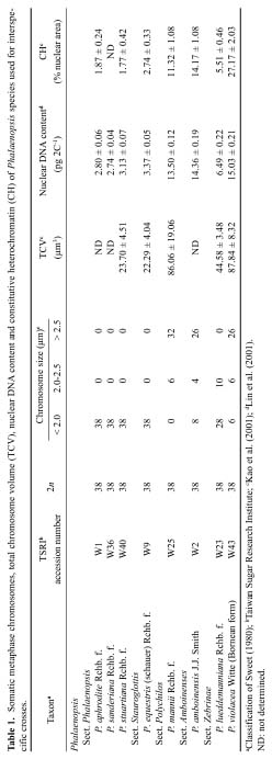
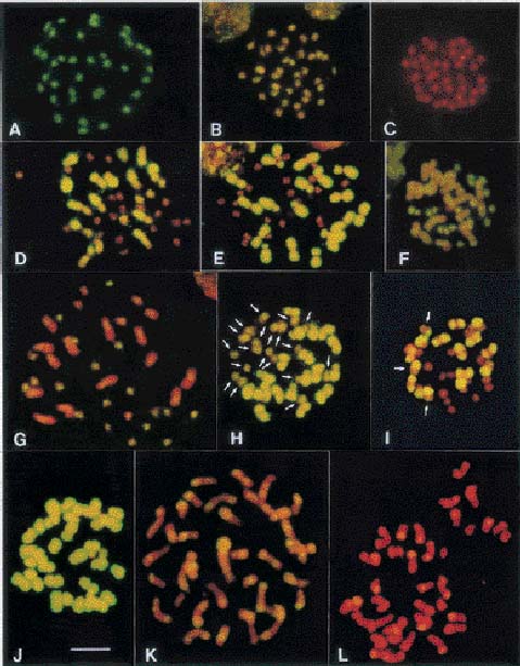
Abstract. All Phalaenopsis species have the same chromosome number (2n = 2x = 38), but the sizes of their genomes vary markedly. In this study, we investigated genome organization and relationships of Phalaenopsis species by genomic in situ hybridization (GISH) of seven interspecific hybrids derived from crosses between species with similar or dissimilar sizes of genomes. In the hybrid P. aphrodite × P. sanderiana, in which both parents possess small genomes, the two parental chromosome sets could not be distinguished by the strength and distribution of hybridization signals. Similar results were obtained from the hybrid P. mannii × P. violacea, in which both parents have large genomes. These results suggest that the two parents of these hybrids have similar genomes. In hybrids in which one parent possesses a large and the other parent a small genome, such as P. amboinensis × P. stuartiana, the two parental chromosome sets could readily be distinguished by GISH with or without the application of blocking DNA. Examination of hybridization signals on chromosomes revealed that species with large genomes have much more repetitive sequences, both in type and amount; however, species with small genomes do have their own specific sequences. In general, genome relationships obtained from GISH are in agreement with those from traditional genome analysis of other investigators. This study demonstrates that GISH is a useful tool for investigating genome organization and relationships of plant species, especially when analysis of meiotic behavior is technically difficult.

Keywords: Genomic in situ hybridization; Genome organization; Genome relationship; Phalaenopsis.

Introduction

Genome analysis provides valuable information about species relationships and, therefore, plays an important role in plant breeding programs. Among the traditional methods of genome analysis, studying the meiotic chromosome pairing in F1 hybrids is most reliable and has been frequently used because it gives direct evidence for genome homology between parental species (Singh, 2003).

The genus Phalaenopsis (Orchidaceae) comprises 45 to 63 species and has a wide geographic distribution, ranging from the Himalayas of northern India through Southeast Asia to northern Australia (Sweet, 1980; Christenson, 2001). Species and hybrids in this genus are of high value in floriculture because of their graceful and long-lasting flowers. Although interspecific and intergeneric hybrid

ization has long been used for breeding superior cultivars in Phalaenopsis orchids, so far only one report has investigated meiotic chromosome pairing in F1 hybrids (Arends, 1970). Analysis of chromosome pairing in Phalaenopsis is difficult because: (1) plants are slow-growing and require about two to three years to reach maturity; (2) each plant produces very few flowers, hindering collection of sufficient microsporocytes at the right stages for analysis; (3) microsporocytes are enclosed in a thick callose wall, which hampers stain penetration; and (4) meiotic chromosomes cannot be spread well due to clumping and stickiness.

Two molecular techniques, genomic Southern hybridization and genomic in situ hybridization (GISH), have been developed (Schwarzacher et al., 1989; Anamthawat-Jónsson et al., 1990) which may complement the method of genome analysis by meiotic chromosome pairing. Both techniques use total genomic DNA as a probe, but the hybridization targets are different. In genomic Southern hybridization the target is DNA on Southern blots while in GISH the target is chromosomes prepared from mitotic or meiotic cells. Both techniques detect hybridization of repetitive sequences (Anamthawat-Jónsson et al., 1990), which constitute the main source of genome-sized variation between related species (Flavell, 1982; Kubis et al., 1998).

3Present address: Crop Improvement Division, Tainan District Agricultural Research & Extension Station, Tainan 712, Taiwan, ROC

4Present address: Department of Life Science, National University of Kaohsiung, Kaohsiung 811, Taiwan, ROC

5Present address: Institute of Molecular and Cellular Biology, National Taiwan University, Taipei 106, Taiwan, ROC

*Corresponding author. E-mail: yykao@ntu.edu.tw; +886-2-33662486; Fax: +886-2-33662478.


Botanical Bulletin of Academia Sinica, Vol. 46, 2005

Previous studies have shown that all Phalaenopsis species have the same chromosome number (2n = 2x = 38) (Sagawa, 1962; Sagawa and Shoji, 1968), but the sizes of their chromosomes differ markedly (Shindo and Kamemoto, 1963). Lin et al. (2001) estimated the nuclear DNA content of 18 Phalaenopsis species by flow cytometry and found a 6-fold difference among these species. Kao et al. (2001) studied karyotypes of nine Phalaenopsis species and found a 4-fold difference in total chromosome volume (TCV) and a 15-fold difference in the amount of constitutive heterochromatin (CH) in their nuclei. They further pointed out a positive correlation between nuclear DNA content and TCV, and between nuclear DNA content and the amount of CH. In addition to satellite repeats present in CH, most higher plants also contain large amounts of retrotransposons (Bennetzen, 1996), which disperse throughout the genome and are difficult to detect by traditional cytological techniques.

To understand genome organization and relationships of Phalaenopsis species with varying sizes of genomes, we examined seven representative interspecific hybrids by GISH. The results from these studies are presented herein.

Materials and Methods

Plant Material

The parental species used in this study and their nuclear content and chromosome size are listed in Table 1. Interspecific crosses were made by investigators of the Taiwan Sugar Research Institute, and the hybrids (Table 2) were shipped to National Taiwan University for cytological studies.

Genomic DNA Isolation and Labeling

Total genomic DNA was isolated from young leaves of parental species as described by Suen et al. (1997). When used as a probe, genomic DNA was digested with EcoRI and then labeled with digoxigenin-11-dUTP by nick translation following the manufacturer's instructions (Roche Molecular Biochemicals). Blocking DNA was sheared to 200-300 bp in length by autoclaving (Anamthawat-Jónsson and Heslop-Harrison, 1994).

Chromosome Preparation

Chromosomes for GISH were prepared from root tips as described by Kao et al. (2001). Briefly, excised root tips were split in half longitudinally, and root caps and velamens (layers of dead cells with thick walls) were removed with a sharp scalpel. They were then treated with 2 mM 8-hydroxyquinoline on a rotary shaker (100 cycles min-1) for 4 h at 20°C, fixed in ethanol-glacial acetic acid (3:1) overnight, and stored in 70% ethanol at -20°C until use. The meristematic tissues of root tips were digested with 2% (w/v) cellulase Onozuka R10 (Yakult Honsha, Japan) and 1% (w/v) macerozyme Onozuka R10 (Yakult Honsha, Japan) in citrate buffer (4 mM citric acid and 6 mM sodium citrate, pH 4.8) at 25°C for 2-3 h and squashed


LIN et al. — Genome organization and relationships of Phalaenopsis

in 45% acetic acid on a microscope slide pretreated with Vectabond (Vector Laboratories, UK), and then a coverglass was added to the slide. The slide was placed upside down over four layers of filter papers, and the chromosomes were spread by applying light pressure on the slide. The coverglass was removed with a razor blade after freezing the slide in liquid nitrogen.

Genomic in Situ Hybridization

Chromosome preparations were treated with 100 µg/ml DNase-free RNase for 1 h and post-fixed in 4% paraformaldehyde for 10 min. Chromosomal DNA was denatured in 70% formamide, 2× SSC (20× SSC: 3 M NaCl, 0.3 M tri-sodium citrate) at 70°C for 2.5 min and dehydrated through an ethanol series at 4°C. The hybridization mixture consisted of 50% formamide, 10% dextran sulfate, 2× SSC, 0.1% SDS, 3 ng/µl of probe DNA, and blocking DNA at concentrations of 50- and 100-fold that of the probe DNA. Hybridization was carried out at 37°C overnight. Slides were washed in 20% formamide, 0.1× SSC at 42°C for 10 min, in 2× SSC at 42°C for 10 min, and in 2× SSC at room temperature for 3 × 5 min. Labeled probe was detected with fluorescein-conjugated antibodies (Roche Molecular Biochemicals), and chromosomes were counterstained with propidium iodide (PI). Slides were visualized under an Olympus AX70 fluorescence microscope with appropriate filter sets, and the images were photographed on Fujicolor Supera 200 ASA print film.

Results and Discussion

Hybrid Between Species with Small Genomes

As listed in Table 1, the two parents of the hybrid P. aphrodite × P. sanderiana both have low nuclear DNA contents (Lin et al., 2001) and small metacentric chromosomes with large blocks of pericentromeric heterochromatin (Kao et al., 2001). When labeled genomic DNA from P. sanderiana was used as a probe for hybridization with this hybrid, the two parental chromosome sets could not be distinguished based on the strength and distribution of hybridization signals (Table 2). In the absence of blocking DNA from P. aphrodite, both parents showed strong signals along the length of their chromosomes (Figure 1A). Addition of blocking DNA to the hybridization solution reduced the intensity and areas of labeling, but the two parental chromosome sets were still indistinguishable (Figure 1B-C). These results suggest that the two species have similar types and amounts of repetitive sequences.

Hybrids Between Species with Small and Large Genomes

GISH was performed in hybrids P. amboinensis × P. stuartiana, P. violacea × P. equestris, P. mannii × P. stuartiana and P. mannii × P. aphrodite, in which one parent possesses a small genome and exclusively small chromosomes while the other parent possesses a large genome


Botanical Bulletin of Academia Sinica, Vol. 46, 2005

Figure 1. Genomic in situ hybridization in Phalaenopsis interspecific hybrids. Somatic metaphase chromosomes prepared from the hybrids were probed with FITC-labeled genomic DNA from one parent in the presence or absence of unlabeled (blocking) DNA from the other parent. Chromosomes were counterstained with PI. A-C, Hybrid P. aphrodite × P. sanderiana chromosomes probed with P. sanderiana DNA in the absence (A) and presence of 50× (B) and 100× (C) blocking DNA; D, Hybrid P. violacea × P. equestris chromosomes probed with P. violacea DNA in the absence of blocking DNA; E, Hybrid P. mannii × P. aphrodite chromosomes probed with P. mannii DNA in the absence of blocking DNA; F, Hybrid P. violacea × P. equestris chromosomes probed with P. equestris DNA in the absence of blocking DNA; G, Hybrid P. amboinensis × P. stuartiana chromosomes probed with P. stuartiana DNA in the presence of 50× blocking DNA; H & I, Hybrid P. mannii × P. lueddemanniana chromosomes probed with P. mannii DNA in the absence (H) and presence of 50× (I) blocking DNA; J-L, Hybrid P. mannii × P. violacea chromosomes probed with P. mannii DNA in the absence (J) and presence of 50× (K) and 100× (L) blocking DNA. Scale bar = 10 µm.


LIN et al. — Genome organization and relationships of Phalaenopsis

twofold. Phalaenopsis lueddemanniana has 28 small and 10 medium chromosomes while P. mannii possesses 6 medium and 32 large chromosomes (Table 1). When labeled genomic DNA from P. mannii was used as a probe for hybridization with this hybrid in the absence of blocking DNA, hybridization signals were detected along chromosomes of both parents; however, signals on P. lueddemanniana chromosomes appeared to be slightly weaker than on P. mannii chromosomes (Figure 1H, arrows). Addition of 50× unlabeled P. lueddemanniana DNA to the hybridization mixture blocked hybridization of repetitive sequences common to both parents, and as a result, differences between the two parental chromosome sets became more apparent. However, arms of some P. mannii chromosomes fluoresced similarly as did the chromosomes of P. lueddemanniana (Figure 1I, arrows), suggesting segmental homology between chromosomes of these two species.

Hybrid Between Species with Large Genomes

The two parents of the hybrid P. mannii × P. violacea have similar 2C DNA values, and both possess mostly large chromosomes (Table 1). When total DNA from either parent was used as a probe for hybridization with this hybrid, the parental chromosomes could not be distinguished by the strength and distribution of hybridization signals (Table 2). In the absence of blocking DNA, strong signals were uniformly distributed on chromosomes of both parents (Figure 1J). The strength and areas of labeling decreased with the increase of the concentration of blocking DNA in the hybridization mixture (Figure 1K, L).

Comparison of the GISH Results with Meiotic Chromosome Pairing

Meiotic behavior of seven F1 hybrids from crosses between Phalaenopsis species with similar or dissimilar sizes of genomes was studied by Arends (1970). A comparison of our GISH results with those of Arends revealed that hybrids between species with similar sizes of genomes generally showed normal chromosome pairing, and the parental chromosome sets could not be distinguished by GISH while hybrids from crosses between species with pronouncedly dissimilar sizes of genomes showed poor pairing, and the parental chromosome sets could be readily distinguished by GISH even in the absence of blocking DNA (Figure 1, Table 2). One exception to this generality was the hybrid P. amboinensis × P. sanderiana, in which the sizes of parental genomes differed approximately 5-fold (Lin et al., 2001), but meiotic chromosome pairing was fairly high as an average of 12.13 bivalents was found at metaphase I (Arends, 1970). Further studies are needed to clarify this inconsistency.

Normal chromosome pairing in hybrids with similar sizes of genomes may be explained by the fact that the two parents have similar types and amounts of repetitive DNA sequences, as evidenced from the indistinguishability of the two parental chromosome sets by GISH. On the other hand, low bivalent formation in hybrids with pronouncedly

and various numbers of large and medium chromosomes in addition to a few small ones (Table 1). In general, results obtained from these hybrids were similar (Table 2). When total DNA from the species with a large genome was used as a probe in the absence of blocking DNA, all chromosomes belonging to the large genome showed strong hybridization signals except for the centromeric regions while chromosomes belonging to the small genome had no or very weak signals in the centromeric regions (Figure 1D, E). The strong signals were from hybridization of repetitive sequences in the large genome. Absence of signals in the centromeric regions of large chromosomes may be attributed to the extended nature of DNA in these regions, as they formed constrictions when other parts of chromosomes were condensed. The strength and distribution of hybridization signals in these hybrids indicate that species with large genomes contain much more repetitive sequences than species with small genomes.

When total DNA from species with a small genome was used as a probe in the absence of blocking DNA, strong signals dispersed on chromosomes of the species with a small genome, and weak signals clustered in the heterochromatin regions of chromosomes of the species with a large genome (Figure 1F). The observation that small chromosomes showed stronger signals than did large chromosomes suggests that although species with small genomes contain less repetitive sequences, they nevertheless have their own specific sequences. The weakness of signals in the heterochromatin regions of large chromosomes suggests that these regions contain other satellite sequences not present in the probe from species with small genomes. Huang (1999) isolated a family of tandem repeats, (GTAAGCC)13-22, from P. violacea. Southern hybridization indicated that this family was present only in species with large genomes, and fluorescence in situ hybridization (FISH) localized it to the heterochromatin regions of large chromosomes.

Addition of 50× unlabeled blocking DNA from species with a large genome to the hybridization solution blocked hybridization of repetitive sequences common to both parents. As a result, the strength and areas of hybridization signals on chromosomes of both parents declined. However, in contrast to this trend, signals in the centromeric regions of small chromosomes became more apparent (Figure 1G), suggesting that these regions contain abundant tandem repeats not present, or present in much smaller amounts, in species with large genomes. Some signals in these regions may result from hybridization of microsatellite sequences as Li (2000) demonstrated by FISH that these regions contained abundant (GA)n sequences. Microsatellite sequences have also been found in the centromeric regions of barley chromosomes (Hudakova et al., 2001).

Hybrid Between Species with Medium and Large Genomes

Nuclear DNA contents of the two parents of the hybrid P. mannii × P. lueddemanniana differ approximately


Botanical Bulletin of Academia Sinica, Vol. 46, 2005

dissimilar sizes of genomes may be attributed to the disparity in the amount of repetitive sequences between parental genomes. As we pointed out earlier, this disparity would affect structural homology of the parental chromosomes and consequently reduce their ability to pair and recombine (Kao et al., 2001)

Arends (1970) found complete bivalent formation in the hybrid P. lueddemanniana × P. mannii, in which the DNA content of the male parent is approximately twice of that of the female parent (Lin et al., 2001). They further pointed out that some of the bivalents were heteromorphic, possibly resulting from pairing the large chromosomes of the male parent and the small chromosomes of the female parent. Our GISH results indicated that arms of some P. mannii chromosomes fluoresced similarly as P. lueddemanniana chromosomes (Figure 1I, arrows) when DNA from the former was the probe, and 50× unlabeled DNA from the latter was the block. This finding suggests segmental homology between chromosomes of the two parents and provides an explanation for the formation of heteromorphic bivalents in the hybrid.

Aggarwal et al. (1997) assigned new genomes to two Oryza species complexes based on differences in the strength of hybridization signals produced from genomic Southern hybridization. One advantage of GISH over genomic Southern hybridization is that it provides information about chromosomal locations of repetitive sequences. GISH should be a valuable tool for studying genome organization and relationships between plant species, especially when analysis of meiotic chromosome pairing is technically difficult.

Acknowledgements. This work was supported by National Science Council Grants NSC88-TSC-B-002-001 to C. C. Chen and NSC89-TSC-7-002-003 to Y. Y. Kao.

Literature Cited

Aggarwal, R.K., D.S. Brar, and G.S. Khush. 1997. Two new genomes in the Oryza complex identified on the basis of molecular divergence analysis using total genomic DNA hybridization. Mol. Gen. Genet. 254: 1-12.

Anamthawat-Jónsson, K. and J.S. Heslop-Harrison. 1994. Establishing relationships between closely related species using total genomic DNA as a probe. Methods Mol. Biol. 50: 209-225.

Anamthawat-Jónsson, K., T. Schwarzacher, A.R. Leitch, and J.S. Heslop-Harrison. 1990. Discrimination between closely related Triticeae species using genomic DNA as a probe. Theor. Appl. Genet. 79: 721-728.

Arends, J.C. 1970. Cytological observations on genome homol

ogy in eight interspecific hybrids of Phalaenopsis. Genetica 41: 88-100.

Bennetzen, J.L. 1996. The contributions of retroelements to plant genome organization, function and evolution. Trends Microbiol. 4: 347-353.

Christenson, E.A. 2001. Phalaenopsis: a Monograph. Timber Press, Portland, Oregon.

Flavell, R. 1982. Sequence amplification, deletion and rearrangement: major sources of variation during species divergence. In G.A. Dover and R.B. Flavell (eds.), Genome Evolution. Academic Press, London, pp. 301-323.

Huang, C.H. 1999. Isolation and characterization of two repetitive DNA sequences in Phalaenopsis orchids. Master thesis, Institute of Botany, National Taiwan University, Taipei, Taiwan.

Hudakova, S., W. Michalek, G.G. Presting, R. ten Hoopen, K. dos Santos, Z. Jasencakova, and I. Schubert. 2001. Sequence organization of barley centromeres. Nucl. Acids Res. 29: 5029-5035.

Kao, Y.Y., S.B. Chang, T.Y. Lin, C.H. Hsieh, Y.H. Chen, W.H. Chen, and C.C. Chen. 2001. Differential accumulation of heterochromatin as a cause for karyotype variation in Phalaenopsis orchids. Ann. Bot. 87: 387-395.

Kubis, S., T. Schmidt, and J.S. Heslop-Harrison. 1998. Repetitive DNA elements as a major component of plant genomes. Ann. Bot. 82(Suppl. A): 45-55.

Li, Y.H. 2000. Investigation on the physical distribution of microsatellites in Phalaenopsis orchids by fluorescence in situ hybridization. Master thesis, Institute of Botany, National Taiwan University, Taipei, Taiwan.

Lin, S., H.C. Lee, W.H. Chen, C.C. Chen, Y.Y. Kao, Y.M. Fu, Y.H. Chen, and T.Y. Lin. 2001. Nuclear DNA contents of Phalaenopsis species and Doritis pulcherrima. J. Amer. Soc. Hort. Sci. 126: 195-199.

Sagawa, Y. 1962. Cytological studies of the genus Phalaenopsis. Amer. Orchid Soc. Bull. 31: 459-465.

Sagawa, Y. and T. Shoji. 1968. Chromosome numbers in Phalaenopsis and Doritis. Bul. Pacific Orchid Soc. 26: 9-13.

Schwarzacher, T., A.R. Leitch, M.D. Bennett, and J.S. Heslop-Harrison. 1989. In situ localization of parental genomes in a wide hybrid. Ann. Bot. 64: 315-324.

Shindo, K. and H. Kamemoto. 1963. Karyotype analysis of some species of Phalaenopsis. Cytologia 28: 390-398.

Singh, R.J. 2003. Plant cytogenetics, 2nd edn. CRC Press, Boca Raton, Florida, pp. 277-306.

Suen, D.F., C.K. Wang, R.F. Lin, Y.Y. Kao, F.M. Lee, and C.C. Chen. 1997. Assignment of DNA markers to Nicotiana sylvestris chromosomes using monosomic alien addition lines. Theor. Appl. Genet. 94: 331-337.

Sweet, H.R. 1980. The genus Phalaenopsis. California, Pomona, Day Printing Corporation.


LIN et al. — Genome organization and relationships of Phalaenopsis