Botanical Studies (2009) 50: 379-387.
MOLECULAR BIOLOGY
Cloning and characterization of a cDNA encoding the endo-1,4-p-D-glucanase gene expressed in rapidly growing tuber and leaf of taro (Colocasia esculenta var. esculenta)
Da-Gin LIN1,2,3,4, Jia-Yu CHANG2, Shiow-Ying CHIOU3, Jih-Min SUNG3, Chii-Ling JEANG2, and Chang-Sheng WANG1'*
^Department of Agronomy, National Chung Hsing University, 250 Kuo Kuang Road, Taichung, 40227, Taiwan 2Department of Food Science and Biotechnology, National Chung Hsing University, 250 Kuo Kuang Road, Taichung, 40227, Taiwan
^Department of Food Science and Applied Biotechnology, Hung-Kuang University, No. 34 Chung-Chi Rd., Sha-lu, Taichung Coimty 407, Taiwan
4Department of Biotechnology, Transworld Institute of Technology, 1221 Zheng Na Rd., Touliu Township, Yunlin County 640, Taiwan
(Received June 10, 2008; Accepted April 9, 2009)
ABSTRACT. A cDNA for endo-p-1,4-glucanase (EGase) was isolated by RT-PCR, and rapid amplification of cDNA ends reaction from taro leaves (Colocasia esculenta var. esculenta). This single copy gene has 2,185 bps and an open reading frame (ORF) of 1,854 bps. A polypeptide of 618 residues was deduced from the ORF, with a calculated molecular mass of 68,434 Da and theoretical pi of 8.8. The remarked activities of the EGase on CMC plate revealed this enzyme to be involved in cellulose metabolism in taro. Taro EGase was identified as a kind of membrane-anchored EGase. Phylogenetic analysis showed it is a member of the y subfamily and an ortholog of the Arabidopsis KORRIGAN gene (KOR), proposed to be involved in cytokinesis, pectin metabolism in the primary cell wall, and cell elongation. Transcripts of taro EGase are highly accumulated in 300 g tubers, in the upper part of 1,000 g tubers, and in rolled leaves undergoing rapid growth, indicating that taro EGase is involved in the regulation of plant growth. The expression pattern of soluble starch synthase III was similar to that of EGase during rapid growth of taro plant tubers. These results suggested that taro EGase' an ortholog of KOR in Arabidopsis, may play an important role in the rapid growth in taro.
Keywords: Cell wall assembly and taro (Colocasia esculenta var. esculenta); Endo-1,4-p-glucanases; KORRI-GAN; Rapid elongation.
Abbreviations: CEL, cellulase; CMC, carboxymethylcellulose; EGase, Endo-1,4-p-glucanases; KOR, KORRIGAN; ORF, open reading frame.
INTRODUCTION
Endo-1,4-p-D-glucanases (EGases, EC 3.2.1.4) are the enzymes produced in insect, nematodes, crayfish, bacteria, fungi, and plants that hydrolyze polysaccharide containing a 1,4-p-D-glucan backbone (Henrissat et al., 1989; Beguin, 1990; Watanabe et al., 1998; Byrne et al., 1999; Rosso et al., 1999). It also was grouped as cellulase which plays an essential role in the mechanism of cellulose degradation and shows potentiality to modify cellulose-containing materials (Bisaria and Mishra, 1989; Beguin, 1990; Beguin and Aubert, 1994).
Sequence analysis demonstrated that all cloned EGases
*Corresponding authors: E-mail: wangchansen@nchu.edu. tw; Tel: 886-4-22851436; Fax: 886-4-22851436.
can be classified into twelve families (Knowles et al., 1987; Henrissat and Bairoch, 1996). All microbial EGases are found in all the twelve families. They can degrade crystalline cellulose, and were intensively studied and used in industrial processes. On the other hand, plant EGases form a separated subclass, family 9, lack a cellulose-binding domain, and are unable to hydrolyze crystalline cellulose (Beguin, 1990; Levy et al., 2002). Furthermore, higher plants often contain multiple EGases. In tomato, for example, seven EGase cDNAs with different expression patterns have been characterized (Lashbrook et al.,
1994; Del Campillo and Bennett, 1996; Brummell et al.,
1997a; Brummell et al., 1997b; Catala et al., 1997). The Arabidopsis EGase family includes 25 members (del Campillo, 1999; Levy et al., 2002). It is still not known to what extent different family members vary in their in vivo functions.
380
Botanical Studies, Vol. 50, 2009
The expressions of most plant EGases are tightly regulated, suggesting that they play some critical roles in plant development. Some EGases, i.e. avocado CEL1, pepper CX1 and tomato Cel2, are expressed specifically during fruit ripening (Christoffersen et al. , 1 984; Lashbrook et al., 1994; Ferrarese et al., 1995). The EGases, i.e. bean BAC1, soybean SAC1 and elder JET1, are found to be associated with abscission (Tucker et al., 1988; Kemmerer and Tucker, 1994; Taylor et al., 1994). Some EGases are proposed to be involved in cell elongation and growth rate, like Arabidopsis Cel1, which is highly expressed in the elongating stem and root, and tomato Cel4, which is highly expressed in the growing etiolated hypocotyls, pistils, and expanding leaves (Milligan and Gasser, 1995; Brummell et al., 1997a; Sham et al., 1997).
Another group of membrane-anchor EGases, located at the plasma membrane, is also required for normal cellulose formation (Brummell et al., 1997a; Nicol et al., 1998; Del Campillo, 1999; Molhoj et al., 2001a). This group includes the Arabidopsis KOR gene, which was identified by the dwarf mutant KORRIGAN and found to be involved in cytokinesis and cell elongation (Nicol et al., 1998; Lane et al., 2001). Moreover, KOR has been postulated to trim sterol residues from nascent glucan or out-of-register glucan as a primer for the elongation of microfibril (Peng et al., 2002). However, the KOR protein could not be associated with cellulose synthase complexes during microfibril elongation (Zuo et al., 2000; Szyjanowicz et al., 2004), and kor mutants contain normal amounts of sterol glycoside (Robert et al., 2004). This suggested that the action of KOR might be indirect during microfibril elongation, and the role of KOR requires more experimental evidence to be defined.
Although plant EGases were reported to be related to ripening, abscission, and cell wall metabolism, the detailed molecular mechanisms of these enzymes are still unclear. Transformation of Arabidopsis Cel1 conferred on plants an altered structure or morphology, i.e. improved growth rate, a greater biomass, or modified fibers (Shoseyov et al., 2001). Cloning of EGases will not only be critical then for studying these subjects, it is also likely to be useful in the molecular breeding of crops. In our previous work, a partial EGase cDNA of 1.8 kb was obtained unpurposely from taro (Colocasia esculenta var. esculenta) during the gene cloning process for soluble starch synthase. To further understand its functions, the full-length cDNA was isolated, characterized, and expressed, and the temporal expression profiles in the tuber and leaf tissues were also described.
MATERIALS AND METHODS Plant material
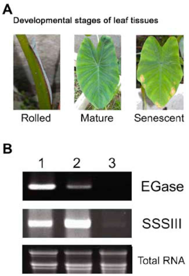
Taro (Colocasia esculenta var. esculenta) tubers were harvested during developmental stages based on their
fresh weight (i.e. 100 g, 300 g, 600 g, and 1000 g) and leaf
growth (i.e., young [rolled], mature, and senescent). The
collected leaves and tubers were frozen immediately in liquid nitrogen, lyophilized, and then stored at -20°C until required.
RNA isolation
RNA was prepared by the method described previously (Lin and Jeang, 2005). The quality and quantity of RNA were determined by spectrophotometry and agarose gel electrophoresis (Sambrook and Russell, 2001). Finally, RNA was stored at -70°C until required. The mRNA for gene cloning was purified with OligotexTM (Qiagen, Valencia, CA) according to the manufacturer's protocol.
Taro EGase cDNA cloning
A s c hema tic re pres entatio n o f our clo ni ng strategy is shown in Figure 1A. The full-length cDNA of taro EGase was cloned by combining RT-PCR and 5'-RACE. 3'-RACE using the primers
P 1 ( G C C G T T G G A C T T AT C G T G C ) a n d P 2 (GAC TC GAGT C GAC A T C G) using the method
described by Frohman et al. (1 988). The template used in 5'-RACE was prepared with oligo-dT primer by a SMART™ RACE cDNA amplification kit (BD Biosciences Clontech, Palo Alto, CA) following the
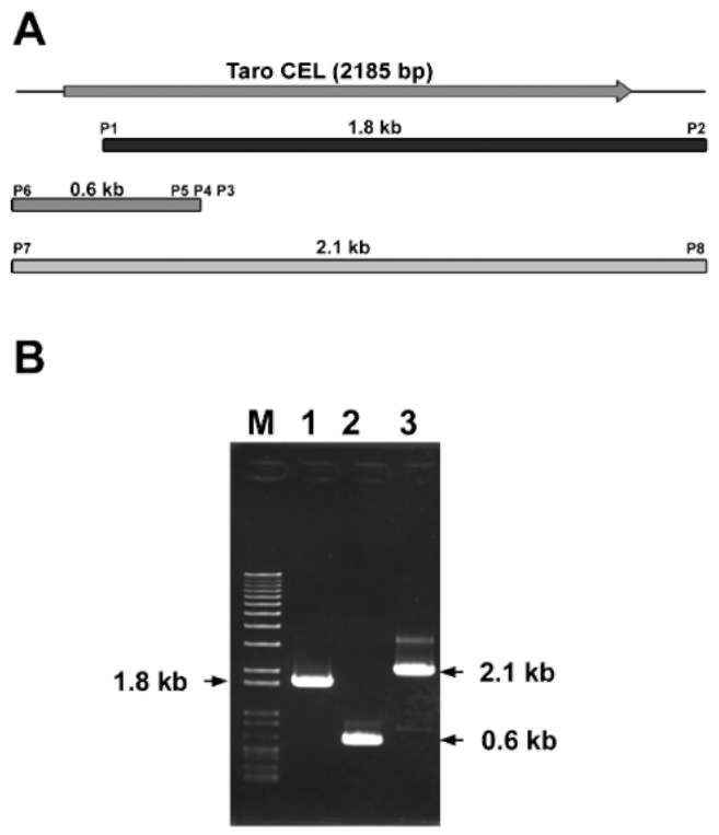
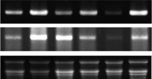
Figure 1. (A) Strategy for taro EGase cDNA cloning. The topmost diagram shows the full length cDNA. The boxes represent cDNAs amplified by RT-PCR and RACE; (B) Agarose gel electrophoresis of RACE and RT-PCR products of EGase. The partial EGase cDNAs were amplified by RT-PCR using P1 and P2 primers (lane 1); The product of 5' SMART- RACE was amplified using P3, P4, P5 and P6 primers (lane 2); The full-length of EGase cDNA was generated by RT-PCR using P7 and P8 primers (lane 3); Lane M: 100 bp ladder (MBI).
LIN et al. ― Cloning and characterization of taro EGase
381
manufacturer's procedure. Then it was subjected to PCR
with primers P4 (CCCCTCCAGGAGACGTTGTTG T), P5 (CCCAGA GCGCTGGGCGTTGAA), and P6 (AAGCAGTGGTATCAACGCAGAGTA). The final
full length cDNA of taro EGase was amplified with
the primers P7 (GAGGAGGAGCTGGTTGAAGGT) and P8 (CCAGCCTTTGGAATTGGGAACA) using
Platinum Pfx DNA polymerase (Invitrogen) following the manufacturer's protocol. All PCR products were sequenced after cloning into pGEM-T-Easy vector (Promega, Madison, WI), and pGEGa, pGEG5' , and pGEGFL were generated, respectively, in this cloning process. The sequences of taro EGase have been deposited in GenBank under Accession No. EF552587.
Expression, production, and purification of recombinant EGase in E. coli
wrap and imaged by a Typhoon 9200 Imager (Amersham Biosciences).
RESULTS
Cloning and expression of taro EGase
A partial EGase cDNA of 1.8 kb (Figure 1 B) was unpurposely obtained during the gene cloning process for soluble starch synthase. In order to obtain the full-length cDNA, a 5'-terminal cDNA of 0.6 kb was generated by the primers, designed according to the cDNA sequence of the
1.8 kb fragment by SMART-RACE (Figure 1B). Finally,
a near full-length EGase cDNA (2.2 kb) was generated by the primers, designed according to the RT-PCR and RACE products, with Pfx DNA polymerase, which possesses a proofreading function (Figure 1B). All products of RT-PCR and RACE were cloned into pGEM-T Easy vector to generate pGEGa, pGEG5', and pGEGFL. The sequences of all products were also confirmed by sequencing and application of the "BlastX" program. Sequence analysis demonstrated that the related overlap regions of these products matched perfectly, indicating that these products arose from the same transcript.
Direct evidence that the clone EGase encoded one p-1, 4-glucanase was obtained by expressing the gene in E. coli BL21. After Congo red staining, the activities of p-1, 4-glucanase were detected on a CMC plate, where the clear halo could be observed around the loading site of the purified recombinant EGase (Figure 2). This indicated that the taro EGase is an active one and can participate in
The 1.8 kb fragment, encoding the entire coding region of taro Egase, was PCR amplified from pGEGFL using
primers sets P9 (ATGTTCGGGCACAACCCCT)-P10
GGT), with the blunt end / EcoRI site introduced. The product was then cloned into pET29a vector (Novagen, San Diego, CA) to generate pETEG. The inserted gene was driven by T7lac promoter, and fused with a His- tag at its 3' end. The entire taro EGase was produced by 2 mM isopropyl-p-D-thiogalactoside (IPTG) induction from E. coli BL21 (DE3) harbored with pETEG. The cultured cells expressing pETEG were induced by 2 mM IPTG at 37°C for 5 h before being harvested, sonicated, and centrifuged to remove cell debris and yield a crude enzyme extract. The recombinant EGase protein was further purified by Ni- NTA column (Qiagen) according to the manufacturer's method for the determination of cellulase activities.
Zymogram assay
Zymogram assay for the recombinant EGase was performed by the method of Loopstra et al., 1998. The recombinant EGase was loaded on a 1% agarose plate containing 0.2% carboxymethylcellulose (CMC, Sigma) and stained by Congo red (1 mg/ mL) after incubation at 37°C for 72 h. The protein concentration was determined by the Lowry method (Lowry et al., 1951).
Southern blot analysis
Genomic DNA was prepared by the CTAB based method (Murray and Thompson, 1980). Taro genomic DNAs (10 fig), digested completely with restriction endonucleases, were electrophoresized on 1.0% agarose gel and blotted onto Hybond-N+ membranes (Amersham Biosciences). Nucleic acid hybridization was conducted by the method described previously (Wang et al., 1994). The
1,010 bp fragment of taro Egase (1,009-2,018 bps) labeled
with a-32P- dATP using the Prime-It II random prime labeling system (Stratagene, La Jolla, CA) was denatured and applied to the hybridization reaction. After stringent washing, the membranes were covered with polyethylene
Figure 2. Different amounts of Ni-NTA purified protein were applied onto 1% agarose plates containing 0.2% CMC and stained with Congo red. Spot (a) represents 200 (ig of purified enzyme from the cell harbored with pET29a, and spots (b) to (C) represent 50 (ig, 100 (ig and 200 (ig of purified enzyme from the cell harbored with pETEG.
382
Botanical Studies, Vol. 50, 2009
cellulose metabolism in taro. Similar results have been observed in tomato's Cel3, which encodes an EGase
(Brummell et al., 1997b).
T he yield of recombinant protein was too low to allow further determination of its biochemical properties. This low yield might been caused by the codon usage or secondary structure of mRNA (Lee et al., 1987; Sorensen et al., 1989). For our purpose, we subcloned the ORF of EGase into pGEX-4T-1 expression vector (Amersham Biosciences) and expressed in E. coli Rosetta (Novagen) to improve the yield. More recombinant protein was observed than in the expression system, E. coli BL21/pETEG (data not shown). The GST-EGase fusion protein will be produced and purified, and then a detailed characterization of the expressed proteins can be described.
Characterization of taro EGase cDNA
The taro EGase cDNA is 2,185 bps in length and contains a coding region of 1,854 bps flanked by the 5'- and 3'- untranslated regions of 153 and 177 bp, respectively (Figure 3). The open reading frame, beginning with an ATG codon at position 154-157 and ending with the TGA codon at position 2,008-2,010, encodes 618 residues protein with a theoretical molecular mass of
68,434 Da and pI of 8.8.
The critical motifs of the taro EGase were analyzed by the programs "InterPro Scan" and "ScanProsite" on the ExPASY website (Figure 3). The results displayed several unique features of this protein. A "transmembrane region" signature was present between amino acids 75 to 95. This motif has often been found in other membrane-anchored EGases (Brummell et al. , 1 997b). This suggests that EGase is a membrane-anchored enzyme involved in cellulose biosynthesis during plant cell wall assembly (Libertini et al., 2004). The signatures o f " G l yc os yl hydro l as e fa mil y 9 " ( Pfa ms Acc. No. PF00759) and "GLYCOSYL_HYDROL_F9_2" were found between amino acids 111 to 586 and 559 to 577, respectively (Figure 3), indicating that the taro EGase is a member of the glycoside hydrolase family 9, which is reported to possess the activity of endoglucanase or cellobiohydrolase (Henrissat and Bairoch, 1993, 1996). A "six-hairpin glycosidase" signature (Pfams Acc. No. SSF48208), located between amino acids 108 to 591, was proposed to be the catalytic domain of EGase (Itoh et al., 2006). In addition, the EGase also harbored 8 putative N-glycosylation sites, located at Asn 136, 152, 218, 324, 345, 408, 425 and 567, and a Pro- rich region located in the C- terminal region.
The taro EGase displays an identity of 76, 78, 78, 78,
79 and 82% and a similarity of 86, 87, 88, 88, 90 and 91%
with the membrane-anchored EGase from bread wheat, tomato, pear, barley, aspen tree, and cotton, respectively. This suggests that the EGase is highly conserved in plant species and has an important function in the growth and development of plants.
To date, 25 Arabidopsis EGases have been published
Figure 3. The nucleotide sequences of taro EGase cDNA. The "transmembrane— region" signature (at amino acids 75-95) and "GLYCOSYL_HYDROL F9_2" signature (at amino acids 559-577) are underlined. The "Six-hairpin glycosidase" signature (at amino acids 108-591) is shown in italic. The proline rich region was showed in boxed. The potential poly-adenylation sites are under double line.
in the database and grouped into three subfamilies, the a, p and y subfamilies, respectively, and the putative function of these subfamilies also have been described (Levy et al., 2002). In this work, the phylogenetic analysis shows that the taro EGase belongs to the y subfamily and that it is closely related to the KOR and KOR3 genes of Arabidopsis (Figure 4). The amino acid sequences of taro EGase displayed 76% and 70% identity with Arabidopsis KOR and KOR3, respectively, suggesting that the taro EGase is an ortholog of Arabidopsis KOR and may have a similar function in plants.
Determination of copy number of EGase in taro
After full-length EGase cDNA cloning, Southern hybridization was applied to examine the copy number of EGase in the taro genome (Figure 5). Genomic DNAs digested with BamHI-, BglII-, EcoRI- and EcoRV- were
LIN et al. ― Cloning and characterization of taro EGase
383
1 2 3 4 5 6
Total RNA
Figure 6. Analysis of the expression of taro EGase in tubers and leaves by RT-PCR. Lane 1: 100 g tuber; lane 2: 300 g tuber; lane 3: 600 g tuber; lane 4: the upper part of 1000 g tuber; lane 5: the bottom part of 1000 g tuber; and lane 6: the rolled leaf of the plant with 1000 g tubers.
probed with cEGase3', fragments of 3.9 kb, 7 kb, 13 kb, and 15 kb were observed, respectively, except that two hybridization signals at 8 kb and 0.6 kb were shown in the HindIII - digested sample. The 0.6 kb fragment matched well with the predicted size in the restriction map of taro EGase cDNA, and this indicates that the EGase is a single copy gene in the taro genome.
Expression of EGase gene during development in taro
The temporal expression of taro EGase during tuber development was monitored by RT-PCR (Figure 6). The EGase transcript was expressed in all the developmental stages of tuber with the highest levels in the 300 g tubers, in the upper part of the 1,000 g tubers and in the rolled leaves. Relatively low expression was found in the bottom part of the 1,000 g tubers. In general, the 300 g tubers and the upper portion of larger tubers undergo more rapid elongation than do tubers in other developmental stages. A similar temporal expression pattern also was observed during leaf development (Figure 7). The EGase transcript was found in all stages of leaf development, with the highest levels in the rolled young leaves undergoing rapid growth, and lower levels in the mature and senescent leaves. Based on the expression patterns of taro EGase in leaf and tuber, it is reasonable to infer that the EGase may play an important role in the cell elongation of taro.
DISCUSSION
A taro EGase cDNA of 2.2 kb was isolated using a RACE cloning strategy in this work. It encodes an EGase which bears a high similarity (86-91%) to the membrane-anchored ones from other species. A transmembrane region was also found to be located between amino acids 75 to 95 by an "InterPro Scan" analysis. Phylogenetic analysis revealed that 25 members of the Arabidopsis EGase family could be divided into the a, p and y subfamilies (Libertini et al., 2004). The taro EGase was found to belong to the y subfamily, one group of the membrane-anchor EGases. These results indicated that the
Figure 4. Relationship of the primary amino acid sequences between Arabidopsis and taro EGase. The dendrogram was generated by the program DRAWTREE in SDSC work bench web site (http://seqtool.sdsc.edu/CGI/BW.cgi). All sequences of EGase were obtained from GenBank/EMBLE/DDBJ.
Figure 5. Southern blot analysis of taro genomic DNA. Ten micrograms genomic DNA was digested with restriction enzyme and subjected to Southern blot analysis. Lane 1: Bam HI; lane 2: Bgl II; lane 3: EcoRI; lane 4: EcoRV; and lane 5: HindIII digested. Hybridization was carried out using the 32P- labeled 1.0 kb of cEGase3' amplified from pGEMTEGFL.
384
Botanical Studies, Vol. 50, 2009
The Arabidopsis KOR gene has been identified by the dwarf mutant KORRIGAN and found to be involved in cytokinesis, pectin metabolism in the primary cell wall, and cell elongation (Nicol et al., 1998; His et al., 2001; Lane et al., 2001). On the other hand, the oilseed rape Cel16, encodes a KOR-like EGase and has been proposed to be involved in cell division and in cell wall assembly during growth (Molhoj et al., 2001b).
Southern blotting confirmed that only one copy of the EGase is present in the taro genome. This observation is unique because several copies of KOR-like EGases have been reported to exist in the A. thaliana or O. sativa genomes (Nicol et al., 1998; Molhoj et al., 2001a; Yoshida et al., 2006). This finding indicated that the cellulose metabolisms of Arabidopsis, rice, and taro may have significant differences.
The transcripts of taro EGase were highly accumulated in 300 g tubers, the upper part of 1000 g tubers, and rolled leaves. The 300 g and 1000 g taro plant tubers form about 6 and 8 months after planting, respectively, and tubers displayed a linear growth rate under the planting conditions in Taiwan. At the same time, the rolled leaves of taro have almost reached their maximum leaf area. These findings suggested that taro EGase is highly expressed in tissues undergoing rapid growth, indicating that the taro EGase is involved in the regulation of plant growth. A similar observation was also made about another non-KOR like EGase, Arabidopsis Cel1 (Milligan and Gasser, 1995; Brummell et al., 1997a; Shani et al., 1997). Curiously, the temporal expression profile of the EGase resembled that of soluble starch synthase III. This revealed that cellulose and starch metabolisms may undergo the same regulation in taro plants. However, this needs to be confirmed by more experimental evidence.
This is the first report showing that the expression a taro EGase, an ortholog of Arabidopsis KOR, is involved in the regulation of plant growth and linked with starch metabolism. While multiple EGases have been identified in higher plants, the biochemical and physiological function of each individual EGase remains to be investigated. Identification in this study of the EGase gene, which encodes a novel endo-1,4-p-D-glucanase accumulating in tissues undergoing rapid growth, provides an opportunity to fill in the gaps and define its precise functional role in the metabolism of cellulose in taro.
Acknowledgements. This manuscript is dedicated to Professor Chii-Ling Jeang (1942-2007), of the Department of Food Science and Biotechnology, National Chung Hsing University, Taichung, Taiwan, for her initiation of this research. We also thank the National Science Council of Taiwan for its financial support (NSC91-2313-B-005-079).
Figure 7. Analysis of the expression of taro EGase in leaf of
the plant with 1000 g tubers by RT-PCR. Lane 1: the rolled leaf;
lane 2: the mature leaf; and lane 3: the senescent leaf.
cloned EGase is a membrane-anchored one that exists in taro plants. The specific function of membrane-anchored EGases is currently unknown, but they are located at the plasma membrane and participate in cellulose metabolism at the inner layers of the cell wall (Brummell et al., 1997b; Nicol et al., 1998; Del Campillo, 1999; Molhoj et al.,
2001a).
The phylogenetic analysis revealed that, except for one member of the y subfamily, the taro EGase was clustered more closely with Arabidopsis KOR and KOR3. A proline rich region was also found in the C-terminal of taro EGase and conserved in all KOR-like EGases. These results suggested that the taro EGase is a KOR-like enzyme. Furthermore, alignment analysis demonstrated
that it shares more identity with KOR (76%) than KOR3
(70%). On the other hand, the temporal expression profiles showed that the transcripts of taro EGase could be found in all tissues during various developmental stages. A similar expression pattern was also found in Arabidopsis KOR (Molhoj et al., 2001a). These findings strongly suggested that taro EGase is an ortholog of Arabidopsis KOR and may play some similar roles.
LITERATURE CITED
Beguin, P. 1990. Molecular biology of cellulose degradation.
Annu. Rev. Microbiol. 44: 219-248.
LIN et al. ― Cloning and characterization of taro EGase
385
Beguin, P. and J.P. Aubert. 1994. The biological degradation of cellulose. FEMS Microbiol. Rev. 13: 25-58.
Bisaria, V. S. and S. Mishra. 1 989. Regulatory aspects of cellulase biosynthesis and secretion. Crit. Rev. Biotechnol. 9: 61-103.
Brummell, D.A., C.R. Bird, W. Schuch, and A.B. Bennett.
1997a. An endo-1,4-p-glucanase expressed at high levels in rapidly expanding tissues. Plant Mol. Biol. 33: 87-95.
Brummell, D.A., C. Catala, C.C. Lashbrook, and A.B. Bennett. 1997b. A membrane-anchored E-type endo-1,4-p-glucanase is localized on Golgi and plasma membranes of higher plants. Proc. Natl. Acad. Sci. USA 94: 4794-4799.
Byrne, K.A., S.A. Lehnert, S.E. Johnson, and S.S. Moore. 1999. Isolation of a cDNA encoding a putative cellulase in the red claw crayfish Cherax quadricarinatus. Gene 239: 317-324.
Catala, C., J.K. Rose, and A.B. Bennett. 1997. Auxin regulation and spatial localization of an endo-1,4-p-D-glucanase and
a xyloglucan endotransglycosylase in expanding tomato
hypocotyls. Plant J. 12: 417-426. Christoffersen, R.E., M.L. Tucker, and G.G. Laties. 1984.
C e l lulase gene expre s s io n in repining avoc ado fruit: the accumulation of cellulase mRNA and proteins as demonstrated by cDNA hybridization and immunodetection.
Plant Mol. Biol. 3: 385-391.
Del Campillo, E. 1999. Multiple endo-1,4-P-D-glucanase (cellulase) genes in Arabidopsis. Curr. Top. Dev. Biol. 46:
39-61.
Del Campillo, E. and A.B. Bennett. 1996. Pedicel breakstrength and cellulase gene expression during tomato flower
abscission. Plant Physiol. 111: 813-820.
Ferrarese, L., L. Trainotti, P. Moretto, P. Polverino de Laureto, N. Rascio, and G. Casadoro. 1995. Differential ethylene-inducible expression of cellulase in pepper plants. Plant
Mol. Biol. 29: 735-747. Frohman, M.A., M.K. Dush, and G.R. Martin. 1988. Rapid
production of full-length cDNAs from rare transcripts: amplification using a single gene-specific oligonucleotide primer. Proc. Natl. Acad. Sci. USA 85: 8998-9002.
Henrissat, B. and A. Bairoch. 1 993. New families in the classification of glycosyl hydrolases based on amino acid sequence similarities. Biochem. J. 293 (Pt 3): 781-788.
Henrissat, B. and A. Bairoch. 1996. Updating the sequence-based classification of glycosyl hydrolases. Biochem. J. 316
(Pt 2): 695-696.
Henrissat, B., M. Claeyssens, P. Tomme, L. Lemesle, and J.P. Mornon. 1989. Cellulase families revealed by hydrophobic cluster analysis. Gene 81: 83-95.
His, I. , A. Driouich, F. Nicol, A. Jauneau, and H. Hofte. 2001. Altered pectin composition in primary cell walls of korrigan, a dwarf mutant of Arabidopsis deficient in a membrane-bound endo-1,4-p-glucanase. Planta 212: 348-358.
Itoh, T., W. Hashimoto, B. Mikami, and K. Murata. 2006. Crystal
structure of unsaturated glucuronyl hydrolase complexed
with substrate: molecular insights into its catalytic reaction
mechanism. J. Biol. Chem. 281: 29807-29816.
Kemmerer, E.C. and M.L. Tucker. 1994. Comparative study of cellulases associated with adventitious root initiation, apical buds, and leaf, flower, and pod abscission zones in soybean. Plant Physiol. 104: 557-562.
Knowles, J., P. Lehtovaara, and T. Teeri. 1987. Cellulase families and their genes. Trends Biotechnol. 5: 255- 261.
Lane, D.R., A. Wiedemeier, L. Peng, H. Hofte, S. Vernhettes,
T. Desprez, C.H. Hocart, R.J. Birch, T.I. Baskin, J.E.
Burn, T. Arioli, A.S. Betzner, and R. E. Williamson. 2001. Temperature-sensitive alleles of RSW2 link the KORRIGAN endo-1,4-p-glucanase to cellulose synthesis and cytokinesis in Arabidopsis. Plant Physiol. 126:
278-288.
Lashbrook, C.C. , C. Gonzalez-Bosch, and A. B. Bennett. 1994. Two divergent endo-p-1,4-glucanase genes exhibit overlapping expression in ripening fruit and abscising flowers. Plant Cell 6: 1485-1493.
Lee, N., S.Q. Zhang, J. Cozzitorto, J.S. Yang, and D. Testa. 1987. Modification of mRNA secondary structure and alteration of the expression of human interferon alpha 1 in
Escherichia coli. Gene 58: 77-86.
Levy, I., Z. Shani, and O. Shoseyov. 2002. Modification of polysaccharides and plant cell wall by endo-1,4-p-glucanase and cellulose-binding domains. Biomol. Eng. 19: 17-30.
Libertini, E. , Y. Li, and S. J. McQueen-Mason. 2004.
Phylogenetic analysis of the plant endo-p-1,4-glucanase
gene family. J. Mol. Evol. 58: 506-515.
Lin, D.G. and C.L. Jeang. 2005. Cloning, expression, and characterization of soluble starch synthase I cDNA from taro (Colocasia esculenta var. esculenta). J. Agric. Food Chem. 53: 7985-7990.
Loopstra, C.A., A. Mouradov, A. Vivian-Smith, T.V. Glassick,
B.V. Gale, S.G. Southerton, H. Marshall, and R.D. Teasdale.
1998. Two pine endo-p-1,4-glucanases are associated with rapidly growing reproductive structures. Plant Physiol. 116:
959-967.
Lowry, O.H., N.J. Rosebrough, A.L. Farr, and R.J. Randall. 1951. Protein measurement with the Folin phenol reagent. J.
Biol. Chem. 193: 265-275.
Milligan, S.B. and C.S. Gasser. 1995. Nature and regulation of pistil-expressed genes in tomato. Plant Mol. Biol. 28:
691-711.
Molhoj, M., B. Jorgensen, P. Ulvskov, and B. Borkhardt. 2001a. Two Arabidopsis thaliana genes, KOR2 and KOR3, which encode membrane-anchored endo-1,4-p-D-glucanases, are differentially expressed in developing leaf trichomes and
their support cells. Plant Mol. Biol. 46: 263-275. Molhoj, M., B. Johansen, P. Ulvskov, and B. Borkhardt. 2001b.
Expression of a membrane-anchored endo-1,4-p-glucanase from Brassica napus, orthologous to KOR from Arabidopsis thaliana, is inversely correlated to elongation in light-grown
plants. Plant Mol. Biol. 45: 93-105.
386
Botanical Studies, Vol. 50, 2009
Murray, M.G. and W.F. Thompson. 1980. Rapid isolation of high molecular weight plant DNA. Nucl. Acids Res. 8: 4321-4325.
Nicol, F., I. His, A. Jauneau, S. Vernhettes, H. Canut, and H. Hofte. 1998. A plasma membrane-bound putative endo-1,4-p-D-glucanase is required for normal wall assembly and cell
elongation in Arabidopsis. EMBO J. 17: 5563-5576.
Peng, L. , Y. Kawagoe, P. Hogan, and D. Delmer. 2002. Sitosterol-p-glucoside as primer for cellulose synthesis in plants. Science 295: 147-150.
Robert, S., G. Mouille, and H. Hofte. 2004. The mechanism and
regulation of cellulose synthesis in primary walls: lessons from cellulose- deficient Arabidopsis mutants. Cellulose 11: 351-364.
Rosso, M.N., B. Favery, C. Piotte, L. Arthaud, J.M. De Boer, R.S. Hussey, J. Bakker, T.J. Baum, and P. Abad. 1999.
Isolation of a cDNA encoding a p-1,4-endoglucanase in the root-knot nematode Meloidogyne incognita and expression analysis during plant parasitism. Mol. Plant Microbe.
Interact. 12: 585-591.
Sambrook, J. and D.W. Russell. (eds.) 2001. Molecular Cloning: A Laboratory Manual, 3rd edn, Cold Spring Harbor Laboratory, New York.
Shani, Z., M. Dekel, G. Tsabary, and O. Shoseyov. 1997.
Cloning and characterization of elongation specific endo-1,4-p-glucanase (cel1) from Arabidopsis thaliana. Plant
Mol. Biol. 34: 837-842.
Shoseyov, O., Z. Shani, and E. Shpigel. 2001. Transgenic Plants
of Altered Morphology (US6184440).
Sorensen, M.A., C.G. Kurland, and S. Pedersen. 1989. Codon usage determines translation rate in Escherichia coli. J.
Mol. Biol. 207: 365-377.
Szyjanowicz, P.M., I. McKinnon, N.G. Taylor, J. Gardiner, M.C. Jarvis, and S.R. Turner. 2004. The irregular xylem 2 mutant is an allele of korrigan that affects the secondary cell wall of Arabidopsis thaliana. Plant J. 37: 730-740.
Taylor, J.E., S.A. Coupe, S. Picton, and J.A. Roberts. 1994.
Characterization and accumulation pattern of an mRNA encoding an abscission-related p-1,4-glucanase from leaflets of Sambucus nigra. Plant Mol. Biol. 24: 961-964.
Tsabary, G., Z. Shani, L. Roiz, I. Levy, J. Riov, and O. Shoseyov. 2003. Abnormal 'wrinkled' cell walls and retarded development of transgenic Arabidopsis thaliana plants expressing endo-1,4-p-glucanase (cell) antisense. Plant Mol.
Biol. 51: 213-224.
Tucker, M.L., R. Sexton, E. Del Campillo, and L.N. Lewis. 1988. Bean abscission cellulase: characterization of a cDNA clone and regulation of gene expression by ethylene and
auxin. Plant Physiol. 88: 1257-1262. Wang, C.S., J.J. Todd, and L.O. Vodkin. 1994. Chalcone
synthase mRNA and activity are reduced in yellow soybean seed coats with dominant I alleles. Plant Physiol. 105:
739-748.
Watanabe, H., H. Noda, G. Tokuda, and N. Lo. 1998. A cellulase
gene of termite origin. Nature 394: 330-331.
Yoshida, K., N. Imaizumi, S. Kaneko, Y. Kawagoe, A. Tagiri, H. Tanaka, K. Nishitani, and K. Komae. 2006. Carbohydrate-binding module of a rice endo-p-1,4-glycanase, OsCel9A, expressed in auxin-induced lateral root primordia, is post-
translationally truncated. Plant Cell Physiol. 47: 1555-1571.
Zuo, J., Q.W. Niu, N. Nishizawa, Y. Wu, B. Kost, and N.H. Chua. 2000. KORRIGAN, an Arabidopsis endo-1,4-p-
glucanase, localizes to the cell plate by polarized targeting and is essential for cytokinesis. Plant Cell 12: 1137-1152.
LIN et al. ― Cloning and characterization of taro EGase
387
檳榔心芋內切型纖維素分解酵素基因之選殖及其在快速生長葉
片與塊莖之表現分析
林大鈞1,2,3,4  張嘉佑2  邱秀英3  宋濟民3  蔣啟玲2  王強生1
1國立中興大學農藝學系

2國立中興大學食品暨應用生物科技學系

3弘光科技大學食品暨應用生物科技學系

4環球技術學院生物科技學系
本研究利用RT-PCRrapid amplification of cDNA ends (RACE)的方法,自檳榔心芋葉片選殖內切
型纖維素分解酵素(EGase) cDNA EGase 以單一拷貝基因存在芋基因體中,其cDNA1,854 bp ,包
618個胺基酸之閱讀框架,理論分子量為 68,434 Da pI 值為 8.8 。此一芋EGase重組蛋白質在 CMC
平板培養基上,具有明顯P-1,4-glucanase活性,顯示其具纖維素代謝的功能。親緣分析結果顯示,芋
EGasey 亞家族成員之一,並與阿拉伯芥KORRIGAN基因(KOR)互為異種同源基因。推論芋EGase
基因應參與細胞的胞質分裂、主細胞壁的基丁質代謝及細胞的伸展等生理現象。另一方面,此EGase
300公克塊莖、1000公克塊莖上部及捲葉等快速生長的組織中大量表現,此一表現模式與快速生長
的芋塊莖中之可溶性澱粉合成酶III基因相似,這些結果顯示,芋EGase基因應在芋的快速生長機制中
扮演重要的角色。
關鍵詞:(Xolocasia esculenta var. esculenta);內切型纖維素分解酵素;KORRIGAN ;快速生長;植
物細胞壁組裝。