Botanical Studies (2010) 51: 37-44.
BIOCHEMISTRY
Monodehydroascorbate reductase cDNA from sweet potato: expression and kinetic studies
Chih-Yu HUANG1,5, Lisa WEN2'5, Rong-Huay JUANG3,5, Dey-Chyi SHEU4, and Chi-Tsai LIN1*
1Institute of Bioscience and Biotechnology and Center for Marine Bioscience and Biotechnology, National Taiwan Ocean University, 2 Pei-Ning Rd, Keelung 202, Taiwan
2Department of Chemistry, Western Illinois University, 1 University Circle, Macomb, IL 61455-1390, USA
3Department of Biochemical Science and Technology, National Taiwan University, Taipei 106, Taiwan
4Department of Bioengineering, Tatung University, Taipei 104, Taiwan 5Footnote: contributed equally to this work.
(Received February 2, 2009; Accepted May 14, 2009)
ABSTRACT. A cDNA encoding a putative monodehydroascorbate reductase (MDAR) was cloned from sweet potato. The deduced protein showed high level of sequence homology with MDARs from other plants or related family from bacteria (2380%). A 3-D homology structure was created for this MDAR. Functional sweet potato MDAR was expressed and purified. The purified enzyme showed an active monomelic form on a 10% native PAGE. The protein's half-life of deactivation at 70°C was 12.4 min, and its thermal inactivation rate constant K was 5.6 x 10⁻² min-1. The enzyme was stable in a broad pH range from 6.0-10.0, and in the presence of 0.8 M imidazole. The K m value for monodehydroascorbate (MDA) and NADH were 21.1 and 39.7 [iM, respectively.
Keywords: Sweet potato (Ipomoea batatas [L.] Lam); 3-D homology structure; Expression; Monodehydroascorbate (MDAR).
Abbreviations: AsA, ascorbate; MDA, monodehydroascorbate; MDAR, monodehydroascorbate reductase; 3-D, Three-dimensional; IPTG, isopropyl p-D-thiogalactopyranoside; FR, ferredoxin reductase.
INTRODUCTION
In both plant and animal metabolism, a major area of research focus on ascorbate (AsA) is its antioxidant properties. Evidence has shown that AsA can protect plants and mammals against oxidative stress. In most cases monodehydroascorbate (MDA) is produced as the primary oxidation product of the effect of AsA. In plants, the reaction catalyzed by AsA peroxidase is a major source of MDA, which scavenges hydrogen peroxide (Hossain et al., 1984). AsA oxidase catalyzes the univalent oxidation of AsA to MDA by dioxygen. Superoxide ('(V) and hydroxyl radical can directly oxidize AsA to MDA, other radicals such as aminoxy, peroxy, phenoxy and thiyl radicals also are able to oxidize AsA to generate MDA (Bielski, 1982; Forni et al., 1984). To maintain the antioxidant activity of AsA, the regeneration of AsA from MDA is indispensable. Monodehydroascorbate reductase (MDAR, EC 1.6.5.4) is responsible for the reduction of MDA to AsA and
*Corresponding author: E-mail: B0220@mail.ntou.edu.tw; Tel: 886-2-24622192 ext. 5513; Fax: 886-2-24622320.
increases reduced form of AsA. An important source of AsA is the recycling in which MDAR catalyzes conversion of MDA to AsA using NAD(P)H as a reducing agent.
Sweet potato is widely distributed in Taiwan, and it is considered as one of the cash crops sustaining the agricultural economy. Therefore, this local crop is especially highly regarded in Taiwan. S everal components from sweet potato have been shown to exhibit antioxidative effects (Lin et al., 1995; Huang et al., 2004a, b; Hou et al., 2004, 2005; Huang et al., 2007a; Jiang et al., 2008). Although contents of sweet potato may have potential medical applications (Huang et al., 2007b), few scientific studies have been reported. Humans are unable to synthesize AsA. This motivates us to search active components from tuberous root of sweet potato for use. Here, we report cloning of a putative MDAR cDNA from sweet potato. The coding region of the cDNA was introduced into an E. coli expression system. The MDAR was expressed and showed high levels of activity in catalyzing MDA to AsA. The recombinant enzyme has been purified and its properties investigated.
38
Botanical Studies, Vol. 51, 2010
MATERIALS AND METHODS
RNA extraction from sweet potato and cDNA synthesis
Young growing roots (called pencil roots) of sweet potato [Ipomoea batatas [L.] Lam. cv. Tainong 57] were collected from the local farm in Yang-Ming Mountain (Kindly provided by Prof. Rong-Huay Juang: Department of Biochemical Science and Technology, National Taiwan University, Taiwan). Its root (wet weight 3 g) were frozen in liquid nitrogen and ground to powder in a ceramic mortar. PolyA mRNA (24 |xg) was prepared using Straight A's mRNA Isolation System (Novagen, USA). Three |xg of the mRNA was used for 5 -RACE-Ready cDNA and 3 -RACE-Ready cDNA synthesis using Clontech's SMART RACE cDNA Amplification Kit.
Isolation of MDAR cDNA
Using 5 -RACE-Ready cDNA of sweet potato as a template and UPM (universal primer A mix, purchased from BD biosciences) primer and a degenerate primer (5' GRT CMA CRT GYT CMA CTC 3), a 1,085 bp fragment was amplified by PCR. The degenerate primer was designed based on the conserved sequences of MDAR from Pisum sativum (accession no. AY662655), Solanum lycopersicum (accession no. L41345), Physcomitrella patens (accession no. DQ159869), Brassica oleracea (accession no. AB125636) and Spinacia oleracea (accession no. AB063289). The 1,085 bp fragment was subcloned and sequenced. Based on this DNA sequence, a forward primer (5' CAC AGC
TAG CAT AGC AGC 3') near the 3'end of the 1,085
bp fragment was synthesized. The forward primer allowed sequence extension to 3'end of the 1,085 bp fragment. Using 3'-RACE-Ready cDNA of sweet potato as a template, a 859 bp fragment between this forward primer and UPM primer was amplified by PCR. This DNA fragment was subcloned and sequenced. Sequence analysis revealed that the combined sequence of the 1,085 bp and the 859 bp covered an open reading frame of MDAR cDNA (1,548 bp, GenBank accession no. EF118034). The identity of MDAR cDNA clone was assigned by comparing the DNA sequence and the inferred amino acid sequence in various data banks using the basic local alignment search tool (BLAST).
Bioinformatic analysis
The blast program was used to search homologous protein sequences in the nonredundant database at the National Center for Biotechnology Information, National Institutes of Health (http://www.ncbi.nlm.nih.gov/).
Multiple alignments were constructed using ClustalW2 program (http://www.ebi.ac.uk/Tools/clustalw2/index.html). Structural modeling was carried out by using the SWISS-MODEL program (Arnold
MODEL.html) to create a 3-D homology model based on the known X-ray structure of putidaredoxin reductase (PDB code 1q1r) from Pseudomonas putida (Sano et al., 2004). The modeling data was then superimposed with putidaredoxin reductase (putidaredoxin reductase and MDAR belong to same family, although homology is
low) by DeepView Swiss-PdbViewer v3.7 (http://www.expasy.org/spdbv/) (Arnold et al., 2006).
Preparation of expression vector
The coding region of the MDAR cDNA was amplified using gene specific flanking primers. The 5' upstream primer contains NdeI recognition site (5' CAT ATG GCG GGG AAG TCA TTC AA 3 ') and the 3' downstream primer contains XhoI recognition site (5' CTC GAG GAT CTT AGA GGC AAA GGT GA 3'). Using 0.2 |xg of sweet potato cDNA as a template, and 10 pmole each of 5' upstream and 3' downstream primers, a 1.3 kb fragment was amplified by PCR. The fragment was ligated into pCR4-Topo and transformed into E. coli. Plasmid DNA was isolated from a positive clone and digested with NdeI and XhoI. The digestion products were separated on a 1% agarose gel. The 1.3 kb insert was gel purified and subcloned into Ndel and Xhol sites of pET-20b(+) vector (Novagen). The DNA was then transformed into E. coli BL21(DE3)pLysS.
Expression and purification of the recombinant MDAR
The transformed E. coli BL21(DE3)pLysS cells containing the putative MDAR cDNA were grown at 37°C in 20 mL of Luria Bertani medium containing 50 μg/mL ampicillin until A500reached 0.8. Protein expression was induced by addition of isopropyl β-D-
thiogalactopyranoside (IPTG) to a final concentration of 0.5 mM. The culture was incubated at 25°C for an additional 20 h at shaking under 80 rpm, and then the bacterial cells were harvested by centrifugation. Soluble proteins were extracted from the cell pellet with glass beads as described before (Ken et al., 2005). The His-tagged MDAR protein was purified by Ni-NTA affinity chromatography as per the manufacturer's instruction (Qiagen). The purified enzyme (1.5 mL) was dialyzed against 50 mL of 0.03 x PBS containing 0.5% glycerol at 4°C for 4 h. Fresh 0.03 x PBS containing 0.5% glycerol was changed once during dialysis. After dialysis, the recombinant protein was checked by enzyme assay. Protein concentration was determined by a Bio-Rad Protein Assay Kit (Richmond, CA) using bovine serum albumin as a standard.
Molecular mass determination by ESI Q-TOF
The protein sample (50 ig / 50 iL) in 0.003 x PBS containing 0.05% glycerol was shipped to Yao-Hong Biotechnology Company (Taiwan) for molecular mass determination using a ESI Q-TOF mass spectrometry (Micromass, Manchester, England).
HUANG et al. ― Cloning of monodehydroascorbate reductase
39
MDAR activity assay and kinetic studies
MDAR was assayed by measuring MDA-dependent NADH oxidation, and MDA was generated by the ascorbate/ascorbate oxidase system (Arrigoni et al., 1981) with some modification. Activities of the recombinant sweet potato MDAR were tested at 25oC by monitoring the oxidation of NADH at A340. The kinetic was studied using the recombinant MDAR (0.1 |xg) in a total volume of 100 |xL 100 mM potassium phosphate buffer (pH 8.0) and 0.1 U ascorbate oxidase (Sigma) at different concentrations of AsA (0.05 to 0. 4 mM). The initial rate was followed under the conditions of either constant NADH (0. 2 mM) or constant AsA (0.1 mM), with concentrations of NADH varied from 0.025 to 0.045 mM. The absorbance at 340 nm was recorded between 20 sec to 40 sec if NADH concentration is constant or between 10 sec to 20 sec if AsA concentration is constant. The extinction coefficient of NADH at 340 nm is 6.22 x 10³ M-1cm-1. The Km, V and Kcat were calculated from Lineweaver-Burk plots.
The amount of MDA generated was determined by linking to AsA oxidation. The solution contained 100 mM potassium phosphate buffer (pH 8.0), 0.05 to 0.4 mM AsA and 0.1 U ascorbate oxidase. The absorbance at 265 nm was recorded between 20 sec to 40 sec and the absorbance decrement was taken for MDA generation. The extinction coefficient of AsA at 265 nm is 12.59 mM-1cm-1 (Jiang et al., 2008).
MDAR zymogram on a native gel
Purified MDAR was also tested for diaphorase activity by the staining procedure of Kaplan and Beutler (1967) after nondenaturing PAGE (polyacrylamide gel electrophoresis). Sample containing 2 |xg MDAR enzyme were electrophoresed on a 1 0% native gel for 2.5 h at 100 V. Staining was carried out by immersing the gel for 10 min in dark in a solution of
1.3 mM NADH, 1.2 mM 3-[4,5-dimethylthiazol-2-
yl]-2,5-diphenyltetrazolium bromide, and 0.06 mM 2,6-dichlorophenol-indophenol in 0.25 M potassium phosphate buffer (pH 8. 0). The area and intensity of active bands were measured by a computing densitometer (Kodak Gel Logic 100 imaging system).
Enzyme characterization
To test the stability of MDAR under various conditions. Aliquots of the recombinant MDAR sample were used to study the following: (1) Thermal effect. Enzyme sample was heated to 70°C for 2, 4, 8 or 16 min. (2) pH effect. Enzyme sample was adjusted to desired pH by adding a half volume of buffer with
different pHs: 0.2 M citrate buffer (pH 2.5, or 4.0), 0.2
M potassium phosphate buffer (pH 6.0, 7.0 or 8.0) or 0.2 M CAPS buffer (pH 10.0, or 11.0). Each sample was
incubated at 37oC for 1 h. (3) SDS effect. SDS, a protein
denaturing reagent, was added to the enzyme sample to the levels of 0.5, 1, or 2% and incubated at 37oC for 1
h. (4) Imidazole effect. During protein purification, the MDAR enzyme was eluted with imidazole. Therefore, its effect on MDAR was examined. Imidazole was added to the enzyme sample to the levels of 0.2, 0.4, or 0.8 M and incubated at 37oC for 1 h. (5) Susceptibility to digestive proteases. The enzyme was incubated with one-twentieth its weight of trypsin or chymotrypsin at pH 8.0, 37oC for a period of 10, 20 or 40 min. In the chymotrypsin digestion, CaCl2 was added to 5 mM. Aliquots were removed at various time intervals for analysis. After each treatment, the sample studied by MDAR zymogram staining.
RESULTS
Molecular cloning and 3-D homologous modeling of MDAR
A MDAR cDNA clone was isolated and identified putatively as the sweet potato MDAR with the consensus pattern and sequence homology to the
published MDARs. This cDNA (1,548 bp, GenBank
accession no. EF118034) contains an open reading frame which encodes a protein of 434 amino acid residues with a predicted molecular mass of 47 kDa. Figure 1A shows the amino acid sequence alignment of the putative sweet potato MDAR with other MDAR, or FR (ferredoxin reductase, belonging to same family flavin oxidoreductases with MDAR), or PR (putidaredoxin reductase, also an FAD-enzyme) from
several sources. The MDAR shared 80, 79, 24 and 23% sequence identity with the MDAR from CsMDAR (Cucumis sativus, BAA05408), AtMDAR (Arabidopsis thaliana, Q9LFA3), PsFR (Pseudomonas sp. KKS102 ferredoxin reductase, Q52437), and PpPR (Pseudomonas putida putidaredoxin reductase, P16640), respectively. A structural model of MDAR was created based on the known structure of putidaredoxin reductase (PDB code 1q1r) from Pseudomonas putida via S WIS S-MODEL program and was superimposed to obtain better structure alignment (Figure 1B, C) via DeepView Swiss-PdbViewer v3.7 programs. The secondary structure was predicted by the same program and represented as a helices and P strands (Figure 1B).
Expression and purification of recombinant
MDAR
The cDNA was introduced into the E. coli expression system as described in the Materials and Methods section. The total cellular proteins expressed were analyzed by SDS-PAGE (Figure 2). The recombinant MDAR was expressed in high yield, 0.22 mg per 20 mL of culture. The MDAR fusion protein was purified by Ni-NTA affinity chromatography. The results showed that the MDAR protein was purified to homogeneity in a single step. Only monomeric MDAR was detected on SDS-PAGE if the sample buffer was without reducing reagent and were not heated, suggesting that sweet
40 Botanical Studies, Vol. 51, 2010
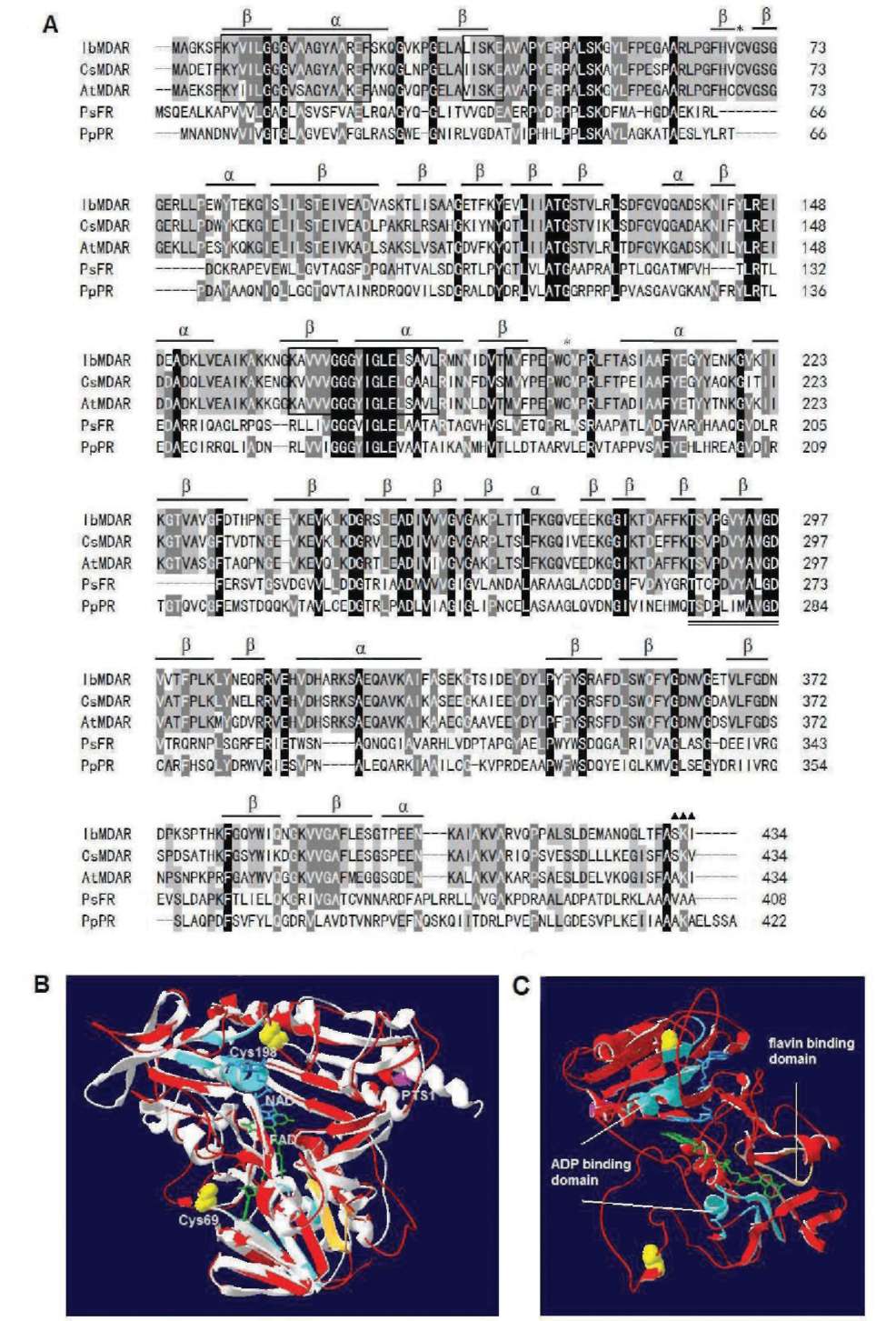
Figure 1. Alignment of the amino acid sequences of MDAR with other organisms and 3-D homology structure. (A) Sequence alignment: IbMDAR (Ipomoea batatas, this study), CsMDAR (Cucumis sativus, BAA05408), AtMDAR (Arabidopsis thaliana, Q9LFA3), PsFR (Pseudomonas sp. KKS102 ferredoxin reductase, Q52437), and PpPR (Pseudomonas putida putidaredoxin reductase, P16640). Identical amino acids in all sequences are shaded black, conservative replacements are shaded gray. Protein secondary structure was predicted by SWISS-MODEL program and represented as a helices and p strands. Stars denote Cys69 and Cys198, triangles denote putative PTS1 (peroxisomal targeting signal) sequence, amino acid domains involved in binding of the ADP moiety of FAD and NAD(P)H are boxed, a conserved 11-amino acid domain (positions 287-297) important in binding of the flavin moiety of FAD is double underlined; (B) 3-D homology structure of MDAR. The structural model of MDAR was created based on the known structure of putidaredoxin reductase (PDB code 1q1r) from Pseudomonas putida via SWISS-MODEL program and was superimposed to obtain better structure alignment and DeepView Swiss-PdbViewer v3.7 programs. Superimposition of MDAR (red) and putidaredoxin reductase (PDB code 1q1r) from Pseudomonas putida (white gray) was shown by using protein solid ribbons. Yellow balls denote Cys69 and Cys198, pink ribbon denotes PTS1 sequence, green denotes FAD, blue denotes NAD; (C) Side view of the same structure. Light blue ribbon denote ADP moiety of FAD and NAD(P)H binding domain, light orange ribbon denotes flavin moiety of FAD binding domain.
HUANG et al. ― Cloning of monodehydroascorbate reductase
41
potato MDAR is monomeric with apparent molecular mass of 47 kDa, as also revealed in zymogram on native PAGE (Figure 4A). This confirms that the active enzyme is monomeric. An ESI Q-TOF of MDAR reveals the presence of only one protein band under the conditions
of 0.003 x PBS (PBS was diluted 330 fold) containing 0.05% glycerol.
Kinetic studies of the purified MDAR
As shown in Figure 3A-B, the Lineweaver-Burk plot of the velocity (1/V) against 1/[MDA] gave the Km = 21.1 |iM, V=244 iM/min, and Kcat = 11750 min-1.
The plot of the velocity (1/V) against 1/[NADH] gave
the Km = 39.7 |iM, Vmax = 286 iM/min, and Kcat = 13768 min-1 for the MDAR, respectively.
Characterization of the purified MDAR
Thermal effect of the recombinant sweet potato MDAR was examined as described in the Materials and Methods section. The enzyme's inactivation kinetics at 70°C fitted the first-order inactivation rate equation ln(E/E) =-K t, where Eand Et represented the original activity and the residual activity after heating for time t, respectively. The thermal inactivation rate constant (Kd) calculated for the enzyme at 70°C was 5.6 x 10⁻² min-1, and the half-life of inactivation was 12.4 min (Figure 4D). The MDAR was stable in a broad pH range from 6.0 to 10.0 (Figure 5A). It lost activity even under 0.5% SDS (Figure 5B). The MDAR showed no decrease in activity with increasing concentration of imidazole to 0.8 M (Figure 5C). The enzyme was resistant to digestion by chymotrypsin (Figure 5D) even at a high enzyme/ substrate (w/w) ratio of 1/20. The results suggested
Figure 2. Purification of the recombinant MDAR. 0.6 mL crude extract was obtained from 20 mL culture. Each sample (15 fiL) was performed on a 10% SDS-PAGE followed by Coomassie blue stain. Lanes M, size markers; 1, crude extract; 2, flowthrough; 3-9, eluted MDAR fractions.
Figure 4. Effect of temperature on the purified MDAR. The enzyme samples heated at 70°C for various time intervals were analyzed by (A) a 10% native PAGE for protein staining; (B) a 10% native PAGE for activity staining; (C) a 10% SDS-PAGE. Staining for protein and activity (2.0 ig /lane). Lanes 1 to 5 (0, 2, 4, 8, 16 min); (D) Plot of thermal inactivation kinetics. The effect of temperature was determined by activity assay. E0 and Et are original activity and residual activity after being heated for different time intervals. The data are mean of three independent experiments.
Figure 3. Double-reciprocal plot of varying AsA, NADH on MDAR activity. The initial rate of the enzymatic reaction was
measured at 0.2 mM NADH with the AsA concentration varied from 0.05 to 0.4 mM as a MDA generation from 16.67 to 73.88 iM in 20 sec to 40 sec (A). At 0.1 mM AsA with the NADH concentration varied from 0.025 to 0.045 mM (B). The Km, Vmax and Kcat were calculated from Lineweaver-Burk plots.
42
Botanical Studies, Vol. 51, 2010
seems likely that the two Cys residues would participate in the transfer of electrons from NADH to catalyze the conversion of MDA to AsA. This assumption is in agreement with Sano et al.'s (1995) report that MDAR from cucumber was inhibited by thiol-modifying reagents. From the structure of Figure 1B, Cys198 (yellow ball) is located near the putative NADH binding domain (blue) as reported in the cucumber MDAR and this Cys residue may really participate in electron transfer through NADH. In addition, from structural point of view that Cys69 and Cys198 are far apart enough and appear to be buried in its 3-D structure and are not expected to form intramolecular disulphide bond or to link the enzyme to form covalent dimers. The results of PAGE analysis (Figures 2, 4A, C) and ESI Q-TOF also suggest the MDAR is existing only as monomer.
It has been reported that trypsin inhibitors (Hou and Lin, 1997), thioredoxin h2 (Huang et al., 2008a),
and defensin (Huang et al., 2008b) of sweet potato also contained MDAR activity, but there were apparent controversies. Enzyme's stability is an important factor for its industrial application. It is evident that the recombinant sweet potato MDAR was stable in a broad pH range from 6-10. It sustained part of the catalytic activity when incubated at 70°C for over ten minutes. The MDAR protein contains potential cleavage sites for trypsin and chymotrypsin, respectively. However, the enzyme appeared to be resistant to digestion by trypsin (data not shown) and chymotrypsin at a high enzyme/ substrate (0.1 fg/2.0 fg) ratio of 1/20 (Figure 5D). This may be due to its rigid or compact structure and limited accessibility of the proteases to the potential cleavage sites under the reaction conditions.
As shown in Table 1, Sano et al. (1995) and Hossain et al. (1984) reported that cucumber and spinach leaf
MDAR had Km values of 4.4 and 7 fM for NADH,
respectively. The sweet potato MDAR has higher Km value for NADH (40 fM). A possible explanation for the discrepancy in their Km values is that sweet potato root is under ground whereas cucumber and spinach leaf are exposed to air, ozone, UV light, among others, and encounter higher oxidative stress. Therefore, the
Figure 5. The stability of purified MDAR under various in vitro treatments. (A) The enzyme samples were incubated in buffers with different pH value at 37°C for 1 h, and then analyzed by activity staining (2.0 fg/lane); (B) The enzyme samples were incubated with SDS of different concentrations at 37°C for 1 h and then subjected to activity staining; (C) The enzyme samples were incubated with different concentrations of imidazole at 37°C for different time intervals and then subjected to activity staining; (D) The enzyme (2.0 fg/lane) activities after treatment with chymotrypsin (0.1 fg in 15 fL proteolysis reaction) measured by activity staining. The data are mean of three independent experiments.
that this MDAR has a rigid structure and the potential cleavage sites were not accessible to the protease under the reaction conditions. 1-Cys peroxiredoxin was used as a positive control in the protease digestion. The peroxiredoxin was degraded within 20 min if treated with either trypsin or chymotrypsin (results not shown).
DISCUSSION
This study reported the first cloning and expression of an important reduction enzyme, MDAR, from sweet potato. The biological active form of the MDAR has been successfully expressed in E. coli. We scanned the MDAR sequence using Swiss-Prot Prosite program to examine potential posttranslational modification sites. Although several potential phosphorylation sites, N-glycosylation sites, and a N-myristoylation site were found, it appears that eukaryotic specific posttranslational modifications are not required for the activity of MDAR as the recombinant protein expressed by E. coli is active.
A lot of oxidoreductases such as dehydroascorbate reductase (Jiang et al., 2008), peroxiredoxins (Wen et al., 2007; Huang et al., 2007), among others, possess Cys residue to catalyze its redox reaction, from the sequence and 3-D structure of MDAR (Figure 1), the enzyme also contains two Cys residues at positions 69 and 198 which are conserved in the enzyme from cucumber. It
Table 1. Kinetic characterization of IbMDAR and other published MDARs.
Km (iM)
Monodehydroascorbate reductase
NADH
Recombinant enzyme of sweet potato (present work)
40
Recombinant enzyme of cucumber (Sano et al., 1995)
4.4
Cucumber fruit (Hossain and Asada, 1985)
4.6
Spinach leaf (Hossain et al., 1984)
7
HUANG et al. ― Cloning of monodehydroascorbate reductase
43
leaf MDAR can use lower concentration of NADH to produce AsA for protection against oxidative stress. However, whether sweet potato contains higher NADH (40 fM) concentration has not been studied in this project.
In conclusion, we have cloned an MDAR cDNA and successfully expressed the enzyme in E. coli. The recombinant enzyme showed high levels of stability under various conditions. The information may be useful in formulating the enzyme as possible antioxidant supplement or construct this cDNA in a vector containing strong promoter, then transfer to other high economic plants for enriching its AsA production.
Acknowledgements. This work was supported partially by the National Science Council of the Republic of China, Taiwan under Grant NSC NSC 97-2313-B-019-001-MY3 to C.-T. Lin.
LITERATURE CITED
Arnold, K., L. Bordoli, J. Kopp, and T. Schwede. 2006. The
SWISS-MODEL workspace: A web-based environment for protein structure homology modelling. Bioinformatics 22: 195-201.
Arrigoni, O., S. Dipiero, and G. Borraccino. 1981. Ascorbate free radical reductase: a key enzyme of the ascorbic acid system. FEBS Lett. 125: 242-244.
Bielski, B. 1982. Ascorbic Acid: chemistry, metabolism and uses. In P.A. Seib and B.M. Tolbert (eds.), American Chem­ical Society. Washington, DC, pp. 81-100.
Forni, L.G., J. Monig, V.O. Mora-Arellano, and R.Wilson. 1984. Thiyl free radicals: direct observations of electron transfer reactions with phenothiazines and ascorbate. J.
Chem. Soc. Perkin Trans. 2: 961-965.
Hossain, M.A., Y. Nakano, and K. Asada. 1984. Monodehydro-ascorbate reductase in spinach chloroplasts and its partici­pation in regeneration of ascorbate for scavenging hydrogen peroxide. Plant Cell Physiol. 25: 385-395.
Hossain, M.A. and K. Asada. 1985. Monodehydroascorbate reductase from cucumber is a flavin adenine dinucleotide
enzyme. J. Biol. Chem. 260: 12920-12926.
Hou, W.C. and Y.H. Lin. 1997. Dehydroascorbate reductase and monodehydroascorbate reductase activities of trypsin inhibitors, the major sweet potato (Ipomoea batatas [L.] Lam) root storage protein. Plant Sci. 128: 151-158.
Hou, W.C., H.J. Chen, C.H. Han, C.Y. Lin, and Y.H. Lin. 2004.
Glutathione peroxidase-like activity of 33 kDa trypsin in­hibitor from roots of sweet potato (Ipomoea batatas (L.) Lam 'Tainong 57'). Plant Sci. 166: 1541-1546.
Hou, W.C., C.H. Han, and H.J. Chen. 2005. Storage proteins of
two cultivars of sweet potato (Ipomoea batatas (L.) Lam) and their protease hydrolysates exhibited antioxidant activ­ity in vitro. Plant Sci. 168: 449-456.
Huang, D.J., H.J. Chen, W.C. Hou, C.D. Lin, and Y.H. Lin.
2004a. Active recombinant thioredoxin h protein with anti-oxidant activities from sweet potato (Ipomoea batatas [L.] Lam 'Tainong 57') storage roots. J. Agri. Food Chem. 52:
4720-4724.
Huang, D.J., C.D. Lin, H.J. Chen, and Y.H. Lin. 2004b. An-tioxidant and antiproliferative activities of sweet potato (Ipomoea batatas [L.] Lam 'Tainong 57') constituents. Bot.
Bull. Acad. Sin. 52: 179-186. Huang, G.J., H.J. Chen, Y.S. Chang, M.J. Sheu, and Y.H. Lin.
2007a. Recombinant sporamin and its synthesized peptides with antioxidant activities in vitro. Bot. Stud. 48: 133-140.
Huang, G.J., M.J. Sheu, H.J. Chen, Y.S. Chang, and Y.H. Lin.
2007b. Growth inhibition and induction of apoptosis in NB4 promyelocytic leukemia cells by trypsin inhibitor from sweet potato storage roots. J. Agric. Food Chem. 55:
2548-2553.
Huang, G.J., H.J. Chen, Y.S. Chang, T.L. Lu, and Y.H. Lin.
2008a. Sweet potato storage root thioredoxin h2 with both dehydroascorbate reductase and monodehydroascorbate reductase activities. Bot. Stud. 49: 1-7.
Huang, G.J., H.C. Lai, Y.S. Chang, M.J. Sheu, T.L. Lu, S. S. Huang, and Y.H. Lin. 2008b. Antimicrobial,
dehydroascorbate reductase, and monodehydroascorbate reductase activities of defensin from sweet potato [Ipomoea batatas (L.) Lam. 'Tainong 57'] storage roots. J. Agric.
Food Chem. 56: 2989-2995. Huang, J. K., C.F. Ken, H.M. Huang, and C.T. Lin. 2007.
Biochemical characterization of a novel 2-Cys peroxiredoxin from Antrodia camphorata. Appl. Microbiol.
Biotechnol. 74: 84-92. Jiang, Y.C., C.Y. Huang, L. Wen, and C.T. Lin. 2008. Dehydro-
ascorbate reductase cDNA from sweet potato (Ipomoea batatas [L.] Lam): expression, enzyme properties, and ki­netic studies. J. Agric. Food Chem. 56: 3623-3627.
Kaplan, J.C. and E. Beutler. 1967. Electrophoresis of red cell NADH and NADPH-diaphorases in normal subjects and patients with congenital methemoglobinemia. Biochem. Biophys. Res. Commun. 29: 605-610.
Ken, C.F., T.M. Hsiung, Z.X. Huang, R.H. Juang, and C.T. Lin. 2005. Characterization of Fe/Mn-superoxide dismutase from diatom Thallassiosira weissflogii: cloning, expression, and property. J. Agric. Food Chem. 53: 1470-1474.
Lin, C.T., M.T. Lin, Y.T. Chen, and J.F. Shaw. 1995. Subunit
interaction enhances enzyme activity and stability of sweet potato cytosolic Cu/Zn-superoxide dismutase purified by a His-tagged recombinant protein method. Plant Mol. Biol.
28: 303-311.
Lin, C.T. and C.Y. Huang. 2006. Characterization of monodehy-droascorbate reductase mRNA from sweet potato. GenBank
Accession EF118034.
Sano, S., C. Miyake, B. Mikami, and K. Asada. 1995. Molecular characterization of monodehydroascorbate radical reductase from cucumber highly expressed in Escherichia coli. J.
Biol. Chem. 270: 21354-21361. Sano, S., Y.N. Kang, H. Shigemizu, N. Morishita, H.J. Yoon, K.
44
Botanical Studies, Vol. 51, 2010
Saito, K. Asada, and B. Mikami. 2004. Crystallization and preliminary crystallographic analysis of monodehydroascor-bate radical reductase from cucumber. Acta Cryst. D Biol.
Crystallogr. 60: 1498-1499.
Wen, L., H.M. Huang, R.H. Juang, and C.T. Lin. 2007. Bio­chemical characterization of 1-Cys peroxiredoxin from Antrodia camphorata. Appl. Microbiol. Biotechnol. 73:
1314-1322.
單去氫抗壞酸還原酶在大腸桿菌中的表現及其生化特性研究
黃智郁1 溫麗莎2 莊榮輝3 許垤棊4 林棋財1
1國立台灣海洋大學生物科技研究所
2美國Western Illinois大學化學系

3國立台灣大學生技系

4大同大學生物工程學系
單去氫抗壞血酸還原酶(monodehydroascorbate reductase, MDAR)在抗氧化中還原單去氫抗壞血酸
(MDA)成抗壞血酸(AsA)。依據甘藷(Ipomoea batatas [L.] Lam)表現庫序列資訊(expressed sequence tag
database)
選殖出MDAR cDNA序列(1,548 bp, EMBL accession no. EF118034)'全長共1,548個核苷酸,
內含轉譯區1302個核苷酸,可轉譯出434個胺基酸的還原酶。經序列比較IbMDAR與其他物種的序列
有很高的相似性,依據已知結構,建立一相似性立體結構(3-D homology structure)。進一歩將其轉譯區
選殖入表現載體pET-20b(+),以大腸桿菌E. coli BL21(DE3)pLysS作爲表現宿主,經親和性管柱純化可
得到具有活性的IbMDAR,其特性在70°C加熱活性降低一半的時間爲12.4分鐘,pH 6.0 ~ 10.0仍然
具有相當的活性。對MDANADHKm値分別爲21.139.7 iM
關鍵詞:甘藷(Jpomoea batatas [L.] Lam);相似性立體結構(3-D homology structure);基因表現;
單去氫抗壞血酸還原酶(monodehydroascorbate reductase, MDAR) °